

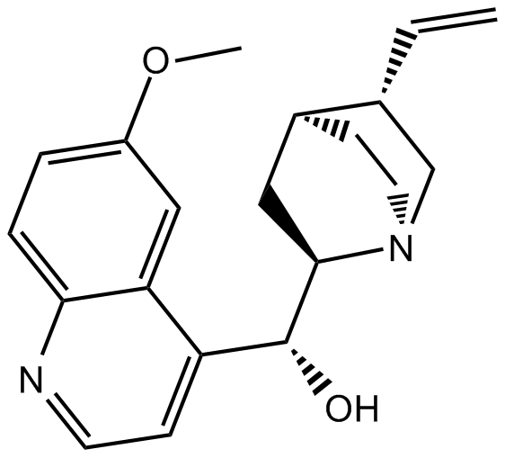
Quinidine (Synonyms: (+)-Quinidine, β-Quinidine) |
カタログ番号GC14264
 One-Click Copy Product Info
|

キニジンは抗不整脈剤です。
Products are for research use only. Not for human use. We do not sell to patients.
Cas No.: 56-54-2
Sample solution is provided at 25 µL, 10mM.
_1-3.$$$39978408@51.jpg');)
_1-9.$$$38538795@65.jpg');)
_1-9.$$$39112701@51.jpg');)
_1-7.$$$37316672@70.jpg');)
_366-372.$$$36198801@70.jpg');)
.$$$38565142@65.jpg');)
_1867-1883.$$$33248023@67.jpg');)
_eads6055.$$$39977546@45.jpg');)
_eadm9805.$$$40080571@45.jpg');)
_eadj3020.$$$39666821@45.jpg');)
_1-20.$$$39757301@28.jpg');)
_1-24.$$$38898113@28.jpg');)
_273-287.$$$36806353@46.jpg');)
_1-16.$$$37156877@44.jpg');)
_1-19.$$$37460805@44.jpg');)
_1291-1307.$$$34518654@46.jpg');)
Quinidine is a class Ia antiarrhythmic agent extracted from cinchona bark, which functions as a K⁺ channel blocker (IC₅₀=19.9µM) and inhibits cytochrome P450db activity. Quinidine reduces automaticity, slows conduction velocity, and prolongs the effective refractory period by inhibiting sodium channels on the cardiomyocyte membrane. Quinidine produces vasodilatory effects through anticholinergic action and α-receptor blockade. Quinidine can be used in research related to atrial fibrillation, atrial flutter, and supraventricular tachycardia[1-4].
In vitro, Quinidine (300µM) was used to treat C6 glioma cells for 48 hours. Quinidine significantly reduced cell proliferation, arrested the cell cycle in the G0/G1 phase, and decreased ornithine decarboxylase (ODC) activity and putrescine concentration[5]. Quinidine (90µM) was used to treat MCF-7 human breast cancer cells for 24–96 hours. Quinidine reduced the expression of the cell cycle marker Ki67 antigen and induced apoptosis and necrosis[6].
In vivo, Quinidine (2.075mg/100g; single dose) was administered intraperitoneally to Swiss mice immediately after Isoproterenol hydrochloride injection. Quinidine significantly reduced the Isoproterenol hydrochloride-induced elevation in serum glutamic-oxaloacetic transaminase (SGOT) levels and cardiac tissue lipid peroxidation, while modulating cardiac antioxidant enzyme activity and counteracting Isoproterenol hydrochloride-induced myocardial ischemic injury[7]. Quinidine (30mg/kg) was administered as a single intraperitoneal injection to NMRl male mice. Quinidine significantly increased the seizure threshold for pentylenetetrazole (PTZ)-induced tonic hind limb extension (THE)[8].
References:
[1] Grace AA, Camm AJ. Quinidine. N Engl J Med. 1998 Jan 1;338(1):35-45.
[2] Alloway JA, Salata MP. Quinidine-induced rheumatic syndromes. Semin Arthritis Rheum. 1995 Apr;24(5):315-22.
[3] Malik C, Ghosh S. Quinidine partially blocks mitochondrial voltage-dependent anion channel (VDAC). Eur Biophys J. 2020 Mar;49(2):193-205.
[4] Yoshitomi S, Takahashi Y, Yamaguchi T, et al. Quinidine therapy and therapeutic drug monitoring in four patients with KCNT1 mutations. Epileptic Disord. 2019 Feb 1;21(1):48-54.
[5] Weiger TM, Colombatto S, Kainz V, et al. Potassium channel blockers quinidine and caesium halt cell proliferation in C6 glioma cells via a polyamine-dependent mechanism. Biochem Soc Trans. 2007 Apr;35(Pt 2):391-5.
[6] Wang S, Melkoumian Z, Woodfork KA, et al. Evidence for an early G1 ionic event necessary for cell cycle progression and survival in the MCF-7 human breast carcinoma cell line. J Cell Physiol. 1998 Sep;176(3):456-64.
[7] Chattopadhyay A, Biswas S, Bandyopadhyay D, et al. Effect of isoproterenol on lipid peroxidation and antioxidant enzymes of myocardial tissue of mice and protection by quinidine. Mol Cell Biochem. 2003 Mar;245(1-2):43-9.
[8] Jamali H, Heydari A. Effect of dextromethorphan/quinidine on pentylenetetrazole- induced clonic and tonic seizure thresholds in mice. Neurosci Lett. 2020 Jun 11;729:134988.
| Cell experiment [1]: | |
Cell lines | C6 glioma cells (rat glioma cell line) |
Preparation Method | C6 glioma cells were maintained in Ham's F-10 medium supplemented with 9% fetal calf serum (FCS) and 1% penicillin/streptomycin at 37°C, 5% CO₂, and 90% humidity. C6 glioma cells were treated with Quinidine at 300µM for 48 hours. |
Reaction Conditions | 300µM; 48h |
Applications | Quinidine significantly reduced cell proliferation with an EC₅₀ of 112µM. Quinidine depolarized the cell membrane by approximately +16.53mV, arrested cells in the G0/G1 phase of the cell cycle, and reduced S-phase. Quinidine decreased intracellular putrescine concentration below detection levels, inhibited ornithine decarboxylase (ODC) activity, reduced polyamine (putrescine) uptake, diminished mitochondrial activity, and increased lactate dehydrogenase (LDH) release. |
| Animal experiment [2]: | |
Animal models | NMRI male mice |
Preparation Method | Mice were intraperitoneally administered a single dose of Quinidine (30mg/kg). Thirty minutes after Quinidine administration, seizures were induced by intravenous infusion of pentylenetetrazole (PTZ) via the tail vein. |
Dosage form | 30mg/kg; i.p.; single injection |
Applications | Quinidine was ineffective in changing the seizure threshold for the onset of myoclonic (MC) twitch. Quinidine significantly increased the threshold dose for the onset of tonic hind limb extension (THE) compared to the saline-treated control group. |
References: | |
| Cas No. | 56-54-2 | SDF | |
| 同義語 | (+)-Quinidine, β-Quinidine | ||
| Chemical Name | (R)-(6-methoxyquinolin-4-yl)((1S,2R,4S,5S)-5-vinylquinuclidin-2-yl)methanol | ||
| Canonical SMILES | O[C@H](C(C1=C2)=CC=NC1=CC=C2OC)[C@@H]3[N@@]4C[C@@H](C=C)[C@@H](CC4)C3 | ||
| Formula | C20H24N2O2 | M.Wt | 324.42 |
| 溶解度 | ≥ 11.95mg/mL in DMSO | Storage | 4°C, protect from light, stored under nitrogen |
| General tips | Please select the appropriate solvent to prepare the stock solution according to the
solubility of the product in different solvents; once the solution is prepared, please store it in
separate packages to avoid product failure caused by repeated freezing and thawing.Storage method
and period of the stock solution: When stored at -80°C, please use it within 6 months; when stored
at -20°C, please use it within 1 month. To increase solubility, heat the tube to 37°C and then oscillate in an ultrasonic bath for some time. |
||
| Shipping Condition | Evaluation sample solution: shipped with blue ice. All other sizes available: with RT, or with Blue Ice upon request. | ||
| Prepare stock solution | |||

|
1 mg | 5 mg | 10 mg |
| 1 mM | 3.0824 mL | 15.4121 mL | 30.8242 mL |
| 5 mM | 616.5 μL | 3.0824 mL | 6.1648 mL |
| 10 mM | 308.2 μL | 1.5412 mL | 3.0824 mL |
Step 1: Enter information below (Recommended: An additional animal making an allowance for loss during the experiment)
 g
μL
g
μL
Step 2: Enter the in vivo formulation (This is only the calculator, not formulation. Please contact us first if there is no in vivo formulation at the solubility Section.)
Calculation results:
Working concentration: mg/ml;
Method for preparing DMSO master liquid: mg drug pre-dissolved in μL DMSO ( Master liquid concentration mg/mL, Please contact us first if the concentration exceeds the DMSO solubility of the batch of drug. )
Method for preparing in vivo formulation: Take μL DMSO master liquid, next addμL PEG300, mix and clarify, next addμL Tween 80, mix and clarify, next add μL saline, mix and clarify.
Method for preparing in vivo formulation: Take μL DMSO master liquid, next add μL Corn oil, mix and clarify.
Note: 1. Please make sure the liquid is clear before adding the next solvent.
2. Be sure to add the solvent(s) in order. You must ensure that the solution obtained, in the previous addition, is a clear solution before proceeding to add the next solvent. Physical methods such as vortex, ultrasound or hot water bath can be used to aid dissolving.
3. All of the above co-solvents are available for purchase on the GlpBio website.
Quality Control & SDS
- View current batch:
- Purity: >98.00% Appearance: A solid
- COA (Certificate of Analysis)
- SDS (Safety Data Sheet)
- Datasheet
Average Rating: 5 (Based on Reviews and 21 reference(s) in Google Scholar.)