

Bufalin (Synonyms: NSC 89595) |
Catalog No.GN10365
 One-Click Copy Product Info
|

Bufalin은 Na+/K+-ATPase 억제제 (α1: Kd = 42.5nM; α2: Kd = 45nM; α3: Kd = 40nM)이다.
Products are for research use only. Not for human use. We do not sell to patients.
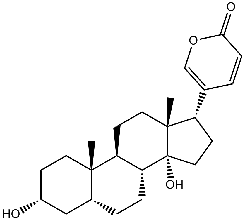
Cas No.: 465-21-4
Sample solution is provided at 25 µL, 10mM.
Bufalin은 Na+/K+-ATPase 억제제 (α1: Kd = 42.5nM; α2: Kd = 45nM; α3: Kd = 40nM)이다. Bufalin은 주로 세포막에서 Na⁺/K⁺-ATPase의 활성을 억제해서 세포내 칼슘 농도를 상승시킨다[2]. Bufalin은 세포 사멸을 유도하고 종양세포의 증식을 억제해서 자가포식을 유발하고 종양 혈관 생성을 억제하는 등 다수의 항종양 기전을 가질 수 있다[3]. Bufalin은 간암, 폐암, 유방암, 백혈병을 포함한 다양한 종양을 치료하그 위해 사용된다[4].
NCI-H640 세포에서 Bufalin (1μM, 2μM, 4μM; 24시간) 처리 후 세포 활성이 현저하게 감소하였다[5]. Ishikawa, SK-OV-3 및 OMC-3 난소암 세포에서 Bufalin (0.1-10ng/mL; 48시간)은 자궁내막암과 난소암 세포의 증식을 억제할 수 있다[6]. 인간 신장암 ACHN 세포에서 Bufalin (0-80nM; 12-72시간)은 PI3K/Akt/mTOR 신호전달 경로를 억제해서 신장세포암의 증식과 전이를 억제한다[7].
키토산에 의해 유도된 발톱 부종 쥐 모델에서 Bufalin (0.15mg/kg, 0.3mg/kg, 0.6mg/kg; ip; 단일 투여) 처리 후 쥐의 발톱 부종이 현저하게 감소했다[8]. HCCLM3 세포 전이성 원위 종양 쥐 모델에서 원위 이식된 종양 조직은 Bufalin (1mg/kg, 1.5mg/kg; ip; 30일) 처리로 인한 괴사와 세포 사멸을 겪었다[9].
References:
[1]. Katz A, Lifshitz Y, Bab-Dinitz E, et al. Selectivity of digitalis glycosides for isoforms of human Na, K-ATPase[J]. Journal of Biological Chemistry, 2010, 285(25): 19582-19592.
[2]. Wu S H, Bau D T, Hsiao Y T, et al. Bufalin induces apoptosis in vitro and has Antitumor activity against human lung cancer xenografts in vivo[J]. Environmental Toxicology, 2017, 32(4): 1305-1317.
[3]. Yin P H, Liu X, Qiu Y Y, et al. Anti-tumor activity and apoptosis-regulation mechanisms of bufalin in various cancers: new hope for cancer patients[J]. Asian Pacific journal of cancer prevention, 2012, 13(11): 5339-5343.
[4]. Lan Y L, Lou J C, Jiang X W, et al. A research update on the anticancer effects of bufalin and its derivatives[J]. Oncology letters, 2019, 17(4): 3635-3640.
[5]. Wu S H, Wu T Y, Hsiao Y T, et al. Bufalin induces cell death in human lung cancer cells through disruption of DNA damage response pathways[J]. The American Journal of Chinese Medicine, 2014, 42(03): 729-742.
[6]. Takai N, Ueda T, Nishida M, et al. Bufalin induces growth inhibition, cell cycle arrest and apoptosis in human endometrial and ovarian cancer cells[J]. International journal of molecular medicine, 2008, 21(5): 637-643.
[7]. Xie J, Lin W, Huang L, et al. Bufalin suppresses the proliferation and metastasis of renal cell carcinoma by inhibiting the PI3K/Akt/mTOR signaling pathway[J]. Oncology letters, 2018, 16(3): 3867-3873.
[8]. Wen L, Huang Y, Xie X, et al. Anti‐inflammatory and antinociceptive activities of bufalin in rodents[J]. Mediators of inflammation, 2014, 2014(1): 171839.
[9]. Zhang Z J, Yang Y K, Wu W Z. Bufalin attenuates the stage and metastatic potential of hepatocellular carcinoma in nude mice[J]. Journal of translational medicine, 2014, 12(1): 57.
| 세포 실험 [1]: | |
세포 라인 | NCI-H640 세포 |
제조 방법 | 12웰 판당 약 2 × 10⁵개의 NCI-H640 세포는 최종 농도가 0 (용매, 0.5% DMSO), 1, 2, 4μM인 Bufalin과 함께 24시간 동안 배양되었습니다. 각 처리로부터의 세포는 PI (5 g/ml)로 염색되었고 우세포 분석법을 통해 분석되었고 세포 생존율이 계산되었습니다. |
반응 조건 | 1μM, 2μM, 4μM; 24h |
응용 분야 | Bufalin 처리 후 세포 활성이 현저하게 감소하였습니다. |
| 동물 실험 [2]: | |
동물 모형 | 키토산에 의해 유도된 발톱 부종 쥐 모델 |
제조 방법 | 항염증 효과는 카라기난 유도된 발톱 부종의 억제를 기반으로 합니다. Bufalin (0.15, 0.3, 0.6mg/kg, ip), 인도메타신 (10mg/kg, ip)및 대체 용매는 카라기난 주사 30분 전에 복강 내 투여되었습니다. 각 쥐는 오른쪽 발톱에 카라기난 (0.1mL, 생리식염수의 1% w/v)의 염상막 주사를 받았습니다. 부종계는 카라기난 주사 전과 후 (1, 2, 4, 6시간) 다양한 시간에 발톱 부종의 부피를 측정하였습니다. 발톱 부종 비율은 카라기난 주사 전에 측정된 발톱 부피의 퍼센트 증가로 계산되었습니다. 쥐의 발은 최종 평가 후에 수집되었습니다. 각 발톱은 발뒤 꿈치뼈 수준에서 잘려서 단백질 자극법으로 사용되었습니다. |
제형 | 0.15mg/kg, 0.3mg/kg, 0.6mg/kg; ip; 단일 주사 |
응용 분야 | Bufalin 처리 후 쥐의 발 부종 부피가 현저하게 감소하였습니다. |
References: | |
| Cas No. | 465-21-4 | SDF | |
| Synonyms | NSC 89595 | ||
| Chemical Name | 5-[(3S,5R,8R,9S,10S,13R,14S,17R)-3,14-dihydroxy-10,13-dimethyl-1,2,3,4,5,6,7,8,9,11,12,15,16,17-tetradecahydrocyclopenta[a]phenanthren-17-yl]pyran-2-one | ||
| Canonical SMILES | CC12CCC(CC1CCC3C2CCC4(C3(CCC4C5=COC(=O)C=C5)O)C)O | ||
| Formula | C24H34O4 | M.Wt | 386.52 |
| Solubility | ≥ 38.7mg/mL in DMSO | Storage | Store at -20°C |
| General tips | Please select the appropriate solvent to prepare the stock solution according to the
solubility of the product in different solvents; once the solution is prepared, please store it in
separate packages to avoid product failure caused by repeated freezing and thawing.Storage method
and period of the stock solution: When stored at -80°C, please use it within 6 months; when stored
at -20°C, please use it within 1 month. To increase solubility, heat the tube to 37°C and then oscillate in an ultrasonic bath for some time. |
||
| Shipping Condition | Evaluation sample solution: shipped with blue ice. All other sizes available: with RT, or with Blue Ice upon request. | ||
| Prepare stock solution | |||

|
1 mg | 5 mg | 10 mg |
| 1 mM | 2.5872 mL | 12.9359 mL | 25.8719 mL |
| 5 mM | 517.4 μL | 2.5872 mL | 5.1744 mL |
| 10 mM | 258.7 μL | 1.2936 mL | 2.5872 mL |
Step 1: Enter information below (Recommended: An additional animal making an allowance for loss during the experiment)
 g
μL
g
μL
Step 2: Enter the in vivo formulation (This is only the calculator, not formulation. Please contact us first if there is no in vivo formulation at the solubility Section.)
Calculation results:
Working concentration: mg/ml;
Method for preparing DMSO master liquid: mg drug pre-dissolved in μL DMSO ( Master liquid concentration mg/mL, Please contact us first if the concentration exceeds the DMSO solubility of the batch of drug. )
Method for preparing in vivo formulation: Take μL DMSO master liquid, next addμL PEG300, mix and clarify, next addμL Tween 80, mix and clarify, next add μL saline, mix and clarify.
Method for preparing in vivo formulation: Take μL DMSO master liquid, next add μL Corn oil, mix and clarify.
Note: 1. Please make sure the liquid is clear before adding the next solvent.
2. Be sure to add the solvent(s) in order. You must ensure that the solution obtained, in the previous addition, is a clear solution before proceeding to add the next solvent. Physical methods such as vortex, ultrasound or hot water bath can be used to aid dissolving.
3. All of the above co-solvents are available for purchase on the GlpBio website.
Quality Control & SDS
- View current batch:
- Purity: >99.50% Appearance: A solid
- COA (Certificate of Analysis)
- SDS (Safety Data Sheet)
- Datasheet
Average Rating: 5 (Based on Reviews and 30 reference(s) in Google Scholar.)