Apatinib Mesylate (Synonyms: YN968D1) |
رقم الكتالوجGC14292 |
يقوم الأباتينيب بحجب انتقال الإشارة الناجمة عن مسار VEGF النازل لتثبيط تشكل الأوعية الدموية الجديدة.
Products are for research use only. Not for human use. We do not sell to patients.
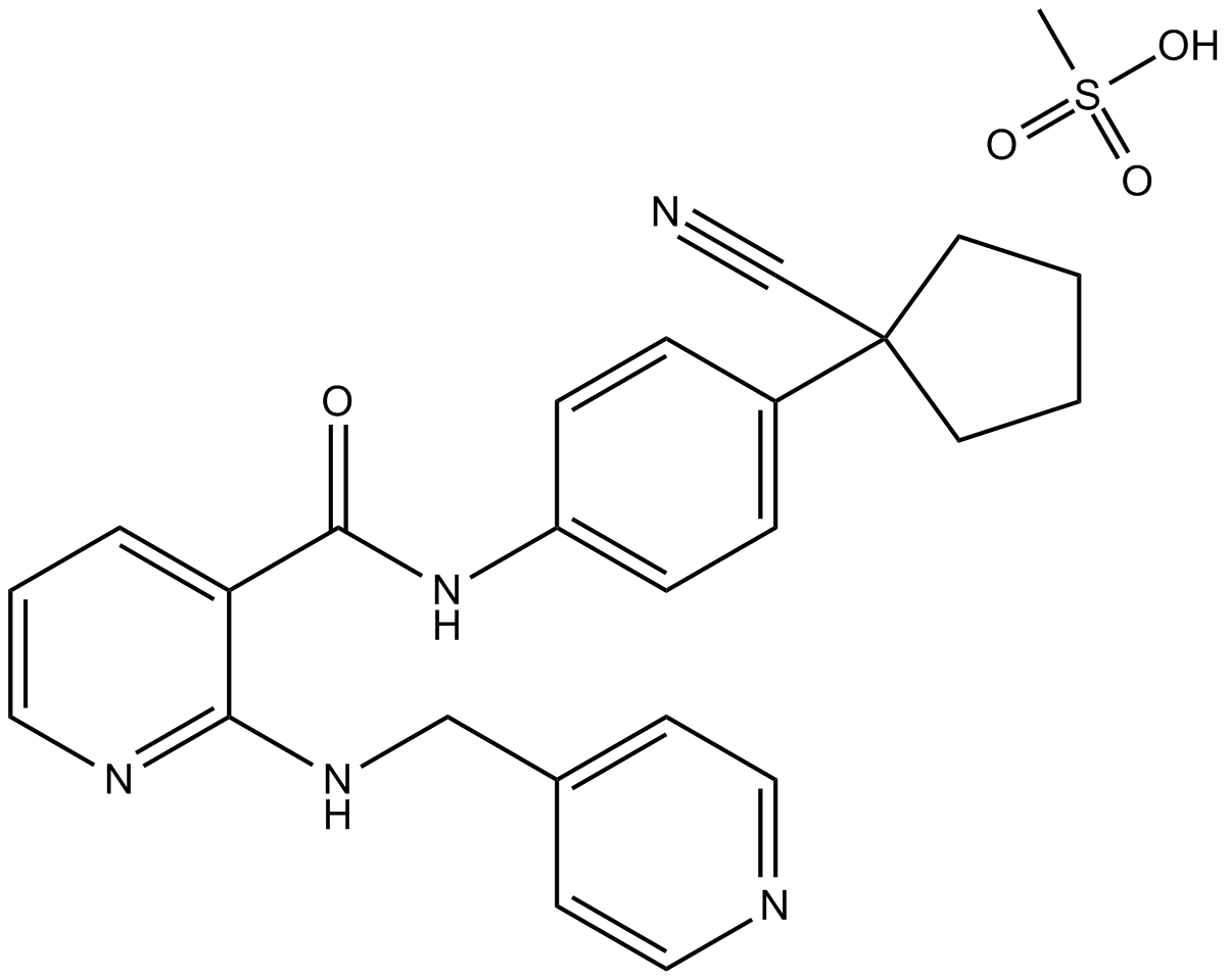
Cas No.: 811803-05-1
Sample solution is provided at 25 µL, 10mM.
Apatinib blocks the downstream signal transduction of VEGF pathway to inhibit neovascularization. Apatinib has showed antitumor effects on many kinds of tumors, such as sarcoma, breast cancer, ovarian cancer, and acute lymphoblastic leukemia (ALL). Moreover, a recent case report provided supporting information for apatinib to treat epithelioid malignant plural mesothelioma.[1]
In vitro experiment indicated that apatinib inhibited cells proliferation in a dose-dependent and time-dependent manner. The IC50 values of apatinib in MPM cells for treatment time 24 h, 48 h, 72 h were 46.34 μM, 45.14 μM, 28.73 μM.[3]
In vivo experiments demonstrated that apatinib inhibits tumor growth and metastasis. The ePCI score of blank control, solvent control and apatinib groups were 9.8 ± 0.9, 10.3 ± 0.9, and 7.3 ± 1.6, respectively. The differences were statistically significant (P = 0.003 for all; P = 0.008, blank control vs. apatinib group; P = 0.001, solvent control vs. apatinib group; P = 0.394, blank control vs. solvent control). The positive rate of Ki-67 in apatinib group was (17.0 ± 8.0)%, which is significantly lower than that in control group (48.1 ±11.5) % (P = 0.000); the positive rate of VEGFR-2 in apatinib group was slightly lower than that in control group, indicating no statistical significance.[1]
References:
[1]. Yang ZR, et al. Apatinib Mesylate Inhibits the Proliferation and Metastasis of Epithelioid Malignant Peritoneal Mesothelioma In Vitro and In Vivo. Front Oncol. 2020 Dec 7;10:585079.
Cell experiment [1]: | |
Cell lines |
Primary cells established by s.c. tumors |
Preparation Method |
Apatinib was dissolved in DMSO to yield a 151 mM stock solution, which was then diluted to the specified concentration in subsequent experiment by using DMEM. |
Reaction Conditions |
Cells were treated with different concentrations of apatinib (0, 12.5, 25, 50 and 100 mM) for 24, 48, and 72 h, respectively. |
Applications |
This could be used to test the ability of apatinib on the inhibitions of MPM Cells’ viability and proliferation. These gradient concentrations of apatinib inhibited cells proliferation; the inhibitory effect of apatinib on MPM cells showed the dose-dependent and time-dependent manner. |
Animal experiment [1]: | |
Animal models |
Specific pathogen free BALB/c nu/nu mice, 4–5 weeks old, 16–18 g |
Preparation Method |
MPM surgical specimens was made to establish patient-derived xenograft (PDX) models in nude mice. Eighteen nude mice were randomly divided into three groups: blank control, solvent control and apatinib groups. Blank control group received no intervention, solvent control group was administrated with 24 μg/g/d 0.5% CMC, and treatment group was treated with 100 μg/g/d apatinib delivered by intra-gastric gavage. The treatment process lasted for 2 weeks. |
Dosage form |
100 μg/g/d; apatinib was diluted in 0.5% Carboxymethyl Cellulose-Na solution |
Applications |
This could be used to test the efficacy and toxicity of apatinib on MPM PDX Model. Results indicated that apatinib did not affect the weight of nude mice, and no adverse effects were observed during the construction of mice models. The positive rate of Ki-67 in apatinib group is significantly lower than that in control group |
References: |
Cas No. | 811803-05-1 | SDF | |
المرادفات | YN968D1 | ||
Chemical Name | N-[4-(1-cyanocyclopentyl)phenyl]-2-(pyridin-4-ylmethylamino)pyridine-3-carboxamide;methanesulfonic acid | ||
Canonical SMILES | CS(=O)(=O)O.C1CCC(C1)(C#N)C2=CC=C(C=C2)NC(=O)C3=C(N=CC=C3)NCC4=CC=NC=C4 | ||
Formula | C25H27N5O4S | M.Wt | 493.58 |
الذوبان | ≥ 49.4mg/mL in DMSO | Storage | Store at -20°C |
General tips | Please select the appropriate solvent to prepare the stock solution according to the
solubility of the product in different solvents; once the solution is prepared, please store it in
separate packages to avoid product failure caused by repeated freezing and thawing.Storage method
and period of the stock solution: When stored at -80°C, please use it within 6 months; when stored
at -20°C, please use it within 1 month. To increase solubility, heat the tube to 37°C and then oscillate in an ultrasonic bath for some time. |
||
Shipping Condition | Evaluation sample solution: shipped with blue ice. All other sizes available: with RT, or with Blue Ice upon request. |
Prepare stock solution | |||
![]() |
1 mg | 5 mg | 10 mg |
1 mM | 2.026 mL | 10.1301 mL | 20.2601 mL |
5 mM | 0.4052 mL | 2.026 mL | 4.052 mL |
10 mM | 0.2026 mL | 1.013 mL | 2.026 mL |
Step 1: Enter information below (Recommended: An additional animal making an allowance for loss during the experiment)
Step 2: Enter the in vivo formulation (This is only the calculator, not formulation. Please contact us first if there is no in vivo formulation at the solubility Section.)
Calculation results:
Working concentration: mg/ml;
Method for preparing DMSO master liquid: mg drug pre-dissolved in μL DMSO ( Master liquid concentration mg/mL, Please contact us first if the concentration exceeds the DMSO solubility of the batch of drug. )
Method for preparing in vivo formulation: Take μL DMSO master liquid, next addμL PEG300, mix and clarify, next addμL Tween 80, mix and clarify, next add μL ddH2O, mix and clarify.
Method for preparing in vivo formulation: Take μL DMSO master liquid, next add μL Corn oil, mix and clarify.
Note: 1. Please make sure the liquid is clear before adding the next solvent.
2. Be sure to add the solvent(s) in order. You must ensure that the solution obtained, in the previous addition, is a clear solution before proceeding to add the next solvent. Physical methods such as vortex, ultrasound or hot water bath can be used to aid dissolving.
3. All of the above co-solvents are available for purchase on the GlpBio website.
Quality Control & SDS
- View current batch:
- Purity: >98.00%
- COA (Certificate Of Analysis)
- SDS (Safety Data Sheet)
- Datasheet
-
Related Biological Data
VEGFA derived from hypoxic PMCs facilitates GC cell adhesion and migration via VEGFR1. a The effect of exogenous VEGFA on cell migration was observed in the presence of Apatinib or Bevacizumab. Representative photographs of migratory cells are shown. Scale bar represents 100 μm.
Apatinib were obtained from GlpBio.
J Exp Clin Canc Res 39.1 (2020): 1-14. PMID: 33081836 IF: 11.163 -
Related Biological Data
Targeting SMYD2 synergistically increases the antiangiogenic activity of apatinib in CRC.IHC staining revealed that both BAY-598 and apatinib treatment reduced CD31 levels, but more satisfactory effects were achieved when the two treatments were combined.
Then, we treated CRC cells with DMSO, BAY-598, apatinib (10 μM), or BAY-598 (10 μM) combined with apatinib(GLPBIO) for 48 h, and prepared TCM according to the standard method.
Angiogenesis (2022): 1-18. PMID: 35503397 IF: 9.5957 -
Related Biological Data
Art-M improves the antitumor effect of apatinib in GC both in vivo and in vitro. (A) Total number of viable cells was counted after treatment of MGC803 and AGS cells with different concentrations of apatinib for 96 h.
Male nude mice carrying GC xenografts were randomly divided into four groups and given Art-M (2 mg/kg; i.p.; once daily), apatinib(GlpBio) (50 mg/kg; p.o.; once daily), or both for 24 days.
Chem-Biol Interact (2023): 110618. PMID: 37394161 IF: 5.1
Average Rating: 5
(Based on Reviews and 39 reference(s) in Google Scholar.)GLPBIO products are for RESEARCH USE ONLY. Please make sure your review or question is research based.
Required fields are marked with *