

Apatinib Mesylate (Synonyms: YN968D1) |
Catalog No.GC14292
 One-Click Copy Product Info
|

Apatinib Mesylate는 강력하고 경구 생체이용률이 있고 ATP 경쟁적인 VEGFR-2 티로신 활성효소 억제제로 세포 없는 환경에서의 IC50 값은 1nM이다.
Products are for research use only. Not for human use. We do not sell to patients.
Cas No.: 811803-05-1
Sample solution is provided at 25 µL, 10mM.
Apatinib Mesylate는 강력하고 경구 생체이용률이 있고 ATP 경쟁적인 VEGFR-2 티로신 활성효소 억제제로 세포 없는 환경에서의 IC50 값은 1nM이다. Apatinib Mesylate는 VEGFR-2의 인산화와 하류의 PLCγ1/ERK1/2, PI3K/AKT 및 STAT3 신호 전달을 억제해서 세포의 증식, 생존, 이동 및 혈관 생성을 제한한다[1][2][4]. Apatinib Mesylate는 종양 혈관 생성, 고형암의 성장 및 이동, 백혈병 생물학, 자식 및 ABC 수송체 매개 다제내성 연구에 사용된다[2][3][4][5][6][7].
체외 실험에서 Apatinib Mesylate는 72시간 동안 VEGF에 의해 자극된 인간 제대정맥 내피세포의 증식을 억제하였고 IC50 값은 0.17μM이었다. 반면 20% 태아 소 혈청에 의해 자극된 증식에 대한 IC50 값은 23.4μM였다[1]. SGC-7901 및 BGC-823 인간 위암 세포에서 Apatinib Mesylate (100, 500, 1000nM; 24시간 처리)는 세포 증식과 VEGF 분비를 감소시켰고 Apatinib Mesylate (500nM; 밤새 처리 후 30분 동안 VEGF 자극)는 VEGFR-2, PLCγ1 및 ERK1/2의 인산화를 감소시켰지만 경로 활성도가 낮은 MGC-803 세포는 현저한 증식 반응을 보이지 않았다[2]. KHOS 및 MG63 인간 골육종 세포에서 Apatinib Mesylate (10μM; 48시간 처리)는 세포 사멸 및 자식 표지자를 증가시키고 G0/G1 세포 주기 정지를 유도해서 STAT3 및 BCL-2 신호 전달을 감소시켰다[3]. Jurkat 및 Nalm6 사람 급성 림프모구성 백혈병 세포에서 Apatinib Mesylate (0, 10, 20, 40μM; 48시간 처리)는 VEGFR-2, ERK, AKT 및 STAT3 경로의 활성화를 용량 의존적으로 감소시켰다[4]. 인간 악성 복막 중피종 세포에서 Apatinib Mesylate (0, 12.5, 25, 50, 100μM; 24, 48, 72시간 처리)는 용량 및 시간 의존적으로 세포 증식을 억제하였고 24, 48, 72시간 시점의 IC50 값은 각각 46.34μM, 45.14μM, 28.73μM이었다[5].
체내 실험에서 CNE-2 인간 비인두암 이종 이식 종양을 이식한 암컷 누드 쥐에게 Apatinib Mesylate (200mg/kg/일; 7일 동안 매일 1회 위관 투여)를 투여한 후 VEGFR-2 양성 종양 세포의 비율이 92.83%에서 54.50%로 감소하고 CD31로 정의된 미세혈관 밀도(MVD)가 8.67에서 5.00으로 감소하였고 종양 성장이 억제되었다[6]. Nalm6 인간 급성 림프모구성 백혈병 이종 이식 종양을 이식한 NODscid-IL2Rg⁻/⁻ 쥐에게 Apatinib Mesylate(100mg/kg/day; 2주 동안 월요일부터 금요일까지 매일 1회 경구 위관 투여)를 투여한 후 말초혈액, 골수 및 비장에 있는 인간 CD45 양성 백혈병 세포가 감소하고 비장 무게가 감소하였다[4]. ABCB1을 과발현하는 KBv200 인간 암세포 누드 쥐 이종 이식 모델에서 Apatinib Mesylate (70mg/kg; 3일마다 1회 경구 투여, 총 4회; 파클리타크셀 투여 1시간 전)는 파클리타크셀의 항종양 활성을 강화해서 52.7%의 종양 성장 억제율을 나타냈고 명백한 체중 감소는 관찰되지 않았다[7].
| 세포 실험 [1]: | |
|
세포주 |
SGC-7901, BGC-823 및 MGC-803 인간 위암세포 |
|
준비 방법 |
세포를 96-웰 판에 웰당 2×10³ 개의 세포로 파종하고 Apatinib Mesylate로 처리하였습니다. 세포 활력은 CCK-8 분석법으로 측정하였고 배양지 내 VEGF 수치는 ELISA로 측정하였습니다. 신호 전달 경로 분석을 위해 SGC-7901 및 BGC-823 세포를 Apatinib Mesylate로 밤새 처리한 후 재조합 인간 VEGF로 자극하였고 p-VEGFR-2, p-PLCγ1 및 p-ERK1/2를 단백질 자극법으로 분석하였습니다. |
|
반응 조건 |
100, 500 및 1000nM 24시간 동안; 500nM를 밤새 처리한 후 30분 동안 VEGF 자극 |
|
적용 |
Apatinib Mesylate는 VEGFR-2 경로 활성도가 높은 SGC-7901 및 BGC-823 세포의 증식과 VEGF 분비를 현저하게 억제하였지만 경로 활성도가 낮은 MGC-803 세포에서는 억제 효과가 나타나지 않았습니다. 500nM의 Apatinib Mesylate는 VEGF에 의해 유도된 VEGFR-2, PLCγ1 및 ERK1/2의 인산화 수준을 기준성 이하로 감소시켰습니다. |
| 동물 실험 [2]: | |
|
동물 모델 |
4주령된 암컷 누드 쥐에 대한 피하 CNE-2 인간 비후두암 이종 이식체 |
|
준비 방법 |
평균 종양 부피가 100mm³에 도달하였을 때 쥐를 무작위 배정하였습니다. Apatinib Mesylate 그룹은 매일 위관 투여를 받았고 대조 용매 그룹은 동일한 경로를 통해 생리식염수를 투여받았습니다. 2일마다 종양 부피와 체중을 기록하였습니다. 치료 종료할 때 종양의 무게를 측정하였고 VEGFR-2 및 CD31 면역 조직 화학법, TUNEL, ¹⁸F-FDG PET/CT를 통해 분석하였습니다. |
|
제형 |
200mg/kg/일;7일 동안 하루에 한 번 위내 투여 |
|
적용 |
Apatinib Mesylate는 VEGFR-2 양성 종양 세포의 비율을 92.83%에서 54.50%로 감소시켰고 CD31로 정의된 미세혈관 밀도를 8.67에서 5.00으로 낮췄습니다. 게다가 종양의 대사 활성과 CNE-2 이종 이식 종양의 성장도 감소되었습니다. |
| Cas No. | 811803-05-1 | SDF | |
| Synonyms | YN968D1 | ||
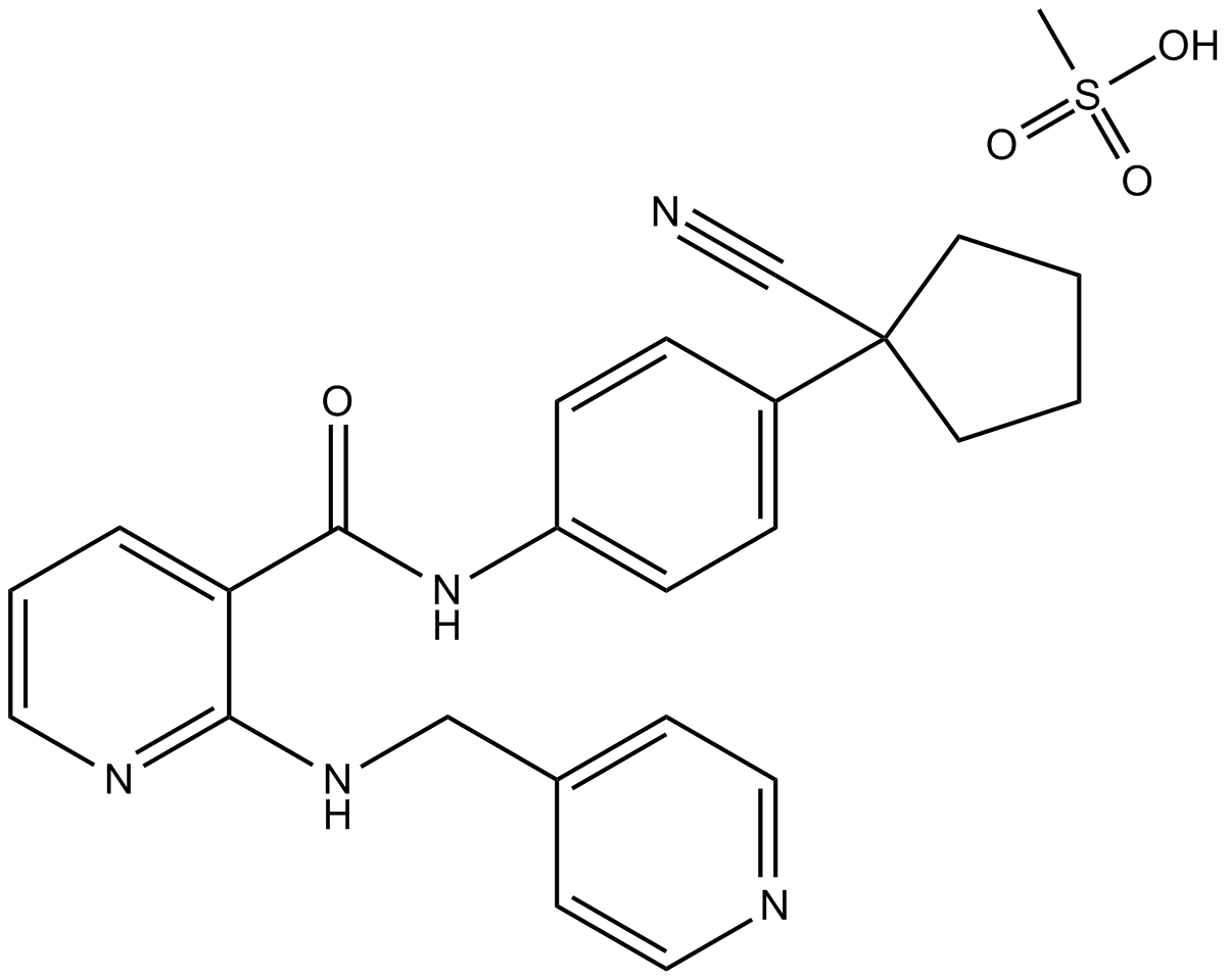
| Chemical Name | N-[4-(1-cyanocyclopentyl)phenyl]-2-(pyridin-4-ylmethylamino)pyridine-3-carboxamide;methanesulfonic acid | ||
| Canonical SMILES | CS(=O)(=O)O.C1CCC(C1)(C#N)C2=CC=C(C=C2)NC(=O)C3=C(N=CC=C3)NCC4=CC=NC=C4 | ||
| Formula | C25H27N5O4S | M.Wt | 493.58 |
| Solubility | ≥ 49.4mg/mL in DMSO | Storage | Store at -20°C |
| Shipping Condition | Evaluation sample solution: shipped with blue ice. All other sizes available: with RT, or with Blue Ice upon request. | ||
| Prepare stock solution | |||

|
1 mg | 5 mg | 10 mg |
| 1 mM | 2.026 mL | 10.1301 mL | 20.2601 mL |
| 5 mM | 405.2 μL | 2.026 mL | 4.052 mL |
| 10 mM | 202.6 μL | 1.013 mL | 2.026 mL |
Step 1: Enter information below (Recommended: An additional animal making an allowance for loss during the experiment)
 g
μL
g
μL
Step 2: Enter the in vivo formulation (This is only the calculator, not formulation. Please contact us first if there is no in vivo formulation at the solubility Section.)
Calculation results:
Working concentration: mg/ml;
Method for preparing DMSO master liquid: mg drug pre-dissolved in μL DMSO ( Master liquid concentration mg/mL, Please contact us first if the concentration exceeds the DMSO solubility of the batch of drug. )
Method for preparing in vivo formulation: Take μL DMSO master liquid, next addμL PEG300, mix and clarify, next addμL Tween 80, mix and clarify, next add μL saline, mix and clarify.
Method for preparing in vivo formulation: Take μL DMSO master liquid, next add μL Corn oil, mix and clarify.
Note: 1. Please make sure the liquid is clear before adding the next solvent.
2. Be sure to add the solvent(s) in order. You must ensure that the solution obtained, in the previous addition, is a clear solution before proceeding to add the next solvent. Physical methods such as vortex, ultrasound or hot water bath can be used to aid dissolving.
3. All of the above co-solvents are available for purchase on the GlpBio website.
Quality Control & SDS
- View current batch:
- Purity: >98.00% Appearance: A solid
- COA (Certificate of Analysis)
- SDS (Safety Data Sheet)
- Datasheet
-
Related Biological Data

VEGFA derived from hypoxic PMCs facilitates GC cell adhesion and migration via VEGFR1. a The effect of exogenous VEGFA on cell migration was observed in the presence of Apatinib or Bevacizumab. Representative photographs of migratory cells are shown. Scale bar represents 100 μm.
Apatinib were obtained from GlpBio.
J Exp Clin Canc Res 39.1 (2020): 1-14. PMID: 33081836 IF: 11.163 -
Related Biological Data

Targeting SMYD2 synergistically increases the antiangiogenic activity of apatinib in CRC.IHC staining revealed that both BAY-598 and apatinib treatment reduced CD31 levels, but more satisfactory effects were achieved when the two treatments were combined.
Then, we treated CRC cells with DMSO, BAY-598, apatinib (10 μM), or BAY-598 (10 μM) combined with apatinib(GLPBIO) for 48 h, and prepared TCM according to the standard method.
Angiogenesis (2022): 1-18. PMID: 35503397 IF: 9.5957 -
Related Biological Data

Art-M improves the antitumor effect of apatinib in GC both in vivo and in vitro. (A) Total number of viable cells was counted after treatment of MGC803 and AGS cells with different concentrations of apatinib for 96 h.
Male nude mice carrying GC xenografts were randomly divided into four groups and given Art-M (2 mg/kg; i.p.; once daily), apatinib(GlpBio) (50 mg/kg; p.o.; once daily), or both for 24 days.
Chem-Biol Interact (2023): 110618. PMID: 37394161 IF: 5.1
Average Rating: 5 (Based on Reviews and 39 reference(s) in Google Scholar.)