

Nile Red
|
Katalog-Nr.GC15539
 One-Click Copy Product Info
|


Nile Red ist ein gängiger Lipidfarbstoff. Es ist hydrophob und metachromatisch, und hat in Wasserlösungen eine geringe Löslichkeit und Fluoreszenz. Es emittiert in hydrophoben Umgebungen Farben von einem matten Rot bis zu einem kräftigen Gelbgold.
Products are for research use only. Not for human use. We do not sell to patients.
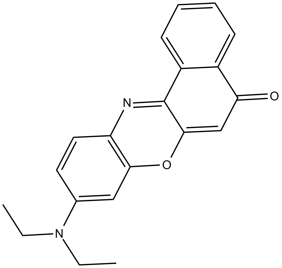
Cas No.: 7385-67-3
Sample solution is provided at 25 µL, 10mM.
Nile Red ist ein gängiger Lipidfarbstoff. Es ist hydrophob und metachromatisch, und hat in Wasserlösungen eine geringe Löslichkeit und Fluoreszenz. Es emittiert in hydrophoben Umgebungen Farben von einem matten Rot bis zu einem kräftigen Gelbgold[1].
Nile red kann Cholesterin im menschlichen Plasma durch Anfärben von Lipidveskeln in glatten Muskelzellen und in kultivierten Makrophagen bei niedriger Zelldichte anfärben. Die Anregungs-/Emissionswellenlängen liegen bei 450 - 500/>528 [2]. Nile red wurde auch zur Forschung der Membranheterogenität[3] und von Liganden-Protein-Wechselwirkungen mit den alternativen Wellenlängen 570/610 [4] sowie zur Forschung von Enzymmechanismen mit den Wellenlängen 550/640 - 660[5] verwendet. Nile red wurde auch erfolgreich zur Färbung intrazellulärer neutraler Lipide, d.h. TAG und Cholesterinester, in Hefen und Pilzen mit gekoppelten Wellenlängen von 488/565 - 585[6] sowie in Mikroalgen mit Wellenlängen von 488 - 525/570 - 600[7] oder zur Färbung der Gesamtlipide mit Wellenlänge von 490/585[8] verwendet.
References:
[1]. Rumin J, Bonnefond H, Saint-Jean B, et al. The use of fluorescent Nile red and BODIPY for lipid measurement in microalgae[J]. Biotechnology for biofuels, 2015, 8(1): 1-16.
[2]. Greenspan P, Mayer E P, Fowler S D. Nile red: a selective fluorescent stain for intracellular lipid droplets[J]. The Journal of cell biology, 1985, 100(3): 965-973.
[3]. Ira and, Krishnamoorthy G. Probing the link between proton transport and water content in lipid membranes[J]. The Journal of Physical Chemistry B, 2001, 105(7): 1484-1488.
[4]. Sackett D L, Wolff J. Nile red as a polarity-sensitive fluorescent probe of hydrophobic protein surfaces[J]. Analytical biochemistry, 1987, 167(2): 228-234.
[5]. Ruvinov S B, Yang X J, Parris K D, et al. Ligand-mediated Changes in the Tryptophan Synthase Indole Tunnel Probed by Nile Red Fluorescence with Wild Type, Mutant, and Chemically Modified Enzymes (∗)[J]. Journal of Biological Chemistry, 1995, 270(11): 6357-6369.
[6]. Kimura K, Yamaoka M, Kamisaka Y. Rapid estimation of lipids in oleaginous fungi and yeasts using Nile red fluorescence[J]. Journal of microbiological methods, 2004, 56(3): 331-338.
[7]. Cooksey K E, Guckert J B, Williams S A, et al. Fluorometric determination of the neutral lipid content of microalgal cells using Nile Red[J]. Journal of microbiological methods, 1987, 6(6): 333-345.
[8]. Lee S J, Yoon B D, Oh H M. Rapid method for the determination of lipid from the green alga Botryococcus braunii[J]. Biotechnology techniques, 1998, 12(7): 553-556.
Der Plan dient nur als Richtlinie. Bitte passen Sie ihn an Ihre spezifischen Bedürfnisse an.
1. Färbelösung herstellen:
(1) Stammlösung herstellen: Nile Red in DMSO lösen und eine Stammlösung mit einer Konzentration von 1 mM herstellen.
Hinweis: Nicht verwendete Stammlösung aliquotieren und im Dunkeln bei -20°C oder -80°C lagern, um wiederholtes Einfrieren und Auftauen zu vermeiden.
(2) Arbeitslösung herstellen: Die Stammlösung mit einem geeigneten Puffer(z.B. serumfreiem Medium oder PBS) verdünnen und eine Arbeitslösung mit einer Konzentration von 0.2-1 μM herstellen.
Hinweis: Bitte passen Sie die Konzentration der Arbeitslösung den jeweiligen Gegebenheiten an und bereiten Sie sie jetzt zu.
2. Färbung der Zellsuspension
(1) Suspensionszellen: 3-5 Minuten bei 1000 g zentrifugieren, den Überstand verwerfen und zweimal je 5 Minuten mit PBS waschen.
(2) Adhärente Zellen: Zweimal mit PBS waschen, Trypsin zur Digestion der Zellen zugeben und nach Abschluss der Digestion 3-5 Minuten bei 1000 g zentrifugieren.
(3) 1 mL Nile-Red-Arbeitslösung zugeben, um die Zellen zu resuspendieren, und 5-30 Minuten im Dunkeln bei Raumtemperatur inkubieren. Die optimale Inkubationszeit variiert je nach Zelltyp; bitte experimentieren Sie entsprechend Ihren spezifischen Anforderungen.
(4) Nach der Inkubation 5 Minuten bei 1000 g zentrifugieren, den Überstand entfernen, PBS zugeben und 2-3 Mal je 5 Minuten waschen.
(5) Zellen in vorgewärmtem serumfreiem Zellkulturmedium oder PBS resuspendieren und mittels Fluoreszenzmikroskopie oder Durchflusszytometrie untersuchen.
3. Zelladhäsionsfärbung
(1) Adhärente Zellen auf sterilen Deckgläschen kultivieren.
(2) Nehmen Sie das Deckglas aus dem Kulturmedium, saugen Sie überschüssiges Kulturmedium ab und legen Sie das Deckglas in eine feuchte Umgebung.
(3) Geben Sie 100 μL Farbstofflösung von einer Ecke des Deckglases hinzu und schütteln Sie es vorsichtig, um alle Zellen gleichmäßig mit dem Farbstoff zu bedecken.
(4) Inkubieren Sie die Zellen 5-30 Minuten lang im Dunkeln bei Raumtemperatur. Die optimale Inkubationszeit variiert je nach Zelltyp. Bitte ermitteln Sie die optimale Zeit entsprechend Ihren experimentellen Anforderungen.
(5) Nach der Inkubation verwerfen Sie die Farbstofflösung und waschen das Deckglas 2-3 Mal mit vorgewärmtem Kulturmedium.
4. Mikroskopische Untersuchung. Bei Triglyceriden(neutralen Lipiden) liegt das Anregungs-/Emissionsmaximum von Nile Red bei 515/585nm; bei Phospholipiden(polaren Lipiden) bei 554nm/638 nm.
Vorsichtsmaßnahmen:
1) Fluoreszierende Farbstoffe weisen alle ein Quenching-Problem auf. Um das Fluoreszenz-Quenching zu verlangsamen, sollte Lichteinwirkung möglichst vermieden werden.
2) Tragen Sie zu Ihrer eigenen Sicherheit und Gesundheit einen Laborkittel und Einweghandschuhe.
| Cas No. | 7385-67-3 | SDF | |
| Chemical Name | 9-(diethylamino)-5H-benzo[a]phenoxazin-5-one | ||
| Canonical SMILES | CCN(C1=CC2=C(N=C3C4=CC=CC=C4C(C=C3O2)=O)C=C1)CC | ||
| Formula | C20H18N2O2 | M.Wt | 318.37 |
| Löslichkeit | DMSO : ≥ 50 mg/mL (157.05 mM) | Storage | Store at 4°C, protect from light |
| General tips | Please select the appropriate solvent to prepare the stock solution according to the
solubility of the product in different solvents; once the solution is prepared, please store it in
separate packages to avoid product failure caused by repeated freezing and thawing.Storage method
and period of the stock solution: When stored at -80°C, please use it within 6 months; when stored
at -20°C, please use it within 1 month. To increase solubility, heat the tube to 37°C and then oscillate in an ultrasonic bath for some time. |
||
| Shipping Condition | Evaluation sample solution: shipped with blue ice. All other sizes available: with RT, or with Blue Ice upon request. | ||
| Prepare stock solution | |||

|
1 mg | 5 mg | 10 mg |
| 1 mM | 3.141 mL | 15.705 mL | 31.41 mL |
| 5 mM | 628.2 μL | 3.141 mL | 6.282 mL |
| 10 mM | 314.1 μL | 1.5705 mL | 3.141 mL |
Step 1: Enter information below (Recommended: An additional animal making an allowance for loss during the experiment)
 g
μL
g
μL
Step 2: Enter the in vivo formulation (This is only the calculator, not formulation. Please contact us first if there is no in vivo formulation at the solubility Section.)
Calculation results:
Working concentration: mg/ml;
Method for preparing DMSO master liquid: mg drug pre-dissolved in μL DMSO ( Master liquid concentration mg/mL, Please contact us first if the concentration exceeds the DMSO solubility of the batch of drug. )
Method for preparing in vivo formulation: Take μL DMSO master liquid, next addμL PEG300, mix and clarify, next addμL Tween 80, mix and clarify, next add μL saline, mix and clarify.
Method for preparing in vivo formulation: Take μL DMSO master liquid, next add μL Corn oil, mix and clarify.
Note: 1. Please make sure the liquid is clear before adding the next solvent.
2. Be sure to add the solvent(s) in order. You must ensure that the solution obtained, in the previous addition, is a clear solution before proceeding to add the next solvent. Physical methods such as vortex, ultrasound or hot water bath can be used to aid dissolving.
3. All of the above co-solvents are available for purchase on the GlpBio website.
Quality Control & SDS
- View current batch:
- Purity: >98.00% Appearance: A solid
- COA (Certificate of Analysis)
- SDS (Safety Data Sheet)
- Datasheet
-
Related Biological Data

24HC induces cholesterol accumulation in multivesicular bodies or late endosomes.C. N2a cells were treated with ETOH, 24HC (5 µM), or 25HC (5 µM) for 24 h, then fixed and stained with Nile red. The cells were then analyzed for PE fluorescence intensity by flow cytometr.
N2a cells were treated with ETOH, 24HC (5 µM), or 25HC (5 µM) for 24 h, then fixed and stained with Nile red(GlpBio).
Redox Biology (2023): 102769. PMID: 37285742 IF: 10.7865 -
Related Biological Data

Effects of OG on lipid synthesis in the damaged epidermal barrier. (A, B) Nile red fluorescence staining and quantitative analysis of neutral lipid in the WT (A) and Dectin-1(B) mice.
The stained slices were then stained with Nile Red Fluorescent Dye Working Solution (Glpbio) for 20 min, and finally mounted with glycerol gelatin and examined with a confocal laser scanning microscope.
Brit J Pharmacol (2023). PMID: 38124222 IF: 7.3003 -
Related Biological Data

Effects of SIP on keratinocyte lipid synthesis. (A) Changes in neutral lipid level of HaCaT cells as examined by Nile red fluorescence staining. The plots beside the images show the quantitation analysis of neutral lipids fluorescence intensity. (B) Detection of lipid content in HaCaT cells by oil-red O.
The slices were incubated with Hoechst solution and then in Nile Red Fluorescent Dye Working Solution (Glpbio).
Frontiers in Pharmacology, 2023, 14. PMID: 36733502 IF: 5.9879 -
Related Biological Data

Oil Red O staining, Nile Red staining, and quantifcation of Oil Red O staining in 3T3-L1 cells treated with Citrus sunki peel extract. (a) 3T3-L1 cells, stained with Oil Red O, were visualized in optical microscopy. (b) 3T3-L1 cells, stained with Nile Red, were visualized in optical microscopy.
Nile Red working solution was prepared by diluting nile red dye (GLPBIO) in PBS to a concentration of 1 μg/mL. Cells were treated with nile red working solution for 30 min at room temperature.
J Food Biochem 2024 (2024). IF: 4 -
Related Biological Data

Downregulation of LIPT1 reduces lipid deposition in HCC cells. A Nile red staining analysis showed a signifcant decrease in lipid content in LIPT1-silenced cells.
Briefy, the transfected cells in 96-well plates (3× 10³ cells/well) were fixed in 4% paraformaldehyde for 30 min and incubated with 0.1 μg/mL Nile red(GlpBio) for 10 min, then washing the plates with PBS twice, followed by DAPI staining for 10 min.
J Cancer Res Clin (2023): 1-17. PMID: 37668796 IF: 3.5999
Average Rating: 5 (Based on Reviews and 32 reference(s) in Google Scholar.)