

AT 56 |
カタログ番号GC11950
 One-Click Copy Product Info
|

AT 56はリポカリン型プロスタグランジンD合成酵素(L-PGDS)の経口活性と選択的な阻害剤であり、IC50値は95µMである。
Products are for research use only. Not for human use. We do not sell to patients.
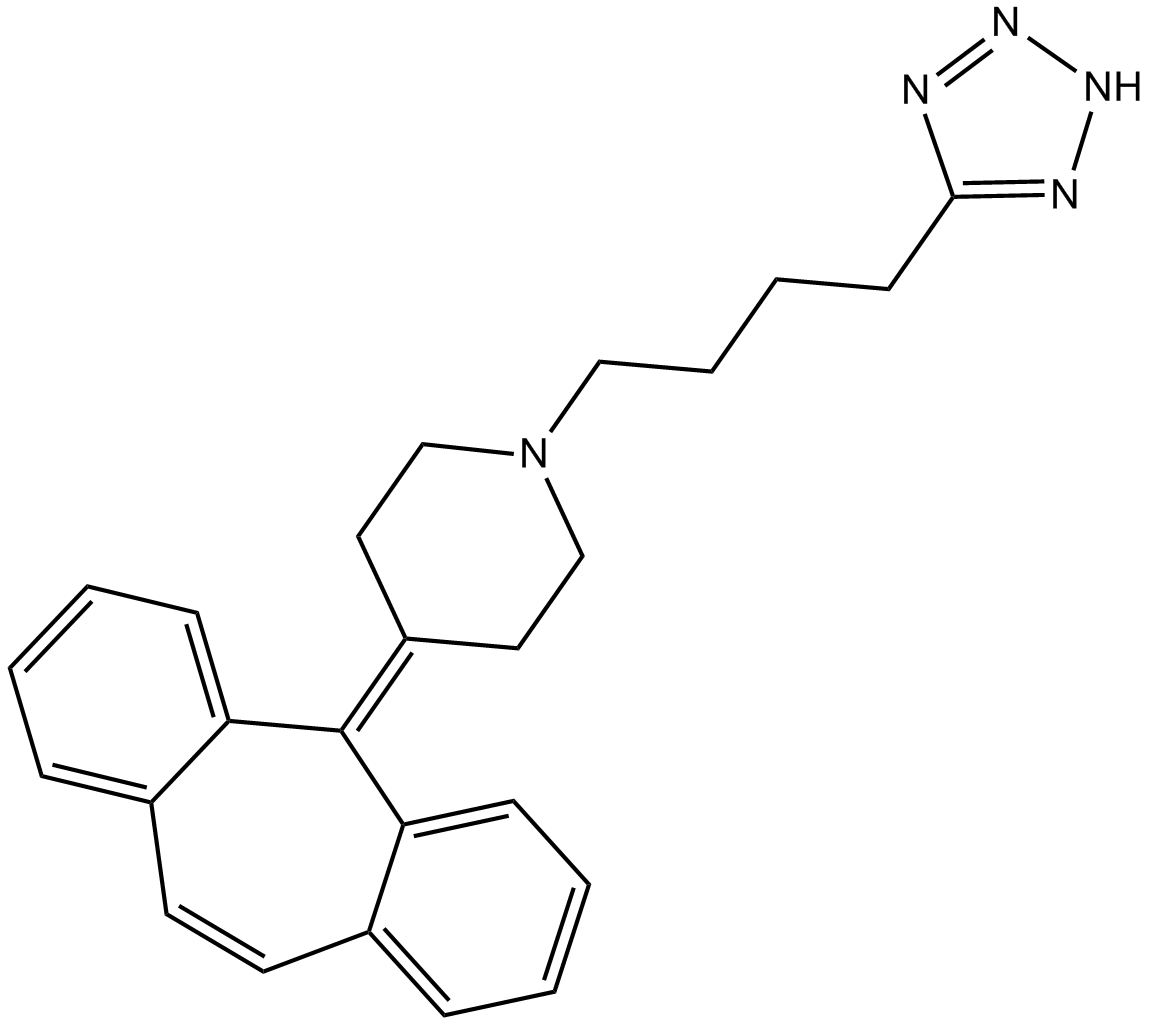
Cas No.: 162640-98-4
Sample solution is provided at 25 µL, 10mM.
_1-3.$$$39978408@51.jpg');)
_1-9.$$$38538795@65.jpg');)
_1-9.$$$39112701@51.jpg');)
_1-7.$$$37316672@70.jpg');)
_366-372.$$$36198801@70.jpg');)
.$$$38565142@65.jpg');)
_1867-1883.$$$33248023@67.jpg');)
_eads6055.$$$39977546@45.jpg');)
_eadm9805.$$$40080571@45.jpg');)
_eadj3020.$$$39666821@45.jpg');)
_1-20.$$$39757301@28.jpg');)
_1-24.$$$38898113@28.jpg');)
_273-287.$$$36806353@46.jpg');)
_1-16.$$$37156877@44.jpg');)
_1-19.$$$37460805@44.jpg');)
_1291-1307.$$$34518654@46.jpg');)
AT 56はリポカリン型プロスタグランジンD合成酵素(L-PGDS)の経口活性と選択的な阻害剤であり、IC50値は95µMである[1]。AT 56はレプチンの産生とPGD2の産生を阻害し、細胞の増殖を影響できる[2]。AT 56は、癌細胞モデルで、内皮間葉転換(EndMT)のプロセスの制御に広く使われている[3]。
体外で、48時間のAT 56(100µM)処理は、瀰漫性大細胞型B細胞リンパ腫(DLBCL)細胞で、細胞の増殖とc-mycの発見を阻害した[4]。5µM AT 56の7日間の処理は、細胞の生存率を影響せずに、野生型オリゴデンドロサイトの成長を著しく阻害した[5]。5µM AT 56の10分間の処理は、血清飢餓条件下のTE671細胞で、PGD2の生産を阻害した[6]。
体内で、30mg/kg/日のAT 56を4週間経口投与した結果、C57BL/6Jマウスの大脳皮質の血管分岐のエタノール誘発の増加を減らし、L-PGDSの発見を減らした[7]。AT 56 (10mg/kg/日)を7日間腹腔内注射した結果、PGD2合成を阻害することで、Clpxfl/fl; Zp3-Creマウスの生殖機能障害を部分的に回復させられる[8]。
References:
[1] Irikura D, Aritake K, Nagata N, et al. Biochemical, functional, and pharmacological characterization of AT-56, an orally active and selective inhibitor of lipocalin-type prostaglandin D synthase[J]. Journal of Biological Chemistry, 2009, 284(12): 7623-7630.
[2] Urade Y. Biochemical and structural characteristics, gene regulation, physiological, pathological and clinical features of lipocalin-type prostaglandin D2 synthase as a multifunctional lipocalin[J]. Frontiers in Physiology, 2021, 12: 718002.
[3] Omori K, Morikawa T, Kunita A, et al. Lipocalin-type prostaglandin D synthase‐derived PGD2 attenuates malignant properties of tumor endothelial cells[J]. The Journal of pathology, 2018, 244(1): 84-96.
[4] Hu S, Ren S, Cai Y, et al. Glycoprotein PTGDS promotes tumorigenesis of diffuse large B-cell lymphoma by MYH9-mediated regulation of Wnt–β-catenin–STAT3 signaling[J]. Cell Death & Differentiation, 2022, 29(3): 642-656.
[5] Pan L, Trimarco A, Zhang A J, et al. Oligodendrocyte-lineage cell exocytosis and L-type prostaglandin D synthase promote oligodendrocyte development and myelination[J]. Elife, 2023, 12: e77441.
[6] Fujimori K, Aritake K, Urade Y. Enhancement of prostaglandin D2 production through cyclooxygenase-2 and lipocalin-type prostaglandin D synthase by upstream stimulatory factor 1 in human brain-derived TE671 cells under serum starvation[J]. Gene, 2008, 426(1-2): 72-80.
[7] Li J, Li C, Subedi U, et al. The Role of Endothelial L-PGDS in the Pro-Angiogenic and Anti-Inflammatory Effects of Low-Dose Alcohol Consumption[J]. Cells, 2024, 13(23): 2007.
[8] Hua R, Hai Z, Gu J, et al. AT-56 RESCUES OOCYTE DYSFUNCTION IN OOCYTE-SPECIFIC KNOCKOUT CLPX MICE BY INHIBITING PGD2 SYNTHESIS[J]. Fertility and Sterility, 2024, 122(4): e171.
| 細胞実験[1]: | |
細胞株 | TE-671細胞 |
準備方法 | TE-671細胞(1×10⁶個の細胞/ウェル)をマルチウェルのプレートに播種した。37°C、5% CO2で、10%の加熱不活化牛胎児血清、4mMのグルタミン、4.5g/lのグルコース、100U/mlのペニシリン、100μg/mlのストレプトマイシン硫酸塩を含有するDulbecco's修飾Eagle培地(DMEM)で培養した。1日間の細胞培養後、様々な用量(0、3、10、30と100μM)でAT 56を添加し、その後、37℃で細胞を10分間インキュベートした。TE-671細胞から放出されたPGD2の量を、酵素結合の免疫吸着法で測定した。 |
反応条件 | 0、3、10、30と100μM;10分間 |
アプリケーション | TE-671細胞内で、AT 56の処理は、PGD2の産生を用量依存的に減らした。 |
| 動物実験 [2]: | |
動物モデル | C57BL/6Jマウス |
準備方法 | 62匹の雄のC57BL/6Jマウス(3か月齢) (25-30g)をランダムに2つのグループに分け、0.7g/kgのエタノール(実験群)または等量の水(対照群)を8週間毎日投与した。対照群と実験群のマウスには、5週目から、AT 56 (30mg/kg/日)を4週間経口投与した。8週間の飼育期の終わりに、マウスを安楽死させ、生理学的条件下で、脳血管密度とL-PGDS発見を測定した。 |
投与形態 | 30mg/kg/日、4週間;経口投与 |
アプリケーション | AT 56の処理は、マウスの大脳皮質と皮質下領域内で、血管分岐のアルコール誘発の増加を著しく減らし、L-PGDSの発見を減らした。 |
References: | |
| Cas No. | 162640-98-4 | SDF | |
| Chemical Name | 1-(4-(2H-tetrazol-5-yl)butyl)-4-(5H-dibenzo[a,d][7]annulen-5-ylidene)piperidine | ||
| Canonical SMILES | N(CC/1)(CCCCC2=NNN=N2)CCC1=C3C4=CC=CC=C4C=CC5=CC=CC=C\35 | ||
| Formula | C25H27N5 | M.Wt | 397.52 |
| 溶解度 | DMF: 30 mg/ml,DMSO: 30 mg/ml,Ethanol: 30 mg/ml,Ethanol:PBS(pH 7.2) (1:1): 0.5 mg/ml | Storage | Store at -20°C |
| General tips | Please select the appropriate solvent to prepare the stock solution according to the
solubility of the product in different solvents; once the solution is prepared, please store it in
separate packages to avoid product failure caused by repeated freezing and thawing.Storage method
and period of the stock solution: When stored at -80°C, please use it within 6 months; when stored
at -20°C, please use it within 1 month. To increase solubility, heat the tube to 37°C and then oscillate in an ultrasonic bath for some time. |
||
| Shipping Condition | Evaluation sample solution: shipped with blue ice. All other sizes available: with RT, or with Blue Ice upon request. | ||
| Prepare stock solution | |||

|
1 mg | 5 mg | 10 mg |
| 1 mM | 2.5156 mL | 12.578 mL | 25.156 mL |
| 5 mM | 503.1 μL | 2.5156 mL | 5.0312 mL |
| 10 mM | 251.6 μL | 1.2578 mL | 2.5156 mL |
Step 1: Enter information below (Recommended: An additional animal making an allowance for loss during the experiment)
 g
μL
g
μL
Step 2: Enter the in vivo formulation (This is only the calculator, not formulation. Please contact us first if there is no in vivo formulation at the solubility Section.)
Calculation results:
Working concentration: mg/ml;
Method for preparing DMSO master liquid: mg drug pre-dissolved in μL DMSO ( Master liquid concentration mg/mL, Please contact us first if the concentration exceeds the DMSO solubility of the batch of drug. )
Method for preparing in vivo formulation: Take μL DMSO master liquid, next addμL PEG300, mix and clarify, next addμL Tween 80, mix and clarify, next add μL saline, mix and clarify.
Method for preparing in vivo formulation: Take μL DMSO master liquid, next add μL Corn oil, mix and clarify.
Note: 1. Please make sure the liquid is clear before adding the next solvent.
2. Be sure to add the solvent(s) in order. You must ensure that the solution obtained, in the previous addition, is a clear solution before proceeding to add the next solvent. Physical methods such as vortex, ultrasound or hot water bath can be used to aid dissolving.
3. All of the above co-solvents are available for purchase on the GlpBio website.
Quality Control & SDS
- View current batch:
- Purity: >98.00% Appearance: A solid
- COA (Certificate of Analysis)
- SDS (Safety Data Sheet)
- Datasheet
Average Rating: 5 (Based on Reviews and 19 reference(s) in Google Scholar.)