

MQAE (Synonyms: MQAE) |
カタログ番号GC30035
 One-Click Copy Product Info
|

MQAEは非比率塩化物イオン(Cl−)消光蛍光センサーとして機能し、細胞内Cl−の濃度([Cl−]i)の測定に使われ、最大の励起/発光波長は350/460nmである。
Products are for research use only. Not for human use. We do not sell to patients.
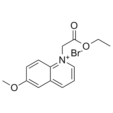
Cas No.: 162558-52-3
Sample solution is provided at 25 µL, 10mM.
MQAEは非比率塩化物イオン(Cl−)消光蛍光センサーとして機能し、細胞内Cl−の濃度([Cl−]i)の測定に使われ、最大の励起/発光波長は350/460nmである[1]。MQAEは、表層蛍光顕微鏡、共焦点顕微鏡、二光子顕微鏡、フローサイトメトリーとその他の生物化学分析実験に適している[1-2]。[Cl−]iの変化と関係なしに、MQAEの濃度が減れば、蛍光の強度は弱まり、MQAEの濃度が増加すれば、蛍光の強度は強まる[2]。MQAEの細胞内分布は均一ではなく、浸透圧ストレスによる細胞体積の変化に影響されない[3]。MQAEは未知の細胞内構造に結合でき、これらの結合のMQAEは、細胞体積が変化する場合でも、気道繊毛細胞で[Cl−]iを測定できるようである[1,4]。MQAEは、ミトコンドリア塩化物イオンの検出用の蛍光プローブの作製に使われることが可能である[5]。MQAEに基づいたプローブの蛍光は、生理pHの範囲では、pHに敏感ではなく、塩化物イオンに消光され、pH 7.0では、Stern-Volmer消光定数は201M-1である[5]。
References:
[1] Ikeuchi Y, Kogiso H, Hosogi S, et al. Measurement of [Cl−]i unaffected by the cell volume change using MQAE-based two-photon microscopy in airway ciliary cells of mice[J]. The journal of physiological sciences, 2018, 68: 191-199.
[2] Andersson C, Roomans G M. Determination of chloride efflux by X‐ray microanalysis versus MQAE‐fluorescence[J]. Microscopy research and technique, 2002, 59(6): 531-535.
[3] Koncz C, Daugirdas J T. Use of MQAE for measurement of intracellular [Cl−] in cultured aortic smooth muscle cells[J]. American Journal of Physiology-Heart and Circulatory Physiology, 1994, 267(6): H2114-H2123.
[4] Miyazaki H, Shiozaki A, Niisato N, et al. Physiological significance of hypotonicity-induced regulatory volume decrease: reduction in intracellular Cl− concentration acting as an intracellular signaling[J]. American Journal of Physiology-Renal Physiology, 2007, 292(5): F1411-F1417.
[5] Park S H, Shin I, Kim Y H, et al. Mitochondrial Cl–-selective fluorescent probe for biological applications[J]. Analytical Chemistry, 2020, 92(18): 12116-12119.
このプロトコールはあくまでもガイドラインであり、ご自分のニーズに合わせてご調整願います。
1.MQAE染色溶液の調製:
5-10mMのMQAE作業溶液は、Krebs-hepes緩衝液(20mM HEPES、128mM NaCl、2.5mM KCl、2.7mM CaCl2、1mM MgCl2、16mMのグルコース;pH 7.4)で調製される。その後、0.22μmのフィルター膜で濾過、殺菌する。実際の条件に応じて、MQAE作業溶液の濃度を調整し、直ちに使用してください。
.2.懸濁細胞の染色(6ウェルプレートのプロトコール)
(1)懸濁細胞について:
1000×gで、細胞懸濁液を3-5分間遠心分離し、注意を払って、上清を吸引除去する。Krebs-Hepes緩衝液で、細胞を二回洗浄し、毎回は5分間洗浄する。その間に、1000×gで遠心分離する。
(2)接着細胞について:
Krebs-Hepes緩衝液で接着細胞を二回すすぐ。トリプシン処理で細胞を剥離し、その後、1000×gで3-5分間遠心分離する。
(3)染料の培養:
細胞ペレットを1mLの染料作業溶液に再懸濁する。室温、暗所で30-60分間培養する(最適の培養時間は細胞の種類により異なる場合がある)。
(4)培養後の処理:
1000×gで5分間遠心分離し、注意を払って上清を除去する。細胞をPBS(毎回5分間)で2-3回洗浄し、その間には遠心分離を行う。
(5)再懸濁と培養:
染色された細胞を無血清培地またはPBSに再懸濁する。顕微鏡の検査を行う。3.接着細胞の染色:
(1)細胞培養物の調製:
希望のコンフルエンスに達するまで、接着細胞を無菌カバーガラスに成長させる。
(2)染色前の処理:
注意を払って、培地を除去し、カバーガラスを加湿チャンバーに置く。
(3)染料の培養:
100μLの染料作業溶液を、カバーガラスの一角から添加し、ゆっくり傾かせ、細胞の単層を溶液が均一にカバーすることを確保する。暗所、室温で30-60分間培養する。
(4)染色後の洗浄:
染料溶液を吸引し、PBSでカバーガラスを2-3回すすぐ(毎回5分間)。
4.顕微鏡の分析:
蛍光顕微鏡またはフローサイトメトリーを使い、染色細胞を検査する。MQAEは、350/460nmの最大励起/発光波長を示す。
References:
[1] Koncz C, Daugirdas J T. Use of MQAE for measurement of intracellular [Cl?] in cultured aortic smooth muscle cells[J]. American Journal of Physiology-Heart and Circulatory Physiology, 1994, 267(6): H2114-H2123.
[2] Miyazaki H, Shiozaki A, Niisato N, et al. Physiological significance of hypotonicity-induced regulatory volume decrease: reduction in intracellular Cl? concentration acting as an intracellular signaling[J]. American Journal of Physiology-Renal Physiology, 2007, 292(5): F1411-F1417.
| Cas No. | 162558-52-3 | SDF | |
| 同義語 | MQAE | ||
| Canonical SMILES | COC1=CC2=CC=C[N+](CC(OCC)=O)=C2C=C1.[Br-] | ||
| Formula | C14H16BrNO3 | M.Wt | 326.19 |
| 溶解度 | DMSO : ≥ 35 mg/mL (107.30 mM) | Storage | Store at -20°C, protect from light |
| General tips | Please select the appropriate solvent to prepare the stock solution according to the
solubility of the product in different solvents; once the solution is prepared, please store it in
separate packages to avoid product failure caused by repeated freezing and thawing.Storage method
and period of the stock solution: When stored at -80°C, please use it within 6 months; when stored
at -20°C, please use it within 1 month. To increase solubility, heat the tube to 37°C and then oscillate in an ultrasonic bath for some time. |
||
| Shipping Condition | Evaluation sample solution: shipped with blue ice. All other sizes available: with RT, or with Blue Ice upon request. | ||
| Prepare stock solution | |||

|
1 mg | 5 mg | 10 mg |
| 1 mM | 3.0657 mL | 15.3285 mL | 30.657 mL |
| 5 mM | 613.1 μL | 3.0657 mL | 6.1314 mL |
| 10 mM | 306.6 μL | 1.5328 mL | 3.0657 mL |
Step 1: Enter information below (Recommended: An additional animal making an allowance for loss during the experiment)
 g
μL
g
μL
Step 2: Enter the in vivo formulation (This is only the calculator, not formulation. Please contact us first if there is no in vivo formulation at the solubility Section.)
Calculation results:
Working concentration: mg/ml;
Method for preparing DMSO master liquid: mg drug pre-dissolved in μL DMSO ( Master liquid concentration mg/mL, Please contact us first if the concentration exceeds the DMSO solubility of the batch of drug. )
Method for preparing in vivo formulation: Take μL DMSO master liquid, next addμL PEG300, mix and clarify, next addμL Tween 80, mix and clarify, next add μL saline, mix and clarify.
Method for preparing in vivo formulation: Take μL DMSO master liquid, next add μL Corn oil, mix and clarify.
Note: 1. Please make sure the liquid is clear before adding the next solvent.
2. Be sure to add the solvent(s) in order. You must ensure that the solution obtained, in the previous addition, is a clear solution before proceeding to add the next solvent. Physical methods such as vortex, ultrasound or hot water bath can be used to aid dissolving.
3. All of the above co-solvents are available for purchase on the GlpBio website.
Quality Control & SDS
- View current batch:
- Purity: >99.50% Appearance: A solid
- COA (Certificate of Analysis)
- SDS (Safety Data Sheet)
- Datasheet
Average Rating: 5 (Based on Reviews and 18 reference(s) in Google Scholar.)