

Coppersensor 1 |
カタログ番号GC62903
 One-Click Copy Product Info
|

Coppersensor 1は、生物サンプル(生細胞を含む)における銅(I)イオン(Cu+)の選択性と敏感な蛍光センサーであり、励起波長は543nmで、発光波長は576nmである。
Products are for research use only. Not for human use. We do not sell to patients.
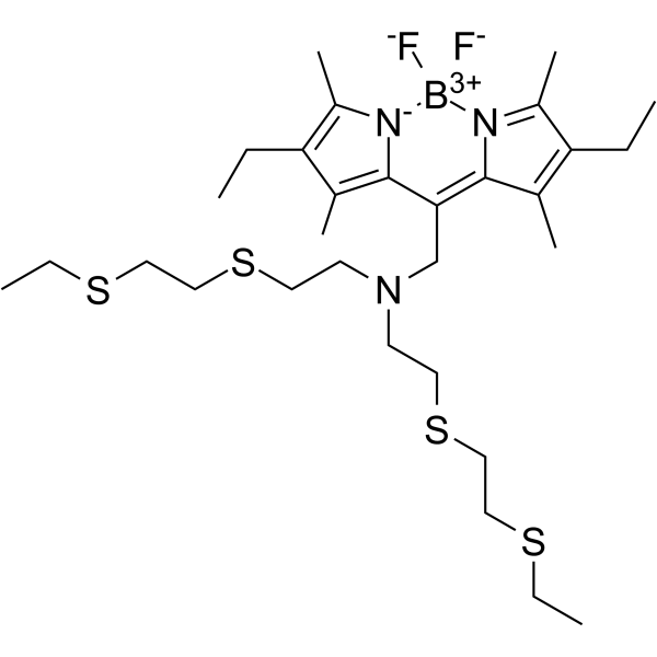
Cas No.: 874748-20-6
Sample solution is provided at 25 µL, 10mM.
Coppersensor 1は、生物サンプル(生細胞を含む)における銅(I)イオン(Cu+)の選択性と敏感な蛍光センサーであり、励起波長は543nmで、発光波長は576nmである[1]。Coppersensor 1は、ボロンジピロメテン(BODIPY)発色団とチオエーテルに富んだ受容体の共役で構成され、Cu+に対しピコモル親和力を持ち、細胞内の様々な金属イオンでCu+に対し高い選択性を持つ。Cu+と結合する時、CS1の蛍光は最大10倍に増える[2]。銅は、鉄と亜鉛に次ぐ三番目に豊富な遷移金属であり、色素の生成、抗酸化、防御とその他の代謝の補因子であるため、生物系で肝心な役割を果たしている[3]。過剰の銅イオンは、活性酸素種(ROS)を生成し、タンパク質、核酸と脂質などの生物分子を破壊し、肝臓、腎臓と消化疾患を誘発し、銅イオンの摂取不足は、脳と神経系の代謝障害を誘発する[4][5]。Coppersensor 1は、癌、抗菌メカニズムと神経変性疾患の研究に応用可能である[6-8]。
References:
[1] Miller, E. W., Zeng, L., Domaille, D. W., & Chang, C. J. (2006). Preparation and use of Coppersensor-1, a synthetic fluorophore for live-cell copper imaging. Nature protocols, 1(2), 824–827.
[2] Zeng, L., Miller, E. W., Pralle, A., Isacoff, E. Y., & Chang, C. J. (2006). A selective turn-on fluorescent sensor for imaging copper in living cells. Journal of the American Chemical Society, 128(1), 10–11.
[3] Li, C., Li, Y., & Ding, C. (2019). The Role of Copper Homeostasis at the Host-Pathogen Axis: From Bacteria to Fungi. International journal of molecular sciences, 20(1), 175.
[4] Scolari Grotto, F., & Glaser, V. (2024). Are high copper levels related to Alzheimer's and Parkinson's diseases? A systematic review and meta-analysis of articles published between 2011 and 2022. Biometals : an international journal on the role of metal ions in biology, biochemistry, and medicine, 37(1), 3–22.
[5] Madsen, E., & Gitlin, J. D. (2007). Copper and iron disorders of the brain. Annual review of neuroscience, 30, 317–337.
[6] Hu, Y., Qian, Y., Wei, J., Jin, T., Kong, X., Cao, H., & Ding, K. (2021). The Disulfiram/Copper Complex Induces Autophagic Cell Death in Colorectal Cancer by Targeting ULK1. Frontiers in pharmacology, 12, 752825.
[7] Santo, C. E., Quaranta, D., & Grass, G. (2012). Antimicrobial metallic copper surfaces kill Staphylococcus haemolyticus via membrane damage. MicrobiologyOpen, 1(1), 46–52.
[8] Eskici, G., & Axelsen, P. H. (2012). Copper and oxidative stress in the pathogenesis of Alzheimer's disease. Biochemistry, 51(32), 6289–6311.
我々の推奨するプロトコールは下記のとおりである。このプロトコールはあくまでもガイドラインであり、特定のニーズに合わせてご調整願います[1]。
1.イメージングのための試薬保存溶液の調製
(1)0.63mgの固体Coppersensor 1をDMSO溶媒の各mlに溶解し、よって1mMのCoppersensor 1貯蔵液(MW 630g/mol)を調製する。
2.生細胞のCoppersensor 1染色:
(1)細胞のイメージング実験には1-2日間の培養期が必要である。ご自分の正常なプロトコールに従い細胞を培養してください。
(2)イメージングの前日に、ポリ-L-リジン(50μg/mL)をコーティングした18mmのカバーガラスの上に細胞を移植する。イメージング用の接着細胞は、50-80%のコンフルエンスまで成長させてください。
(3)細胞をインキュベーターから除去し、3mLのPBS緩衝液を含有する35mmの培養皿にカバーガラスを移した。
(4)培養皿に15μLの1mM Coppersensor 1貯蔵液を添加し、その結果、最終の染料濃度は5μMとなった。徹底的に混合させる。
(染料濃度は高ければ高いほど、背景蛍光レベルは高くなる)
(5)暗所で25または37℃で5-20分間培養する。
3.Coppersensor 1染色細胞のイメージング:
(1)励起波長が543nm、発光波長が576nmの標準共焦点顕微鏡の下で画像化する。(Coppersensor 1は、落射蛍光、共焦点、多光子など、いかなる種類の蛍光顕微鏡でも画像化できる)
注意事項:
①陽性対照群の設置を推奨する。後続する実験を行う前に、細胞を30μMのオレイン酸で8時間培養する。
②蛍光染料は消光しやすい。消光を遅延させるために、できるだけ光から遠ざけてください。
③安全と健康のために、実験中は白衣と使い捨ての手袋をご装着ください。
References:
[1] Miller, E. W., Zeng, L., Domaille, D. W., & Chang, C. J. (2006). Preparation and use of Coppersensor-1, a synthetic fluorophore for live-cell copper imaging. Nature protocols, 1(2), 824–827.
| Cas No. | 874748-20-6 | SDF | |
| Formula | C30H50BF2N3S4 | M.Wt | 629.81 |
| 溶解度 | DMSO : 25 mg/mL (39.69 mM; Need ultrasonic) | Storage | Store at 2-8°C,protect from light |
| General tips | Please select the appropriate solvent to prepare the stock solution according to the
solubility of the product in different solvents; once the solution is prepared, please store it in
separate packages to avoid product failure caused by repeated freezing and thawing.Storage method
and period of the stock solution: When stored at -80°C, please use it within 6 months; when stored
at -20°C, please use it within 1 month. To increase solubility, heat the tube to 37°C and then oscillate in an ultrasonic bath for some time. |
||
| Shipping Condition | Evaluation sample solution: shipped with blue ice. All other sizes available: with RT, or with Blue Ice upon request. | ||
| Prepare stock solution | |||

|
1 mg | 5 mg | 10 mg |
| 1 mM | 1.5878 mL | 7.9389 mL | 15.8778 mL |
| 5 mM | 317.6 μL | 1.5878 mL | 3.1756 mL |
| 10 mM | 158.8 μL | 793.9 μL | 1.5878 mL |
Step 1: Enter information below (Recommended: An additional animal making an allowance for loss during the experiment)
 g
μL
g
μL
Step 2: Enter the in vivo formulation (This is only the calculator, not formulation. Please contact us first if there is no in vivo formulation at the solubility Section.)
Calculation results:
Working concentration: mg/ml;
Method for preparing DMSO master liquid: mg drug pre-dissolved in μL DMSO ( Master liquid concentration mg/mL, Please contact us first if the concentration exceeds the DMSO solubility of the batch of drug. )
Method for preparing in vivo formulation: Take μL DMSO master liquid, next addμL PEG300, mix and clarify, next addμL Tween 80, mix and clarify, next add μL saline, mix and clarify.
Method for preparing in vivo formulation: Take μL DMSO master liquid, next add μL Corn oil, mix and clarify.
Note: 1. Please make sure the liquid is clear before adding the next solvent.
2. Be sure to add the solvent(s) in order. You must ensure that the solution obtained, in the previous addition, is a clear solution before proceeding to add the next solvent. Physical methods such as vortex, ultrasound or hot water bath can be used to aid dissolving.
3. All of the above co-solvents are available for purchase on the GlpBio website.
Quality Control & SDS
- View current batch:
- Purity: >97.00% Appearance: A solid
- COA (Certificate of Analysis)
- SDS (Safety Data Sheet)
- Datasheet
Average Rating: 5 (Based on Reviews and 26 reference(s) in Google Scholar.)