

Digoxin (Synonyms: NSC 95100) |
カタログ番号GC13108
 One-Click Copy Product Info
|

ジゴキシン(Digoxin)は有力なNa+/K+-ATPase阻害剤であり、臨床実験では、不整脈や心不全の治療に使われる。
Products are for research use only. Not for human use. We do not sell to patients.
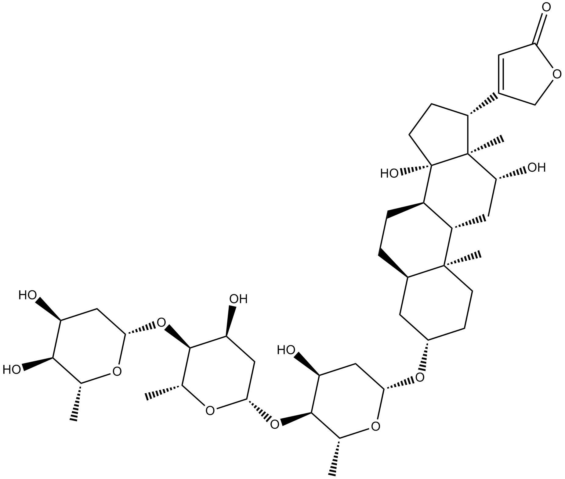
Cas No.: 20830-75-5
Sample solution is provided at 25 µL, 10mM.
ジゴキシン(Digoxin)は有力なNa+/K+-ATPase阻害剤であり、臨床実験では、不整脈や心不全の治療に使われる[1]。ジゴキシンはジギタリス配糖体のクラスに属する。ジギタリス配糖体は心臓の活量と効率を増やし、または心拍と心リズムをコントロールする為に使われている[2]。ジゴキシンはレチノイン酸受容体関連のオーファン核受容体RORγtの転写活性の特定的な阻害剤である[3]。
試験管内実験では、ジゴキシン(0-1µM)でA549とH1299細胞を24または48時間処理した結果、A549とH1299細胞の増殖を用量依存的に阻害しDNAの破壊を誘発し、ROSの生産を増加した[4]。IC50値はそれぞれ0.10と0.12μMである。ジゴキシン(1µM)でT84上皮細胞を72時間処理した結果、P糖タンパク質MDR1の転写レベル(92倍)、総タンパク質(7倍)、頂点MDR1タンパク質(4.7倍)と機能活性(1.75倍)を効果的に誘発した[5]。
生体内実験では、Raji細胞を移植されたマウスにジゴキシン(2mg/kg)を20日間腹腔内注射した結果、66.2%の腫瘍体積を阻害し、マウスには明な毒性効果がなく、腫瘍組織のRaji細胞の分化を減らし、Ki-67とc-mycタンパク質の発見をも減らした[6]。ジゴキシン(1, 2mg/kg)を実験的自己免疫性ぶどう膜炎(EAU)のマウスに14日間静脈内注射したところ、EAUの発展を阻害し、光受容体間レチノイド結合タンパク質(IRBP)に対する細胞免疫反応をも阻害したが、網膜には大きなダメージを与えた[7]。
References:
[1] Ashbrook A W, Lentscher A J, Zamora P F, et al. Antagonism of the sodium-potassium ATPase impairs chikungunya virus infection[J]. MBio, 2016, 7(3): 10.1128/mbio. 00693-16.
[2] Ibrahim N A M. An up-to-date review of digoxin toxicity and its management[J]. Int. J. Res. Pharm. Pharm. Sci, 2019, 4: 59-64.
[3] Huh J R, Leung M W L, Huang P, et al. Digoxin and its derivatives suppress TH17 cell differentiation by antagonizing RORγt activity[J]. Nature, 2011, 472(7344): 486-490.
[4] Wang Y, Ma Q, Zhang S, et al. Digoxin enhances the anticancer effect on non-small cell lung cancer while reducing the cardiotoxicity of adriamycin[J]. Frontiers in pharmacology, 2020, 11: 186.
[5] Haslam I S, Jones K, Coleman T, et al. Rifampin and digoxin induction of MDR1 expression and function in human intestinal (T84) epithelial cells[J]. British journal of pharmacology, 2008, 154(1): 246-255.
[6] Wang T, Xu P, Wang F, et al. Effects of digoxin on cell cycle, apoptosis and NF-κ B pathway in Burkitt’s lymphoma cells and animal model[J]. Leukemia & Lymphoma, 2017, 58(7): 1673-1685.
[7] Hinshaw S J H, Ogbeifun O, Wandu W S, et al. Digoxin inhibits induction of experimental autoimmune uveitis in mice, but causes severe retinal degeneration[J]. Investigative ophthalmology & visual science, 2016, 57(3): 1441-1447.
| 細胞実験[1]: | |
細胞株 | A549とH1299細胞 |
準備方法 | 細胞(3×10⁴ 細胞/mL)を様々な濃度のジゴキシン(0-1µM)で処理した96ウェルのプレートで培養した。各ウェルにMTT(5mg/mL)を追加した後、細胞を更に4時間培養した。 |
反応条件 | 0-1µM; 24または48時間 |
アプリケーション | ジゴキシンはA549とH1299細胞の増殖を用量依存的に阻害し、A549とH1299細胞に対するIC50値はそれぞれ0.10と0.12µMであった。 |
| 動物実験 [2]: | |
動物モデル | 雄のBALB/cヌードマウス |
準備方法 | 異種移植の拒否反応を抑制するためにマウスにシクロホスファミド(75mg/kg)を腹腔内注射した二日後に、四週齢の雄BALB/cヌードマウスの右側面に2×106のRaji細胞(100μLの1640培地とマトリゲル混合物)を皮下注射した。腫瘍の体積が50mm3に達した時、動物をランダムに対照群と実験群に分ける。三週間内、動物にジゴキシン(2mg/kg/日)または溶媒を腹腔内注射した。マウスの腫瘍体積と体重は週に二回監視した。マウスはこの治療の二十日後に殺処分した。腫瘍は収集され、ヘマトキシリンエオシンと免疫組織化学(IHC)の為に4%のパラホルムアルデヒドで固定した。 |
投与形態 | 2mg/kg; i.p.(腹腔内注射) |
アプリケーション | ジゴキシンは腫瘍体積を66.2%阻害し、マウスにはこれといった毒性は与えず、体重にも変化はなかった。ジゴキシンは腫瘍組織のRaji細胞の分布を減少させ、Ki-67とc-mycタンパク質の発見を減少した。 |
References: | |
| Cas No. | 20830-75-5 | SDF | |
| 同義語 | NSC 95100 | ||
| Canonical SMILES | O[C@]1([C@]([C@@H](C2=CC(OC2)=O)CC1)(C)[C@@H]3O)[C@H]4[C@H](C3)[C@](CC[C@@H]5O[C@@H](C[C@@H]6O)O[C@H](C)[C@H]6O[C@@H](C[C@@H]7O)O[C@H](C)[C@H]7O[C@@H](C[C@@H]8O)O[C@H](C)[C@H]8O)(C)[C@@H](C5)CC4 | ||
| Formula | C41H64O14 | M.Wt | 780.94 |
| 溶解度 | ≥ 33.25mg/mL in DMSO | Storage | Store at -20°C,protect from light |
| General tips | Please select the appropriate solvent to prepare the stock solution according to the
solubility of the product in different solvents; once the solution is prepared, please store it in
separate packages to avoid product failure caused by repeated freezing and thawing.Storage method
and period of the stock solution: When stored at -80°C, please use it within 6 months; when stored
at -20°C, please use it within 1 month. To increase solubility, heat the tube to 37°C and then oscillate in an ultrasonic bath for some time. |
||
| Shipping Condition | Evaluation sample solution: shipped with blue ice. All other sizes available: with RT, or with Blue Ice upon request. | ||
| Prepare stock solution | |||

|
1 mg | 5 mg | 10 mg |
| 1 mM | 1.2805 mL | 6.4025 mL | 12.8051 mL |
| 5 mM | 256.1 μL | 1.2805 mL | 2.561 mL |
| 10 mM | 128.1 μL | 640.3 μL | 1.2805 mL |
Step 1: Enter information below (Recommended: An additional animal making an allowance for loss during the experiment)
 g
μL
g
μL
Step 2: Enter the in vivo formulation (This is only the calculator, not formulation. Please contact us first if there is no in vivo formulation at the solubility Section.)
Calculation results:
Working concentration: mg/ml;
Method for preparing DMSO master liquid: mg drug pre-dissolved in μL DMSO ( Master liquid concentration mg/mL, Please contact us first if the concentration exceeds the DMSO solubility of the batch of drug. )
Method for preparing in vivo formulation: Take μL DMSO master liquid, next addμL PEG300, mix and clarify, next addμL Tween 80, mix and clarify, next add μL saline, mix and clarify.
Method for preparing in vivo formulation: Take μL DMSO master liquid, next add μL Corn oil, mix and clarify.
Note: 1. Please make sure the liquid is clear before adding the next solvent.
2. Be sure to add the solvent(s) in order. You must ensure that the solution obtained, in the previous addition, is a clear solution before proceeding to add the next solvent. Physical methods such as vortex, ultrasound or hot water bath can be used to aid dissolving.
3. All of the above co-solvents are available for purchase on the GlpBio website.
Quality Control & SDS
- View current batch:
- Purity: >98.00% Appearance: A solid
- COA (Certificate of Analysis)
- SDS (Safety Data Sheet)
- Datasheet

Average Rating: 5 (Based on Reviews and 16 reference(s) in Google Scholar.)