

Digoxin (Synonyms: NSC 95100) |
Catalog No.GC13108
 One-Click Copy Product Info
|

Digoxin은 강력한 Na+/K+-ATPase 억제제로 임상적으로 부정맥과 심부전을 치료하는데 사용된다.
Products are for research use only. Not for human use. We do not sell to patients.
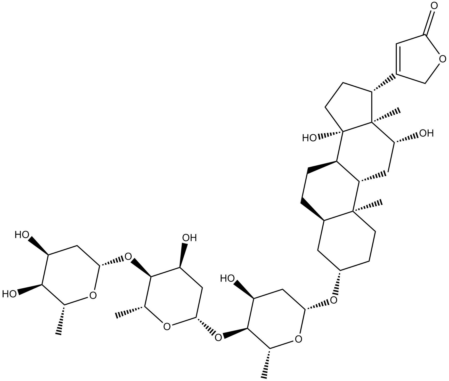
Cas No.: 20830-75-5
Sample solution is provided at 25 µL, 10mM.
Digoxin은 강력한 Na+/K+-ATPase 억제제로 임상적으로 부정맥과 심부전을 치료하는데 사용된다. Digoxin은 심장의 힘과 효율성을 증가시키거나 심박수와 리듬을 조절하기 위해 사용되는 디지털리스 글리코사이드 클래스에 속한다[2]. Digoxin은 비타민A산 수용체와 관련된 고아 핵 수용체 RORγt의 전사활동을 특이적으로 억제하는 인힐레이터이다[3].
체외 실험에서 A549와 H1299 세포에 0-1μM의 Digoxin을 24나 48시간 동안 처리하여 A549와 H1299 세포의 증식을 농도 의존적으로 억제했고 IC50 값이 각각 0.10과 0.12μM였고 DNA 손상과 ROS 생산 증가를 유도했다[4]. Digoxin (1μM)을 T84 상피세포에 72시간 동안 처리하여 P- glycoprotein MDR1의 전사 수준(92배), 총 단백질(7배), 정점 MDR1 단백질(4.7배) 및 기능적 활동(1.75배)을 효과적으로 유도했다[5].
체내 실험에서 라지 세포를 이식한 쥐에복강 주사를 통한 Digoxin (2mg/kg) 치료를 20일 동안 진행하여 종양 부피를 66.2% 억제하고 쥐에 명확한 독성 효과가 없었고 라지 세포가 종양 조직에서의 분포를 줄이고 Ki-67 및 c-myc 단백질의 표현을 낮추었다[6]. 실험적 자가면역성 포도막염 (EAU)가 있는 쥐에 복강 주사를 통한 Digoxin (1, 2mg/kg) 치료를 14일 동안 진행하여 EAU의 발전과 광수용체 간레티노이드 산 결합 단백질 (IRBP)의 세포면역 반응을 억제했고 망막에 심각한 손상을 일으켰다[7].
References:
[1] Ashbrook A W, Lentscher A J, Zamora P F, et al. Antagonism of the sodium-potassium ATPase impairs chikungunya virus infection[J]. MBio, 2016, 7(3): 10.1128/mbio. 00693-16.
[2] Ibrahim N A M. An up-to-date review of digoxin toxicity and its management[J]. Int. J. Res. Pharm. Pharm. Sci, 2019, 4: 59-64.
[3] Huh J R, Leung M W L, Huang P, et al. Digoxin and its derivatives suppress TH17 cell differentiation by antagonizing RORγt activity[J]. Nature, 2011, 472(7344): 486-490.
[4] Wang Y, Ma Q, Zhang S, et al. Digoxin enhances the anticancer effect on non-small cell lung cancer while reducing the cardiotoxicity of adriamycin[J]. Frontiers in pharmacology, 2020, 11: 186.
[5] Haslam I S, Jones K, Coleman T, et al. Rifampin and digoxin induction of MDR1 expression and function in human intestinal (T84) epithelial cells[J]. British journal of pharmacology, 2008, 154(1): 246-255.
[6] Wang T, Xu P, Wang F, et al. Effects of digoxin on cell cycle, apoptosis and NF-κ B pathway in Burkitt’s lymphoma cells and animal model[J]. Leukemia & Lymphoma, 2017, 58(7): 1673-1685.
[7] Hinshaw S J H, Ogbeifun O, Wandu W S, et al. Digoxin inhibits induction of experimental autoimmune uveitis in mice, but causes severe retinal degeneration[J]. Investigative ophthalmology & visual science, 2016, 57(3): 1441-1447.
| 세포 실험 [1]: | |
세포 라인 | A549 및 H1299 세포 |
제조 방법 | 세포 (3×10⁴ 세포/mL)는 96웰 판에서 다양한 농도의 Digoxin (0-1μM)으로 24시간이나 48시간 동안 처리되었습니다. 각 웰에 MTT (5mg/mL)을 추가한 후 세포는 추가로 4시간 더 재배양되었습니다. |
반응 조건 | 0-1µM; 24 또는48시간 동안 |
응용 분야 | Digoxin은 농도 의존적인 방식으로 A549와 H1299 세포의 증식을 억제했고 A549와 H1299 세포의 IC50 값이 각각 0.10과 0.12μM입니다. |
| 동물 실험 [2]: | |
동물 모형 | 수컷 BALB/c 누드 쥐 |
제조 방법 | 200만개의 라지 세포 (100μL의 1640 매디엄과 인공 기저막 혼합물)가 4주령 암컷 BALB/c 무모세포 쥐의 하단부에 피부하로 주입되었고 이틀 후 쥐는 복강 주사를 통해 cyklophosphamide (75mg/kg)를 주입하여 이종 이식 거부 반응을 억제했습니다. 종양 부피가 50mm³에 달했을 때 동물들은 통제군과 치료군으로 무작위 분배되었습니다. 3주 동안 동물들은 복강 주사를 통해 Digoxin (2mg/kg/일)이나 차량으로 치료를 받았습니다. 쥐의 종양 부피와 체중은 매주 두 번 관철했습니다. 치료 20일 후 쥐는 처치되었습니다. 종양은 4%의 파라폼알데히드로 고정되어 헬랭신-이오신 염색과 면역조직화학 (IHC)을 실시했습니다. |
제형 | 2mg/kg; i.p. |
응용 분야 | Digoxin은 종양 부피를 66.2% 억제했고 쥐에게 명확한 독성을 일으키지 않고 체중 변화도 없었습니다. Digoxin은 라지 세포가 종양 조직에서의 분포를 줄였습니다. Digoxin은 Ki-67와 c-myc 단백질의 표현을 낮추었습니다. |
References: | |
| Cas No. | 20830-75-5 | SDF | |
| Synonyms | NSC 95100 | ||
| Canonical SMILES | O[C@]1([C@]([C@@H](C2=CC(OC2)=O)CC1)(C)[C@@H]3O)[C@H]4[C@H](C3)[C@](CC[C@@H]5O[C@@H](C[C@@H]6O)O[C@H](C)[C@H]6O[C@@H](C[C@@H]7O)O[C@H](C)[C@H]7O[C@@H](C[C@@H]8O)O[C@H](C)[C@H]8O)(C)[C@@H](C5)CC4 | ||
| Formula | C41H64O14 | M.Wt | 780.94 |
| Solubility | ≥ 33.25mg/mL in DMSO | Storage | Store at -20°C,protect from light |
| General tips | Please select the appropriate solvent to prepare the stock solution according to the
solubility of the product in different solvents; once the solution is prepared, please store it in
separate packages to avoid product failure caused by repeated freezing and thawing.Storage method
and period of the stock solution: When stored at -80°C, please use it within 6 months; when stored
at -20°C, please use it within 1 month. To increase solubility, heat the tube to 37°C and then oscillate in an ultrasonic bath for some time. |
||
| Shipping Condition | Evaluation sample solution: shipped with blue ice. All other sizes available: with RT, or with Blue Ice upon request. | ||
| Prepare stock solution | |||

|
1 mg | 5 mg | 10 mg |
| 1 mM | 1.2805 mL | 6.4025 mL | 12.8051 mL |
| 5 mM | 256.1 μL | 1.2805 mL | 2.561 mL |
| 10 mM | 128.1 μL | 640.3 μL | 1.2805 mL |
Step 1: Enter information below (Recommended: An additional animal making an allowance for loss during the experiment)
 g
μL
g
μL
Step 2: Enter the in vivo formulation (This is only the calculator, not formulation. Please contact us first if there is no in vivo formulation at the solubility Section.)
Calculation results:
Working concentration: mg/ml;
Method for preparing DMSO master liquid: mg drug pre-dissolved in μL DMSO ( Master liquid concentration mg/mL, Please contact us first if the concentration exceeds the DMSO solubility of the batch of drug. )
Method for preparing in vivo formulation: Take μL DMSO master liquid, next addμL PEG300, mix and clarify, next addμL Tween 80, mix and clarify, next add μL saline, mix and clarify.
Method for preparing in vivo formulation: Take μL DMSO master liquid, next add μL Corn oil, mix and clarify.
Note: 1. Please make sure the liquid is clear before adding the next solvent.
2. Be sure to add the solvent(s) in order. You must ensure that the solution obtained, in the previous addition, is a clear solution before proceeding to add the next solvent. Physical methods such as vortex, ultrasound or hot water bath can be used to aid dissolving.
3. All of the above co-solvents are available for purchase on the GlpBio website.
Quality Control & SDS
- View current batch:
- Purity: >98.00% Appearance: A solid
- COA (Certificate of Analysis)
- SDS (Safety Data Sheet)
- Datasheet

Average Rating: 5 (Based on Reviews and 16 reference(s) in Google Scholar.)