

Ceftriaxone |
カタログ番号GC35648
 One-Click Copy Product Info
|

Ceftriaxoneは、広いスペクトラの殺菌活性を持つβ-ラクタム抗生物質であり、様々なグラム陰性菌とグラム陽性菌に対して良好な抗菌活性を持っている。
Products are for research use only. Not for human use. We do not sell to patients.
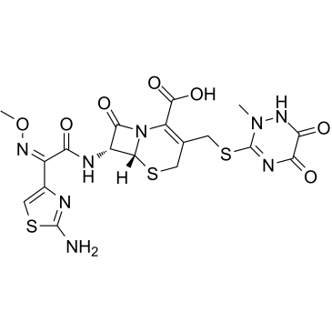
Cas No.: 73384-59-5
Sample solution is provided at 25 µL, 10mM.
Ceftriaxoneは、広いスペクトラの殺菌活性を持つβ-ラクタム抗生物質であり、様々なグラム陰性菌とグラム陽性菌に対して良好な抗菌活性を持っている。Ceftriaxoneは、市中肺炎、髄膜炎や淋病など様々な感染症に使われている。Ceftriaxoneはグリコーゲン合成酵素キナーゼ3β(GSK3β)の共有結合型酵素であり、60分間の前インキュベーション下で、IC50値は0.78μMである。GSK3βはセリン/トレオニンキナーゼであり、幾つかの疾患の有望な創薬標的として見られている。癌において、Ceftriaxoneは抗増殖、細胞周期停止とアポトーシス誘発の特性を発揮する;アルツハイマー病(AD)において、Ceftriaxoneは、タウの過剰リン酸化の阻害に媒介され、アミロイド-βペプチドの産生を減らし、グルタミン酸トランスポーター1を上方制御する[1][2]。
In vitro(体外)実験で、500-1000μMのCeftriaxoneの10日間(A549)、7日間(H520)または14日間(H1650)の処理により、Aurora Bを標的にすることで、コロニー形成を著しく、用量依存的に減らした[2]。LPS誘発性A549とPC3細胞で、Ceftriaxone(100μg/mL;3時間)は、NOD様受容体ピリンドメイン含有タンパク質3(NLRP3)とカスパーゼ-1促進性転写を上方制御したものの、NLRP3タンパク質発現を軽減させた[4]。
In vivo(体内)実験で、A549(57日間)またはH520(32日間)異種移植片を持つ無胸腺ヌードマウスで、Ceftriaxone(100mg/kg;腹腔内投与;週に三回)は、Aurora Bの活性を阻害することで、腫瘍の増殖を著しく遅らせ、腫瘍の大きさを減らした[2]。Ceftriaxone(200mg/kg/日;腹腔内投与;6週間)は、D-ガラクトース(DGL)誘発性肝損傷と腎損傷ラットモデルで、機能指標、酸化ストレスと炎症パラメーターを改善した[3]。
| キナーゼ実験 [1]: | |
|
調製方法 |
Ceftriaxoneを100% (v/v) DMSOに溶解し、10mMの原液(2ml)を調製した。まず、100μMで、GSK3βに対してCeftriaxoneをスクリーニングした。生物学的なアッセイを黒色の384ウェルプレートで行った。Ceftriaxoneの試験を行う為に、100nlの原液(10mM)を5μlのペプチド/キナーゼの混合液、2.4μlのキナーゼ緩衝液(50mM HEPES pH 7.5、0.01% BRIJ-35、10mM MgCl2、1mM EGTA)と2.5μlのATP溶液と混合した。最終的に得られた10μlのキナーゼ反応液には、テストされたセファロスポリン(100μM)、1% (v/v) DMSO、キナーゼペプチド基質と25μM ATP(50mM HEPES pH 7.5、0.01% BRIJ-35、10mM MgCl2と1mM EGTAに溶解)が含まれた。混合液を30秒間振盪して、その後は室温で60分間インキュベートし、キナーゼ反応を完了させた。続いて、5μlの発色試薬溶液を各反応混合液に添加し、30秒間振盪して、室温で更に60分間静置した。最後に、445nmのλexと520nmのλemで、蛍光の発光を測定した。GSK3βを不可逆的に阻害するCeftriaxoneの可能性を評価する為に、ATP溶液を添加する前に、まずは10μMでキナーゼを60分間インキュベートした。 |
|
反応条件 |
100μM;30秒間+60分間、または60分間+60分間 |
|
応用 |
Ceftriaxoneの処理により、60%以上が阻害され、そのIC50値の計算すべく、更なるスクリーニングが検討された。プレインキュベーションなしで、GSK3βに対するIC50値は7.35μMである。GSK3βと60分間プレインキュベートした後、IC50値は0.78μMに低下した。 |
| 細胞実験 [2]: | |
|
細胞株 |
A549、H520とH1650細胞 |
|
調製方法 |
細胞を様々な濃度のCeftriaxoneで10日間(A549)、7日間(H520)または14日間(H1650)で培養し、その後、コロニーを数えた。 |
|
反応条件 |
500または1000μM;7、10または14日間 |
|
応用 |
未処理の対照細胞と比較して、Ceftriaxoneで処理したA549、H520とH1650細胞では、コロニーの形成数が著しく減少した。 |
| 動物実験 [2]: | |
|
動物モデル |
無胸腺ヌードマウス(Cr:NIH(S)、NIH Swissヌードマウス、6–9週齢) |
|
調製方法 |
マウスをランダムに三つのグループに分けた:(i)溶媒群;(ii)100mg/kgのCeftriaxone処理群;(iii)細胞非注射群。100mg/kgのCeftriaxoneで処理したA549またはH520細胞を、最初の二つのグループの各マウスの左側脇腹に皮下接種(2×106個の細胞)した。腫瘍の平均体積が100mm3に達した時、処理を開始した。Ceftriaxone群では、500µlの生理食塩水に溶解した2.5mg Ceftriaxoneを各マウスに、週に三回腹腔内注射(i.p.)した。溶媒群では、500µlの生理食塩水を各マウスに、週に三回腹腔内注射(i.p.)した。動物実験の経過時間は、A549細胞の実験では57日間であり、H520細胞の実験では32日間であった。 |
|
剤形 |
100mg/kg;週に3回;腹腔内注射 |
|
応用 |
溶媒群と比較して、100mg/kg Ceftriaxoneで処理した腫瘍は、増殖が明らかに遅く、腫瘍のサイズも小さかった。溶媒群と比較して、ceftriaxone処理群で、リン酸化ヒストンH3の発現が著しく低下した。ceftriaxoneは、in vivo(体内)で、Aurora Bの活性を阻害することで腫瘍の増殖を抑制した。 |
| Cas No. | 73384-59-5 | SDF | |
| Canonical SMILES | O=C(C(N12)=C(CSC(N(C)NC3=O)=NC3=O)CS[C@]2([H])[C@H](NC(/C(C4=CSC(N)=N4)=N\OC)=O)C1=O)O | ||
| Formula | C18H18N8O7S3 | M.Wt | 554.58 |
| 溶解度 | DMSO:≥100mg/mL | Storage | Store at -20°C |
| General tips | Please select the appropriate solvent to prepare the stock solution according to the
solubility of the product in different solvents; once the solution is prepared, please store it in
separate packages to avoid product failure caused by repeated freezing and thawing.Storage method
and period of the stock solution: When stored at -80°C, please use it within 6 months; when stored
at -20°C, please use it within 1 month. To increase solubility, heat the tube to 37°C and then oscillate in an ultrasonic bath for some time. |
||
| Shipping Condition | Evaluation sample solution: shipped with blue ice. All other sizes available: with RT, or with Blue Ice upon request. | ||
| Prepare stock solution | |||

|
1 mg | 5 mg | 10 mg |
| 1 mM | 1.8032 mL | 9.0158 mL | 18.0317 mL |
| 5 mM | 360.6 μL | 1.8032 mL | 3.6063 mL |
| 10 mM | 180.3 μL | 901.6 μL | 1.8032 mL |
Step 1: Enter information below (Recommended: An additional animal making an allowance for loss during the experiment)
 g
μL
g
μL
Step 2: Enter the in vivo formulation (This is only the calculator, not formulation. Please contact us first if there is no in vivo formulation at the solubility Section.)
Calculation results:
Working concentration: mg/ml;
Method for preparing DMSO master liquid: mg drug pre-dissolved in μL DMSO ( Master liquid concentration mg/mL, Please contact us first if the concentration exceeds the DMSO solubility of the batch of drug. )
Method for preparing in vivo formulation: Take μL DMSO master liquid, next addμL PEG300, mix and clarify, next addμL Tween 80, mix and clarify, next add μL saline, mix and clarify.
Method for preparing in vivo formulation: Take μL DMSO master liquid, next add μL Corn oil, mix and clarify.
Note: 1. Please make sure the liquid is clear before adding the next solvent.
2. Be sure to add the solvent(s) in order. You must ensure that the solution obtained, in the previous addition, is a clear solution before proceeding to add the next solvent. Physical methods such as vortex, ultrasound or hot water bath can be used to aid dissolving.
3. All of the above co-solvents are available for purchase on the GlpBio website.
Quality Control & SDS
- View current batch:
- Purity: >98.00% Appearance: A solid
- COA (Certificate of Analysis)
- SDS (Safety Data Sheet)
- Datasheet
Average Rating: 5 (Based on Reviews and 15 reference(s) in Google Scholar.)