

β,β-Dimethylacrylshikonin |
カタログ番号GC37982
 One-Click Copy Product Info
|

β,β-Dimethylacrylshikoninは、ムラサキ(Lithospermum officinale)から抽出されたナフトキノン誘導体である。
Products are for research use only. Not for human use. We do not sell to patients.
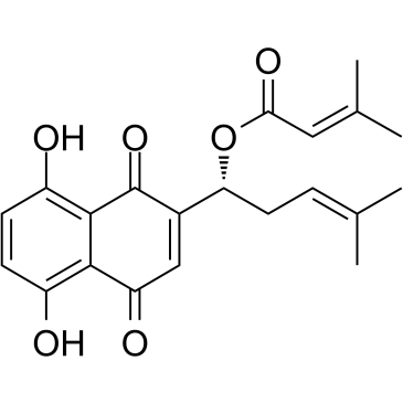
Cas No.: 24502-79-2
Sample solution is provided at 25 µL, 10mM.
β,β-Dimethylacrylshikoninは、ムラサキ(Lithospermum officinale)から抽出されたナフトキノン誘導体である[1]。β,β-Dimethylacrylshikoninは核因子κB (NF-κB)とマイトジェン活性化タンパク質キナーゼ(MAPK)シグナル伝達経路を阻害することで、抗炎症、抗腫瘍と抗酸化作用を発揮する[2, 3]。
体外では、β,β-Dimethylacrylshikonin (15μM)でA549細胞を24時間処理した結果、細胞内で、ミトコンドリア膜電位の低下と、サイトクロムcの放出を誘発し、膜電位は81.9%から33.2%に減った[4]。β,β-Dimethylacrylshikonin (0-15μg/mL)でヒト胃癌SGC-7901細胞を24時間と48時間処理した結果、用量と時間依存的に細胞生存率を阻害し、アポトーシスを誘発し、XIAP、cIAP-2とBcl-2の発見を下方制御し、BakとBaxの発見を上方制御し、ミトコンドリア膜電位の喪失と、サイトクロムcの放出に導く[5]。β,β-Dimethylacrylshikonin (2.5, 5, 10μg/mL)でヒト大腸癌HCT-116細胞を12-48時間処理した結果、用量と時間依存的に細胞の成長を阻害し、アポトーシスと細胞周期停止を誘発する[6]。
体内では、HCT-116異種移植マウスにβ,β-Dimethylacrylshikonin (0.3, 0.6, 1.2mg/kg)を静脈内注射した結果、腫瘍の成長を著しく阻害し、Bcl-2の発見を下方制御し、腫瘍組織でBaxの発見を上方制御した[6]。
References:
[1] Han J, Weng X, Bi K. Antioxidants from a Chinese medicinal herb–Lithospermum erythrorhizon[J]. Food chemistry, 2008, 106(1): 2-10.
[2] Guo C, He J, Song X, et al. Pharmacological properties and derivatives of shikonin—A review in recent years[J]. Pharmacological research, 2019, 149: 104463.
[3] Boulos J C, Rahama M, Hegazy M E F, et al. Shikonin derivatives for cancer prevention and therapy[J]. Cancer letters, 2019, 459: 248-267.
[4] Wang H, Ma X. β, β-Dimethylacrylshikonin induces mitochondria-dependent apoptosis of human lung adenocarcinoma cells in vitro via p38 pathway activation[J]. Acta Pharmacologica Sinica, 2015, 36(1): 131-138.
[5] Shen X J, Wang H B, Ma X Q, et al. β, β-Dimethylacrylshikonin induces mitochondria dependent apoptosis through ERK pathway in human gastric cancer SGC-7901 cells[J]. 2012.
[6] Fan Y, Jin S, He J, et al. Effect of β, β-dimethylacrylshikonin on inhibition of human colorectal cancer cell growth in vitro and in vivo[J]. International Journal of Molecular Sciences, 2012, 13(7): 9184-9193.
| 細胞実験[1]: | |
細胞株 | A549細胞 |
準備方法 | β,β-Dimethylacrylshikonin (15μM)のある/ない条件下で、細胞(5×103細胞/ウェル)を24時間処理し、その後は37℃で、JC-1で15分間染色した。ミトコンドリア膜電位をフローサイトメトリーで検出した。 |
反応条件 | 15μM;24時間 |
アプリケーション | β,β-Dimethylacrylshikoninで処理した後、膜電位は81.9%から33.2%に減少した。 |
| 動物実験 [2]: | |
動物モデル | 雄のヌードマウス |
準備方法 | HCT-116細胞(1×10⁷の細胞/mL, 0.2mL/マウス)を、マウスの左上脇腹領域に皮下注射した。マウスをランダムに4つのグループに分けた。各グループのマウスを毎日、β,β-Dimethylacrylshikonin (0.3, 0.6, 1.2mg/kg)で処理し、または等量の生理食塩水(陰性対照群)を隔日腹腔内投与した。全てのマウスをHCT-116細胞の接種の13日後に殺処分した。腫瘍の最大径と最小径を測定し、腫瘍体積を計算した。 |
投与形態 | 0.3, 0.6, 1.2mg/kg;腹腔内投与 |
アプリケーション | β,β-Dimethylacrylshikoninの処理は、マウスで腫瘍の成長を著しく阻害し、腫瘍組織におけるBcl-2の発見を下方制御し、Baxの発見を上方制御した。 |
References:
[1]Wang H, Ma X. β, β-Dimethylacrylshikonin induces mitochondria-dependent apoptosis of human lung adenocarcinoma cells in vitro via p38 pathway activation[J]. Acta Pharmacologica Sinica, 2015, 36(1): 131-138.
[2]Fan Y, Jin S, He J, et al. Effect of β, β-dimethylacrylshikonin on inhibition of human colorectal cancer cell growth in vitro and in vivo[J]. International Journal of Molecular Sciences, 2012, 13(7): 9184-9193.
| Cas No. | 24502-79-2 | SDF | |
| Canonical SMILES | O=C1C2=C(O)C=CC(O)=C2C(C=C1[C@@H](C/C=C(C)/C)OC(/C=C(C)/C)=O)=O | ||
| Formula | C21H22O6 | M.Wt | 370.4 |
| 溶解度 | DMSO: 25 mg/mL (67.49 mM) | Storage | 4°C, protect from light |
| General tips | Please select the appropriate solvent to prepare the stock solution according to the
solubility of the product in different solvents; once the solution is prepared, please store it in
separate packages to avoid product failure caused by repeated freezing and thawing.Storage method
and period of the stock solution: When stored at -80°C, please use it within 6 months; when stored
at -20°C, please use it within 1 month. To increase solubility, heat the tube to 37°C and then oscillate in an ultrasonic bath for some time. |
||
| Shipping Condition | Evaluation sample solution: shipped with blue ice. All other sizes available: with RT, or with Blue Ice upon request. | ||
| Prepare stock solution | |||

|
1 mg | 5 mg | 10 mg |
| 1 mM | 2.6998 mL | 13.4989 mL | 26.9978 mL |
| 5 mM | 540 μL | 2.6998 mL | 5.3996 mL |
| 10 mM | 270 μL | 1.3499 mL | 2.6998 mL |
Step 1: Enter information below (Recommended: An additional animal making an allowance for loss during the experiment)
 g
μL
g
μL
Step 2: Enter the in vivo formulation (This is only the calculator, not formulation. Please contact us first if there is no in vivo formulation at the solubility Section.)
Calculation results:
Working concentration: mg/ml;
Method for preparing DMSO master liquid: mg drug pre-dissolved in μL DMSO ( Master liquid concentration mg/mL, Please contact us first if the concentration exceeds the DMSO solubility of the batch of drug. )
Method for preparing in vivo formulation: Take μL DMSO master liquid, next addμL PEG300, mix and clarify, next addμL Tween 80, mix and clarify, next add μL saline, mix and clarify.
Method for preparing in vivo formulation: Take μL DMSO master liquid, next add μL Corn oil, mix and clarify.
Note: 1. Please make sure the liquid is clear before adding the next solvent.
2. Be sure to add the solvent(s) in order. You must ensure that the solution obtained, in the previous addition, is a clear solution before proceeding to add the next solvent. Physical methods such as vortex, ultrasound or hot water bath can be used to aid dissolving.
3. All of the above co-solvents are available for purchase on the GlpBio website.
Quality Control & SDS
- View current batch:
- Purity: >98.00% Appearance: A solid
- COA (Certificate of Analysis)
- SDS (Safety Data Sheet)
- Datasheet
Average Rating: 5 (Based on Reviews and 33 reference(s) in Google Scholar.)