

Dihydrotanshinone I (Synonyms: DHTS, Dihydrotanshinone I) |
Katalog-Nr.GN10617
 One-Click Copy Product Info
|

Dihydrotanshinone I is the principal lipophilic phenanthioquinone compound found in Salvia miltiorrhiza Bunge, with antitumor properties.
Products are for research use only. Not for human use. We do not sell to patients.
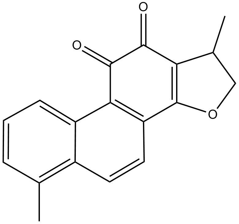
Cas No.: 87205-99-0
Sample solution is provided at 25 µL, 10mM.
_1-3.$$$39978408@51.jpg');)
_1-9.$$$38538795@65.jpg');)
_1-9.$$$39112701@51.jpg');)
_1-7.$$$37316672@70.jpg');)
_366-372.$$$36198801@70.jpg');)
.$$$38565142@65.jpg');)
_1867-1883.$$$33248023@67.jpg');)
_eads6055.$$$39977546@45.jpg');)
_eadm9805.$$$40080571@45.jpg');)
_eadj3020.$$$39666821@45.jpg');)
_1-20.$$$39757301@28.jpg');)
_1-24.$$$38898113@28.jpg');)
_273-287.$$$36806353@46.jpg');)
_1-16.$$$37156877@44.jpg');)
_1-19.$$$37460805@44.jpg');)
_1291-1307.$$$34518654@46.jpg');)
Dihydrotanshinone I is the principal lipophilic phenanthioquinone compound found in Salvia miltiorrhiza Bunge, with antitumor properties [1]. Dihydrotanshinone I mainly induced intrinsic apoptosis mediated by regulation of Bcl-2 family proteins and activation of caspases, which included decreasing mitochondria membrane potential, inhibiting expression of anti-apoptotic protein such as Bcl-xL, Bcl-2[2]. Dihydrotanshinone I has been widely used to inhibit the growth of Gram-positive bacteria and to suppress the expression of pro-inflammatory genes[3].
In vitro, Dihydrotanshinone I significantly inhibited the proliferation of U-2 OS cells, HeLa cells, and NRK-49F cells after 24 hours of treatment, with IC50 values of 3.83μM, 15.48μM, and 25.00μM, respectively[4]. Treatment with 20μM Dihydrotanshinone I for 24 hours significantly induced apoptosis in HCT116 cells, accompanied by an increase in caspase-3/9 levels and the degradation of p62[5]. Treatment of MDA-MB-468 cells with 1μM Dihydrotanshinone I for 24 hours resulted in decreased expression of mesenchymal markers (such as vimentin, N-cadherin and active β-catenin), and inhibition of colony formation[6].
In vivo, Dihydrotanshinone I treatment via oral administration at a dose of 25mg/kg/day for three consecutive days alleviated the pathological changes of colitis in mice induced by dextran sulfate sodium salt (DSS), and inhibited the secretion of inflammatory cytokines[7]. Intraperitoneal injection of Dihydrotanshinone I (10mg/kg) was administered three times a week for 4 weeks, which inhibited the tumor growth in the MDA-MB-231 cell-xenograft mouse model[8].
References:
[1] Yue J, Hao D, Wang Y, et al. The multifaceted mechanisms of Dihydrotanshinone I in the treatment of tumors[J]. Biomedicine & Pharmacotherapy, 2024, 175: 116635.
[2] Chen X, Yu J, Zhong B, et al. Pharmacological activities of dihydrotanshinone I, a natural product from Salvia miltiorrhiza Bunge[J]. Pharmacological Research, 2019, 145: 104254.
[3] Jiang Z, Gao W, Huang L. Tanshinones, critical pharmacological components in Salvia miltiorrhiza[J]. Frontiers in Pharmacology, 2019, 10: 202.
[4] Fan L, Peng C, Zhu X, et al. Dihydrotanshinone I Enhances Cell Adhesion and Inhibits Cell Migration in Osteosarcoma U− 2 OS Cells through CD44 and Chemokine Signaling[J]. Molecules, 2022, 27(12): 3714.
[5] Wang L, Hu T, Shen J, et al. Dihydrotanshinone I induced apoptosis and autophagy through caspase dependent pathway in colon cancer[J]. Phytomedicine, 2015, 22(12): 1079-1087.
[6] Kashyap A, Umar S M, JR A D, et al. Dihydrotanshinone-I modulates epithelial mesenchymal transition (EMT) thereby impairing migration and clonogenicity of triple negative breast cancer cells[J]. Asian Pacific Journal of Cancer Prevention: APJCP, 2021, 22(7): 2177.
[7] Guo Y, Wu X, Wu Q, et al. Dihydrotanshinone I, a natural product, ameliorates DSS-induced experimental ulcerative colitis in mice[J]. Toxicology and applied pharmacology, 2018, 344: 35-45.
[8] Tsai S L, Suk F M, Wang C I, et al. Anti-tumor potential of 15, 16-dihydrotanshinone I against breast adenocarcinoma through inducing G1 arrest and apoptosis[J]. Biochemical pharmacology, 2007, 74(11): 1575-1586.
| Cell experiment [1]: | |
Cell lines | HCT116 cells |
Preparation Method | HCT116 cells were cultured in RPMI 1640 medium supplemented with 10% fetal bovine serum (FBS), 100mg/l penicillin G and 100U/ml streptomycin, under conditions of 37℃ and 5% CO2. The cells were inoculated into 24-well plates and cultured at 37℃ overnight. Cells were treated with different concentrations of Dihydrotanshinone I (0.325, 6.25, 12,5, and 20µM) for 24 hours. The 0.1% (v/v) DMSO solution was used as the solvent control group. After 24 hours, the apoptosis of the cells was detected. |
Reaction Conditions | 0.325, 6.25, 12,5, and 20µM; 24h |
Applications | Dihydrotanshinone I treatment induced apoptosis in HCT116 cells in a dose-dependent manner. |
| Animal experiment [2]: | |
Animal models | Male Balb/cAnN-Foxn1 nude mice |
Preparation Method | Male Balb/cAnN-Foxn1 nude mice (4-5 weeks old) were housed in a standard animal room for 1-2 weeks before the experiment. Each group consisted of 5 mice, and 5×10⁶ MDA-MB-231 cells were subcutaneously injected between the scapulae of each mouse. One week after transplantation, the mice were intraperitoneally injected with 40μl DMSO (control group) or 10mg/kg Dihydrotanshinone I three times a week for 4 weeks. At the end of the experiment, the mice were euthanized by cervical dislocation, and tumor specimens were collected, photographed, and weighed. |
Dosage form | 10mg/kg; three times a week for 4 weeks; i.p. |
Applications | Dihydrotanshinone I treatment reduced the tumor growth in MDA-MB-231 cell-xenograft mouse models. |
References: | |
| Cas No. | 87205-99-0 | SDF | |
| Überlieferungen | DHTS, Dihydrotanshinone I | ||
| Chemical Name | (1R)-1,6-dimethyl-1,2-dihydronaphtho[1,2-g][1]benzofuran-10,11-dione | ||
| Canonical SMILES | CC1COC2=C1C(=O)C(=O)C3=C2C=CC4=C3C=CC=C4C | ||
| Formula | C18H14O3 | M.Wt | 278.3 |
| Löslichkeit | DMF: 0.2 mg/ml,DMSO: 0.2 mg/ml | Storage | Store at -20°C |
| General tips | Please select the appropriate solvent to prepare the stock solution according to the
solubility of the product in different solvents; once the solution is prepared, please store it in
separate packages to avoid product failure caused by repeated freezing and thawing.Storage method
and period of the stock solution: When stored at -80°C, please use it within 6 months; when stored
at -20°C, please use it within 1 month. To increase solubility, heat the tube to 37°C and then oscillate in an ultrasonic bath for some time. |
||
| Shipping Condition | Evaluation sample solution: shipped with blue ice. All other sizes available: with RT, or with Blue Ice upon request. | ||
| Prepare stock solution | |||

|
1 mg | 5 mg | 10 mg |
| 1 mM | 3.5932 mL | 17.9662 mL | 35.9324 mL |
| 5 mM | 718.6 μL | 3.5932 mL | 7.1865 mL |
| 10 mM | 359.3 μL | 1.7966 mL | 3.5932 mL |
Step 1: Enter information below (Recommended: An additional animal making an allowance for loss during the experiment)
 g
μL
g
μL
Step 2: Enter the in vivo formulation (This is only the calculator, not formulation. Please contact us first if there is no in vivo formulation at the solubility Section.)
Calculation results:
Working concentration: mg/ml;
Method for preparing DMSO master liquid: mg drug pre-dissolved in μL DMSO ( Master liquid concentration mg/mL, Please contact us first if the concentration exceeds the DMSO solubility of the batch of drug. )
Method for preparing in vivo formulation: Take μL DMSO master liquid, next addμL PEG300, mix and clarify, next addμL Tween 80, mix and clarify, next add μL saline, mix and clarify.
Method for preparing in vivo formulation: Take μL DMSO master liquid, next add μL Corn oil, mix and clarify.
Note: 1. Please make sure the liquid is clear before adding the next solvent.
2. Be sure to add the solvent(s) in order. You must ensure that the solution obtained, in the previous addition, is a clear solution before proceeding to add the next solvent. Physical methods such as vortex, ultrasound or hot water bath can be used to aid dissolving.
3. All of the above co-solvents are available for purchase on the GlpBio website.
Quality Control & SDS
- View current batch:
- Purity: >99.00% Appearance: A solid
- COA (Certificate of Analysis)
- SDS (Safety Data Sheet)
- Datasheet
Average Rating: 5 (Based on Reviews and 30 reference(s) in Google Scholar.)