

Kojic acid (Synonyms: NSC 1942) |
Katalog-Nr.GC32365
 One-Click Copy Product Info
|

KojisÄure ist eine natÜrliche Substanz, die von Aspergillus oryzae produziert wird und auch als Antioxidans und Strahlenschutzmittel verwendet wird.
Products are for research use only. Not for human use. We do not sell to patients.
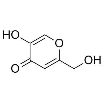
Cas No.: 501-30-4
Sample solution is provided at 25 µL, 10mM.
Kojic acid is a fungus-derived tyrosinase inhibitor with metal-chelating activity (IC50=182.7μM). Kojic acid inhibits the conversion of L-tyrosine to L-DOPA to reduce melanin production and exerts antibacterial effects by enhancing antioxidant activity. Kojic acid can be used for research on pigmentation disorders such as melasma, skin whitening, food anti-browning, and antibacterial preservation[1-4].
In vitro, treatment of A375 cells with 8μg/mL Kojic acid for 24h downregulated tumor suppressor-related genes including APOBEC1, ARHGEF16, CD22, FGFR3, GALNT1, UNC5C, and ZNF146[5]. Treatment of corneal endothelial cells with 1mM Kojic acid for 7 days enhanced cell migration and downregulated galectin 8 and laminin α1 expression[6]. Treatment of HaCaT and SCC-13 cells transfected with pNF-κB-SEAP-NPT plasmid with 10mM Kojic acid inhibited NF-κB activation in the cells[7].
In vivo, intra-articular injection of 10μL 20μM Kojic acid into the knee joint twice weekly for 8 weeks in a mouse model of arthritis reduced cartilage destruction and osteophyte formation, reversed decreases in subchondral bone BV/TV and Tb.Th, increased aggrecan expression, decreased MMP13 expression, and lowered OARSI scores[8]. Subcutaneous injection of 75mg/kg or 300mg/kg Kojic acid 27h before 8Gy (lethal dose) gamma irradiation increased survival rates and promoted recovery of white blood cells, red blood cells, hemoglobin, hematocrit, and platelets in C57BL/6 mice[9]. Oral administration of 50mg/kg/day Kojic acid every other day for 3 weeks in LPS-induced C57BL/6N mice decreased expression of TLR4, Iba-1, GFAP, p-JNK, p-NF-κB, TNF-α, and IL-1β in the brain, reduced LPO and ROS levels, increased Nrf2 and HO-1, and reduced neuronal shrinkage in the cortex and hippocampal DG, CA1, and CA3 regions[10].
References:[1] Saeedi M, Eslamifar M, Khezri K. Kojic acid applications in cosmetic and pharmaceutical preparations. Biomed Pharmacother. 2019 Feb;110:582-593.
[2] Suryadi H, Irianti MI, Septiarini TH. Methods of Random Mutagenesis of Aspergillus Strain for Increasing Kojic Acid Production. Curr Pharm Biotechnol. 2022;23(4):486-494.
[3] Sharma S, Singh S, Sarma SJ. Challenges and advancements in bioprocess intensification of fungal secondary metabolite: kojic acid. World J Microbiol Biotechnol. 2023 Mar 30;39(6):140.
[4] Zilles JC, Dos Santos FL, Kulkamp-Guerreiro IC, et al. Biological activities and safety data of kojic acid and its derivatives: A review. Exp Dermatol. 2022 Oct;31(10):1500-1521.
[5] Cheng S-L, Liu R H, Sheu J-N, et al. Toxicogenomics of Kojic Acid on Gene Expression Profiling of A375 Human Malignant Melanoma Cells. Biol Pharm Bull. 2006 Apr;29(4):655-69.
[6] Wei X, Luo D, Yan Y, et al. Kojic acid inhibits senescence of human corneal endothelial cells via NF-κB and p21 signaling pathways. Exp Eye Res. 2019;180:174-183.
[7] Li J, Yu G, He Z, et al. Kojic acid alleviates osteoarthritis by attenuating inflammation and restoring impaired autophagy through regulating NF-kB and PI3K/AKT/mTOR signaling pathways: An in vivo and in vitro study. Int Immunopharmacol. 2025;147:114022.
[8] Zhong C-C, Xu Z, Gan J, et al. Acute Ongoing Nociception Delays Recovery of Consciousness from Sevoflurane Anesthesia via a Midbrain Circuit. J Neurosci. 2024 Aug 21;44(34):e0740242024.
[9] Wang K, Liu C, Di C, et al. Kojic Acid Protects C57BL/6 Mice from Gamma-irradiation Induced Damage. Asian Pac J Cancer Prev. 2014;15(1):291-297.
[10] Ali W, Choe K, Park JS, et al. Kojic acid reverses LPS-induced neuroinflammation and cognitive impairment by regulating the TLR4/NF-kB signaling pathway. Front Pharmacol. 2024 Aug 9;15:1443552.
| Cell experiment [1]: | |
|
Cell lines |
A375 cells (human malignant melanoma cell line) |
|
Preparation Method |
A375 cells were maintained in Dulbecco's Modified Eagle's Medium (DMEM) supplemented with 10% fetal bovine serum, 1.5g/L sodium bicarbonate, 1mM sodium pyruvate and 1% antibiotic-antimycotic at 37°C, 5% CO2. A375 cells were treated with 8μg/ml Kojic acid for 24h. After treatment, RNA was extracted and gene expression profiling was analyzed by DNA microarray and validated by real-time quantitative PCR. |
|
Reaction Conditions |
8μg/mL; 24h |
|
Applications |
Kojic acid changed gene expression profiling in A375 cells, with 136 up-regulated genes and 225 down-regulated genes. Kojic acid down-regulated APOBEC1, ARHGEF16, CD22, FGFR3, GALNT1, UNC5C and ZNF146 tumor suppressor genes. |
| Animal experiment [2]: | |
|
Animal models |
C57BL/6 mice |
|
Preparation Method |
Mice underwent destabilized medial meniscus (DMM) surgery on the right knee joint. Seven days after surgery, mice received intra-articular injection of Kojic acid (10μL; 20μM) two times per week over an eight-week span. Mice were sacrificed after 8 weeks for analysis. |
|
Dosage form |
20μM in 10μL; intra-articular injection; two times per week for 8 weeks |
|
Applications |
Kojic acid treatment reduced osteophyte formation, alleviated cartilage destruction, reversed DMM-induced decreases in bone volume/tissue volume and trabecular thickness, increased aggrecan expression, decreased MMP13 expression, and lowered OARSI score. |
| References: [1] Cheng S-L, Liu R H, Sheu J-N, et al. Toxicogenomics of Kojic Acid on Gene Expression Profiling of A375 Human Malignant Melanoma Cells. Biol Pharm Bull. 2006 Apr;29(4):655-69. [2] Li J, Yu G, He Z, et al. Kojic acid alleviates osteoarthritis by attenuating inflammation and restoring impaired autophagy through regulating NF-kB and PI3K/AKT/mTOR signaling pathways: An in vivo and in vitro study. Int Immunopharmacol. 2025;147:114022. | |
| Cas No. | 501-30-4 | SDF | |
| Überlieferungen | NSC 1942 | ||
| Canonical SMILES | O=C1C=C(CO)OC=C1O | ||
| Formula | C6H6O4 | M.Wt | 142.11 |
| Löslichkeit | DMSO: ≥ 100 mg/mL (703.68 mM); Water: 50 mg/mL (351.84 mM) | Storage | Store at RT |
| Shipping Condition | Evaluation sample solution: shipped with blue ice. All other sizes available: with RT, or with Blue Ice upon request. | ||
| Prepare stock solution | |||

|
1 mg | 5 mg | 10 mg |
| 1 mM | 7.0368 mL | 35.184 mL | 70.368 mL |
| 5 mM | 1.4074 mL | 7.0368 mL | 14.0736 mL |
| 10 mM | 703.7 μL | 3.5184 mL | 7.0368 mL |
Step 1: Enter information below (Recommended: An additional animal making an allowance for loss during the experiment)
 g
μL
g
μL
Step 2: Enter the in vivo formulation (This is only the calculator, not formulation. Please contact us first if there is no in vivo formulation at the solubility Section.)
Calculation results:
Working concentration: mg/ml;
Method for preparing DMSO master liquid: mg drug pre-dissolved in μL DMSO ( Master liquid concentration mg/mL, Please contact us first if the concentration exceeds the DMSO solubility of the batch of drug. )
Method for preparing in vivo formulation: Take μL DMSO master liquid, next addμL PEG300, mix and clarify, next addμL Tween 80, mix and clarify, next add μL saline, mix and clarify.
Method for preparing in vivo formulation: Take μL DMSO master liquid, next add μL Corn oil, mix and clarify.
Note: 1. Please make sure the liquid is clear before adding the next solvent.
2. Be sure to add the solvent(s) in order. You must ensure that the solution obtained, in the previous addition, is a clear solution before proceeding to add the next solvent. Physical methods such as vortex, ultrasound or hot water bath can be used to aid dissolving.
3. All of the above co-solvents are available for purchase on the GlpBio website.
Quality Control & SDS
- View current batch:
- Purity: >99.50% Appearance: A solid
- COA (Certificate of Analysis)
- SDS (Safety Data Sheet)
- Datasheet
Average Rating: 5 (Based on Reviews and 30 reference(s) in Google Scholar.)