

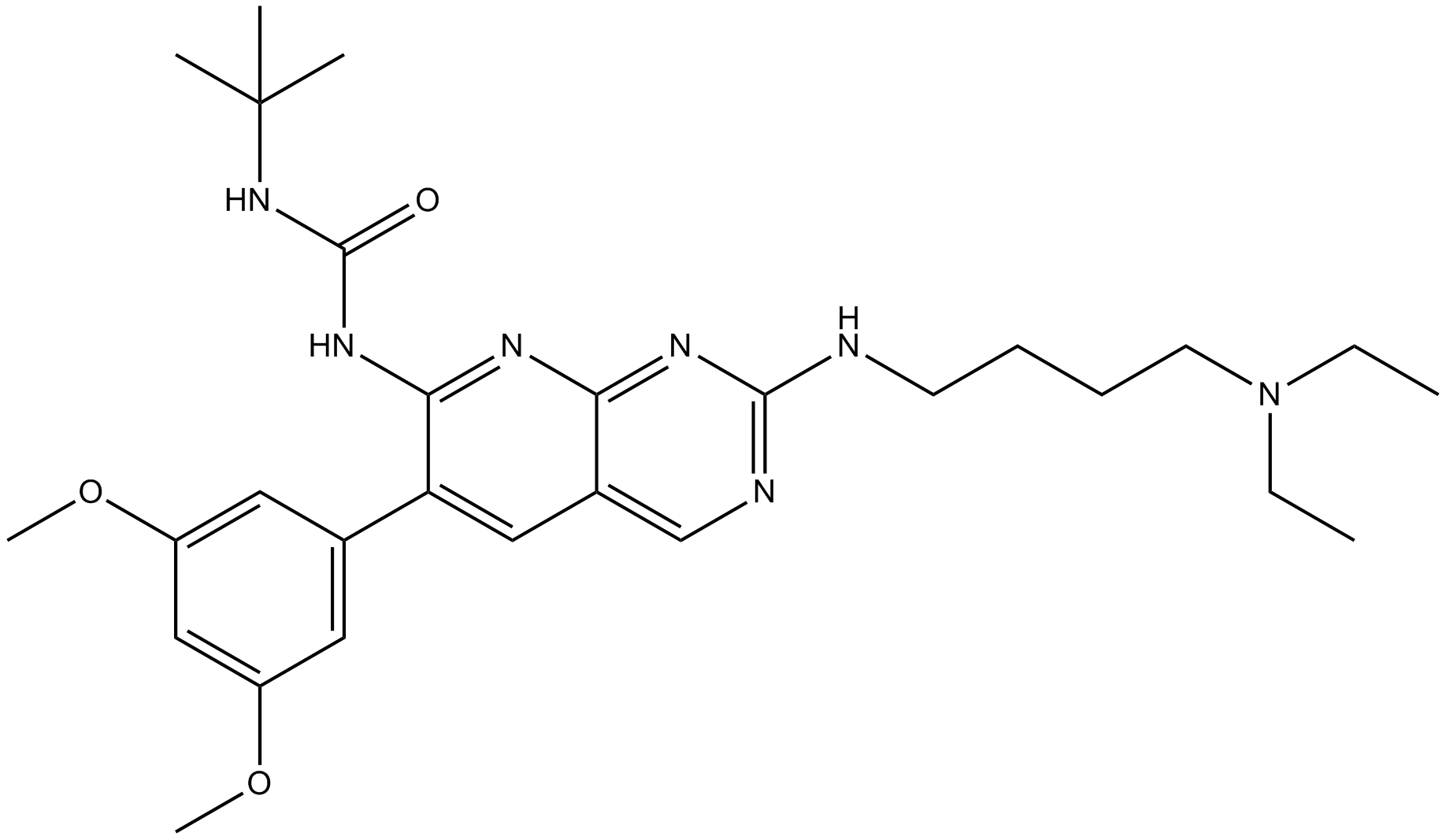
PD173074 (Synonyms: PD 173074,PD-173074) |
Katalog-Nr.GC17943
 One-Click Copy Product Info
|

PD173074 is an ATP-competitive FGFR1 and VEGFR2 inhibitor with an IC50 value of 26nM and 100-200nM, respectively, showing higher selectivity for FGFR1 over PDGFR and c-Src.
Products are for research use only. Not for human use. We do not sell to patients.
Cas No.: 219580-11-7
Sample solution is provided at 25 µL, 10mM.
PD173074 is an ATP-competitive FGFR1 and VEGFR2 inhibitor with an IC50 value of 26nM and 100-200nM, respectively, showing higher selectivity for FGFR1 over PDGFR and c-Src[1]. PD173074 weakly inhibits the activities of Src, InsR, EGFR, PDGFR, MEK, and PKC[2].
in vitro, PD173074 (0.5, 1, 10, 20 and 50μM; 48h) blocks G1/S transition via CUL3-mediated ubiquitin protease in HepG2 and Hep3B cells[3]. PD173074 (0.25 or 1µmol/L, 20µL/well; 4h)reverses MRP7 (ABCC10)-mediated multidrug resistance[4]. PD173074 (0-100μM; 30min) binds to the C-terminus of oncofetal architectural chromatin factor high-mobility group AT-hook 2 (HMGA2) and modulates its DNA-binding and transcriptional activation functions[5].
in vivo, PD173074 (50mg/kg; up to 28 days; goral gavage) blocks small cell lung cancer growth in a nude mice model[6]. PD173074 (20 mg/kg; 3 days; i.p)block FGFR-dependent urothelial carcinoma growth in a Balb/c mice model[7]. PD173074 (20mg/kg; once per day; 10 days; i.p) improves antitumor immunity and impairs breast cancer metastasis in a Balb/c mice model[8].
References:
[1] Dimitroff, C J et al. “Anti-angiogenic activity of selected receptor tyrosine kinase inhibitors, PD166285 and PD173074: implications for combination treatment with photodynamic therapy.” *Investigational new drugs* vol. 17,2 (1999): 121-35. doi:10.1023/a:1006367032156
[2] Tan, Li et al. “Development of covalent inhibitors that can overcome resistance to first-generation FGFR kinase inhibitors.” *Proceedings of the National Academy of Sciences of the United States of America* vol. 111,45 (2014): E4869-77. doi:10.1073/pnas.1403438111
[3] Qiao, Chuchu et al. “PD173074 blocks G1/S transition via CUL3-mediated ubiquitin protease in HepG2 and Hep3B cells.” *PloS one* vol. 15,6 e0234708. 18 Jun. 2020, doi:10.1371/journal.pone.0234708
[4] Anreddy, Nagaraju et al. “PD173074, a selective FGFR inhibitor, reverses MRP7 (ABCC10)-mediated MDR.” *Acta pharmaceutica Sinica. B* vol. 4,3 (2014): 202-7. doi:10.1016/j.apsb.2014.02.003
[5] Ahmed, Syed Moiz et al. “The FGFR inhibitor PD173074 binds to the C-terminus of oncofetal HMGA2 and modulates its DNA-binding and transcriptional activation functions.” *FEBS letters* vol. 597,15 (2023): 1977-1988. doi:10.1002/1873-3468.14675
[6] Pardo, Olivier E et al. “The fibroblast growth factor receptor inhibitor PD173074 blocks small cell lung cancer growth in vitro and in vivo.” *Cancer research*vol. 69,22 (2009): 8645-51. doi:10.1158/0008-5472.CAN-09-1576
[7] Lamont, F R et al. “Small molecule FGF receptor inhibitors block FGFR-dependent urothelial carcinoma growth in vitro and in vivo.” *British journal of cancer* vol. 104,1 (2011): 75-82. doi:10.1038/sj.bjc.6606016
[8] Ye, Tinghong et al. “Inhibition of FGFR signaling by PD173074 improves antitumor immunity and impairs breast cancer metastasis.” *Breast cancer research and treatment* vol. 143,3 (2014): 435-46. doi:10.1007/s10549-013-2829-y
| Kinase experiment [1]: | |
Preparation Method | The assay media for TK activity contained 25mM HEPES buffer (pH 7.4), 150mM NaCl, 10mM MnCl2, 0.1mM sodium orthovanadate, 750µg/ml of random co-polymer of glutamic acid and tyrosine (4:1), various concentrations of inhibitor and 60–75ng of enzyme. The reaction (total volume, 100µL) was initiated by the addition of [γ-32P]ATP (50µM ATP containing 0.4µCi [32P]ATP per incubation. Samples were incubated for 10 minutes at 25℃ and terminated by the addition of 30% trichloroacetic acid. |
Reaction Conditions | 10 minutes at 25℃ |
Applications | PD173074 displayed selective inhibitory activity towards FGFR1TK at 26nM. |
| Cell experiment [2]: | |
Cell lines | HepG2, Hep3B, human normal liver cell line HL7702 |
Preparation Method | HepG2, Hep3B or HL7702 cells were seeded into 96-well plate at 5000cells/well. After adherence, the cells were treated with different concentrations of PD173074 (0.5, 1, 10, 20 and 50μM). 48h later, the cells were incubated with MTT solution (500μg/mL) for 4h at 37°C. The formazan was solubilized with DMSO and relative cell viability was measured at 490nm with a microplate reader. Growth inhibition in response to various concentrations of PD173074 was calculated using GraphPad Prism 7.0. |
Reaction Conditions | 0.5, 1, 10, 20 and 50μM; 48h |
Applications | PD173074 significantly arrested HepG2 and Hep3B cells in G1 phase and inhibited cell proliferation. |
| Animal experiment [3]: | |
Animal models | nude mice |
Preparation Method | H510 (1:1 cell suspension) or H69 cells were implanted into the flank of nude mice. When tumors became measurable, 50mg/kg PD173074/mice or equivalent volume of buffer alone were administered daily for 14 or 28d. In addition, mice received or did not receive two doses of 5mg/kg cisplatin. Tumor volume was monitored using a calliper. Animals were sacrificed when tumor burden reached 15mm in any dimension and survival recorded as a Kaplan-Meier plot. Tissues were formalin fixed and paraffin embedded before staining as indicated in the figure legends. For the endomucin experiments, pictures were acquired using a ×10 objective and analyzed using ImageJ. For activated Caspase 3 and cytokeratin 18 scoring, the number of positive cells in five high-power field views/tumor (five tumors per condition) was determined and results represented as bar graphs. The total number of nuclei per field was determined by manual counting using event flagging in Metamorph. |
Dosage form | 50mg/kg;up to 28 days; goral gavage |
Applications | In the H-510 xenograft, tumor growth was impaired similar to that seen with single-agent cisplatin administration, increasing median survival compared with control sham–treated animals. Crucially, the effect of cisplatin was significantly potentiated by coadministration of PD173074. In H-69 xenografts, PD173074 induced complete responses lasting >6 months in 50% of mice. |
References: | |
| Cas No. | 219580-11-7 | SDF | |
| Überlieferungen | PD 173074,PD-173074 | ||
| Chemical Name | 1-tert-butyl-3-[2-[4-(diethylamino)butylamino]-6-(3,5-dimethoxyphenyl)pyrido[2,3-d]pyrimidin-7-yl]urea | ||
| Canonical SMILES | CCN(CC)CCCCNC1=NC2=NC(=C(C=C2C=N1)C3=CC(=CC(=C3)OC)OC)NC(=O)NC(C)(C)C | ||
| Formula | C28H41N7O3 | M.Wt | 523.67 |
| Löslichkeit | ≥ 26.1835mg/mL in DMSO, ≥ 108.4 mg/mL in EtOH with ultrasonic | Storage | Store at -20°C |
| General tips | Please select the appropriate solvent to prepare the stock solution according to the
solubility of the product in different solvents; once the solution is prepared, please store it in
separate packages to avoid product failure caused by repeated freezing and thawing.Storage method
and period of the stock solution: When stored at -80°C, please use it within 6 months; when stored
at -20°C, please use it within 1 month. To increase solubility, heat the tube to 37°C and then oscillate in an ultrasonic bath for some time. |
||
| Shipping Condition | Evaluation sample solution: shipped with blue ice. All other sizes available: with RT, or with Blue Ice upon request. | ||
| Prepare stock solution | |||

|
1 mg | 5 mg | 10 mg |
| 1 mM | 1.9096 mL | 9.548 mL | 19.096 mL |
| 5 mM | 381.9 μL | 1.9096 mL | 3.8192 mL |
| 10 mM | 191 μL | 954.8 μL | 1.9096 mL |
Step 1: Enter information below (Recommended: An additional animal making an allowance for loss during the experiment)
 g
μL
g
μL
Step 2: Enter the in vivo formulation (This is only the calculator, not formulation. Please contact us first if there is no in vivo formulation at the solubility Section.)
Calculation results:
Working concentration: mg/ml;
Method for preparing DMSO master liquid: mg drug pre-dissolved in μL DMSO ( Master liquid concentration mg/mL, Please contact us first if the concentration exceeds the DMSO solubility of the batch of drug. )
Method for preparing in vivo formulation: Take μL DMSO master liquid, next addμL PEG300, mix and clarify, next addμL Tween 80, mix and clarify, next add μL saline, mix and clarify.
Method for preparing in vivo formulation: Take μL DMSO master liquid, next add μL Corn oil, mix and clarify.
Note: 1. Please make sure the liquid is clear before adding the next solvent.
2. Be sure to add the solvent(s) in order. You must ensure that the solution obtained, in the previous addition, is a clear solution before proceeding to add the next solvent. Physical methods such as vortex, ultrasound or hot water bath can be used to aid dissolving.
3. All of the above co-solvents are available for purchase on the GlpBio website.
Quality Control & SDS
- View current batch:
- Purity: >98.00% Appearance: A solid
- COA (Certificate of Analysis)
- SDS (Safety Data Sheet)
- Datasheet
Average Rating: 5 (Based on Reviews and 30 reference(s) in Google Scholar.)