Startseite>>Biochemical Assay Reagents>>PEG300 (Glycols polyethylene)

PEG300 (Glycols polyethylene)

Katalog-Nr.: GC30022

PEG300 (Glykolpolyethylen) (Polyethylenglykol 300), ein neutrales Polymer mit einem Molekulargewicht von 300, ist ein wasserlÖsliches, wenig immunogenes und biokompatibles Polymer, das aus sich wiederholenden Einheiten von Ethylenglykol gebildet wird.

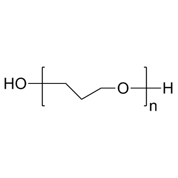
PEG300 (Glycols polyethylene) Chemische Struktur

Größe Preis Lagerbestand Menge
100g
37,00 $
Auf Lager

Kundenbewertungen

Basiert auf Kundenrezensionen.

Tel: (626) 353-8530 E-Mail: sales@glpbio.com

Sample solution is provided at 25 µL, 10mM.

Product Citations

Protocol

Cell experiment [1]:

Cell lines

Caco-2 cells

Preparation Method

When Caco-2 cells were added, 1.5 ml of complete medium was added to the top [apical (AP) compartment] and 2.6 ml to the bottom [basolateral (BL) compartment] of each transwell1. The P-gp activity in these cell monolayers was routinely evaluated by bidirectional transport studies of the P-gp substrate [3H] paclitaxel.

Reaction Conditions

0/20% for 3h

Applications

The AP-to-BL flux of testosterone (0.3 mM) across Caco-2 cell monolayers was determined in the absence and presence of the highest concentration (20%, v/v) of PEG-300 used in these studies to test any possible effects of this excipient on the passive transcellular pathway across Caco2 cells.33 The experimental results suggest that PEG-300 does not alter the passive transcellular pathway across Caco-2 cell monolayers.

References:

[1]. Hugger E D, Audus K L, Borchardt R T. Effects of poly (ethylene glycol) on efflux transporter activity in Caco©\2 cell monolayers[J]. Journal of pharmaceutical sciences, 2002, 91(9): 1980-1990.

Hintergrund

PEG-300, a neutral polymer with a molecular weight of 300, is a water-soluble, low immunogenic and biocompatible polymer formed by repeating units of ethylene glycol [1,2].

Relatively high but clinically achievable PEG-300 levels can inhibit P-gp activity in Caco-2 cell monolayers, thereby enhancing the permeability of anticancer drugs such as paclitaxel and doxorubicin. For example, increasing the concentration of PEG-300 will lead to the increase of Papp and AP to BL of [3H] paclitaxel [P-gp substrate][3-6]12-14,28 and the decrease of Papp and BL to AP. At high concentrations (20%, v/v) of peg-300, it seems that paclitaxel is transported across Caco-2 cell monolayers by simple passive transcellular diffusion. Similar peg induced inhibition of efflux transporters (e.g., P-gp and / or P-gp / MRP activity) was observed in Caco-2 cells, [5] doxorubicin, indicating that PEG induced inhibition of efflux is not a unique phenomenon of paclitaxel.

References:
[1] Lee C C, Su Y C, Ko T P, et al. Structural basis of polyethylene glycol recognition by antibody[J]. Journal of biomedical science, 2020, 27(1): 1-13.
[2] Billingham J, Breen C, Yarwood J. Adsorption of polyamine, polyacrylic acid and polyethylene glycol on montmorillonite: An in situ study using ATR-FTIR[J]. Vibrational Spectroscopy, 1997, 14(1): 19-34.
[3] Krishna R, Mayer L D. Multidrug resistance (MDR) in cancer: mechanisms, reversal using modulators of MDR and the role of MDR modulators in influencing the pharmacokinetics of anticancer drugs[J]. European Journal of Pharmaceutical Sciences, 2000, 11(4): 265-283.
[4] Van Asperen J, Van Tellingen O, Sparreboom A, et al. Enhanced oral bioavailability of paclitaxel in mice treated with the P-glycoprotein blocker SDZ PSC 833[J]. British Journal of Cancer, 1997, 76(9): 1181-1183.
[5] van Asperen J, van Tellingen O, van der Valk M A, et al. Enhanced oral absorption and decreased elimination of paclitaxel in mice cotreated with cyclosporin A[J]. Clinical cancer research: an official journal of the American Association for Cancer Research, 1998, 4(10): 2293-2297.

Chemische Eigenschaften

Cas No. 25322-68-3 SDF
Canonical SMILES OCCCCOC[H].[n]
Formula H(OCH2CH2)nOH M.Wt 300.00
Löslichkeit DMSO : 100 mg/mL (960.15 mM);Water : ≥ 50 mg/mL (480.08 mM) Storage Store at RT
Allgemeine Tipps Um eine höhere Löslichkeit zu erreichen, erwärmen Sie das Röhrchen auf 37°C und schütteln Sie es eine Weile im Ultraschallbad. Die Stammlösung kann mehrere Monate unter -20°C gelagert werden.
Versandbedingungen Probenlösung zur Bewertung: Lieferung mit blauem Eis
Alle anderen verfügbaren Größen: Lieferung mit RT , oder blauem Eis auf Anfrage

In vivo Formulation Calculator (Clear solution)

Step 1: Enter information below (Recommended: An additional animal making an allowance for loss during the experiment)

mg/kg g μL

Step 2: Enter the in vivo formulation (This is only the calculator, not formulation. Please contact us first if there is no in vivo formulation at the solubility Section.)

% DMSO % % Tween 80 % saline
%DMSO %

Calculation results:

Working concentration: mg/ml;

Method for preparing DMSO master liquid: mg drug pre-dissolved in μL DMSO ( Master liquid concentration mg/mL, Please contact us first if the concentration exceeds the DMSO solubility of the batch of drug. )

Method for preparing in vivo formulation: Take μL DMSO master liquid, next addμL PEG300, mix and clarify, next addμL Tween 80, mix and clarify, next add μL saline, mix and clarify.

Method for preparing in vivo formulation: Take μL DMSO master liquid, next add μL Corn oil, mix and clarify.

Note: 1. Please make sure the liquid is clear before adding the next solvent.
2. Be sure to add the solvent(s) in order. You must ensure that the solution obtained, in the previous addition, is a clear solution before proceeding to add the next solvent. Physical methods such as vortex, ultrasound or hot water bath can be used to aid dissolving.
3. All of the above co-solvents are available for purchase on the GlpBio website.

  • Molaritätsrechner

  • Verdünnung-Rechner

Gewicht
=
Konzentration
x
Inhalt
x
MW*


**Bei der Herstellung von Stammlösungen ist immer das chargenspezifische Molekulargewicht von

Berechnen

Research Update

Injectable silk-polyethylene glycol hydrogels

Silk hydrogels for tissue repair are usually pre-formed via chemical or physical treatments from silk solutions. For many medical applications, it is desirable to utilize injectable silk hydrogels at high concentrations (>8%) to avoid surgical implantation and to achieve slow in vivo degradation of the gel. In the present study, injectable silk solutions that formed hydrogels in situ were generated by mixing silk with low-molecular-weight polyethylene glycol (PEG), especially PEG300 and 400 (molecular weight 300 and 400g mol(-1)). Gelation time was dependent on the concentration and molecular weight of PEG. When the concentration of PEG in the gel reached 40-45%, gelation time was less than 30min, as revealed by measurements of optical density and rheological studies, with kinetics of PEG400 faster than PEG300. Gelation was accompanied by structural changes in silk, leading to the conversion from random coil in solution to crystalline β-sheets in the gels, based on circular dichroism, attenuated total reflection Fourier transform infrared spectroscopy and X-ray diffraction. The modulus (127.5kPa) and yield strength (11.5kPa) determined were comparable to those of sonication-induced hydrogels at the same concentrations of silk. The time-dependent injectability of 15% PEG-silk hydrogel through 27G needles showed a gradual increase of compression forces from ?10 to 50N within 60min. The growth of human mesenchymal stem cells on the PEG-silk hydrogels was hindered, likely due to the presence of PEG, which grew after a 5 day delay, presumably while the PEG solubilized away from the gel. When 5% PEG-silk hydrogel was subcutaneously injected in rats, significant degradation and tissue in-growth took place after 20 days, as revealed by ultrasound imaging and histological analysis. No significant inflammation around the gel was observed. The features of injectability, slow degradation and low initial cell attachment suggests that these PEG-silk hydrogels are of interest for many biomedical applications, such as anti-fouling and anti-adhesion.

Pore size of swelling-activated channels for organic osmolytes in Jurkat lymphocytes, probed by differential polymer exclusion

The present study explores the impact of the molecular size on the permeation of low-molecular-weight polyethylene glycols (PEG200-1500) through the plasma membrane of Jurkat cells under iso- and hypotonic conditions. To this end, we analyzed the cell volume responses to PEG-substituted solutions of different osmolalities (100-300 mOsm) using video microscopy. In parallel experiments, the osmotically induced changes in the membrane capacitance and cytosolic conductivity were measured by electrorotation (ROT). Upon moderate swelling in slightly hypotonic solutions (200 mOsm), the lymphocyte membrane remained impermeable to PEG300-1500, which allowed the cells to accomplish regulatory volume decrease (RVD). During RVD, lymphocytes released intracellular electrolytes through the swelling-activated pathways, as proved by a decrease of the cytosolic conductivity measured by electrorotation. RVD also occurred in strongly hypotonic solutions (100 mOsm) of PEG600-1500, whereas 100 mOsm solutions of PEG300-400 inhibited RVD in Jurkat cells. These findings suggest that extensive hypotonic swelling rendered the cell membrane highly permeable to PEG300-400, but not to PEG600-1500. The swelling-activated channels conducting PEG300-400 were inserted into the plasma membrane from cytosolic vesicles via swelling-mediated exocytosis, as suggested by an increase of the whole cell capacitance. Using the hydrodynamic radii R(h) of PEGs (determined by viscosimetry), the observed size-selectivity of membrane permeation yielded an estimate of approximately 0.74 nm for the cut-off radius of the swelling-activated channel for organic osmolytes. Unlike PEG300-1500, the smallest PEG (PEG200, R(h)=0.5 nm) permeated the lymphocyte membrane under isotonic conditions thus leading to a continuous isotonic swelling. The results are of interest for biotechnology and biomedicine, where PEGs are widely used for cryopreservation of cells and tissues.

Chemical Interactions of Polyethylene Glycols (PEGs) and Glycerol with Protein Functional Groups: Applications to Effects of PEG and Glycerol on Protein Processes

In this work, we obtain the data needed to predict chemical interactions of polyethylene glycols (PEGs) and glycerol with proteins and related organic compounds and thereby interpret or predict chemical effects of PEGs on protein processes. To accomplish this, we determine interactions of glycerol and tetraEG with >30 model compounds displaying the major C, N, and O functional groups of proteins. Analysis of these data yields coefficients (α values) that quantify interactions of glycerol, tetraEG, and PEG end (-CH2OH) and interior (-CH2OCH2-) groups with these groups, relative to interactions with water. TetraEG (strongly) and glycerol (weakly) interact favorably with aromatic C, amide N, and cationic N, but unfavorably with amide O, carboxylate O, and salt ions. Strongly unfavorable O and salt anion interactions help make both small and large PEGs effective protein precipitants. Interactions of tetraEG and PEG interior groups with aliphatic C are quite favorable, while interactions of glycerol and PEG end groups with aliphatic C are not. Hence, tetraEG and PEG300 favor unfolding of the DNA-binding domain of lac repressor (lacDBD), while glycerol and di- and monoethylene glycol are stabilizers. Favorable interactions with aromatic and aliphatic C explain why PEG400 greatly increases the solubility of aromatic hydrocarbons and steroids. PEG400-steroid interactions are unusually favorable, presumably because of simultaneous interactions of multiple PEG interior groups with the fused ring system of the steroid. Using α values reported here, chemical contributions to PEG m-values can be predicted or interpreted in terms of changes in water-accessible surface area (ΔASA) and separated from excluded volume effects.

Electroencephalographic effects and serum concentrations after intranasal and intravenous administration of diazepam to healthy volunteers

Aims: To evaluate the electroencephalographic (EEG) effects, blood concentrations, vehicle irritation and dose-effect relationships for diazepam administered nasally.
Methods: The study had a cross-over design with eight healthy volunteers (one drop out). It consisted of four legs with four different administrations: intranasal (i.n.) placebo, 4 mg diazepam i.n., 7 mg diazepam i.n. and 5 mg intravenous (i.v.) diazepam. Polyethylene glycol 300 (PEG300) was used as a vehicle in the nasal formulations to solubilize a clinically relevant dose of diazepam. Changes in N100, P200 and P300 brain event-related potentials (ERP) elicited by auditory stimulation and electroencephalographic beta-activity were used to assess effects on neurological activity.
Results: The mean [95% confidence intervals] differences between before and after drug administration values of P300-N100 amplitude differences were -0.9 [-6.5, 4.7], -6.4 [-10.1, -2,7], -8.6 [-11.4, -5.8] and -9.6 [-12.1, -7.1] for placebo, 4 mg i.n., 7 mg i.n. and 5 mg i.v. diazepam, respectively, indicating statistically significant drug induced effects. The bioavailabilities of 4 and 7 mg i.n. formulations, were found to be similar, 45% [32, 58] and 42% [22, 62], respectively.
Conclusion: The present study indicates that it is possible to deliver a clinically effective nasal dose of diazepam for the acute treatment of epilepsy, using PEG300 as a solubilizer.

Lysine-PEG-modified polyurethane as a fibrinolytic surface: Effect of PEG chain length on protein interactions, platelet interactions and clot lysis

Fibrinolytic polyurethane surfaces were prepared by conjugating lysine to the distal terminus of surface-grafted poly(ethylene glycol) (PEG). Conjugation was through the alpha-amino group leaving the epsilon-amino group free. Lysine in this form is expected to adsorb both plasminogen and t-PA specifically from blood. It was shown in previous work that the PEG spacer, while effectively resisting nonspecific protein adsorption, was a deterrent to the specific binding of plasminogen. In the present work, the effects of PEG spacer chain length on the balance of nonspecific and specific protein binding were investigated. PEG-lysine (PEG-Lys) surfaces were prepared using PEGs of different molecular weight (PEG300 and PEG1000). The lysine-derivatized surfaces with either PEG300 or PEG1000 as spacer showed good resistance to fibrinogen in buffer. The PEG300-Lys surface adsorbed plasminogen from plasma more rapidly than the PEG1000-Lys surface. The PEG300-Lys was also more effective in lysing fibrin formed on the surface. These results suggest that the optimum spacer length for protein resistance and plasminogen binding is relatively short. Immunoblots of proteins eluted after plasma contact confirmed that the PEG-lysine surface adsorbed plasminogen while resisting most of the other plasma proteins. The hemocompatibility of the optimized PEG-lysine surface was further assessed in whole blood experiments in which fibrinogen adsorption and platelet adhesion were measured simultaneously. Platelet adhesion was shown to be strongly correlated with fibrinogen adsorption. Platelet adhesion was very low on the PEG-containing surfaces and neither surface-bound lysine nor adsorbed plasminogen promoted platelet adhesion.

Bewertungen

Review for PEG300 (Glycols polyethylene)

Average Rating: 5 ★★★★★ (Based on Reviews and 31 reference(s) in Google Scholar.)

5 Star
100%
4 Star
0%
3 Star
0%
2 Star
0%
1 Star
0%
Review for PEG300 (Glycols polyethylene)

GLPBIO products are for RESEARCH USE ONLY. Please make sure your review or question is research based.

Required fields are marked with *

You may receive emails regarding this submission. Any emails will include the ability to opt-out of future communications.