

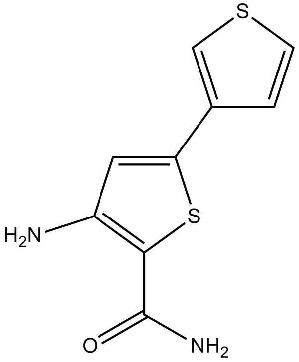
SC-514 (Synonyms: GK 01140) |
Katalog-Nr.GC12372
 One-Click Copy Product Info
|

SC-514 ist ein selektiver IKK-2-Inhibitor (IC50 = 11,2 μM), der andere IKK-Isoformen oder andere Serin-Threonin- und Tyrosinkinasen nicht hemmt.
Products are for research use only. Not for human use. We do not sell to patients.
Cas No.: 354812-17-2
Sample solution is provided at 25 µL, 10mM.
_1-3.$$$39978408@51.jpg');)
_1-9.$$$38538795@65.jpg');)
_1-9.$$$39112701@51.jpg');)
_1-7.$$$37316672@70.jpg');)
_366-372.$$$36198801@70.jpg');)
.$$$38565142@65.jpg');)
_1867-1883.$$$33248023@67.jpg');)
_eads6055.$$$39977546@45.jpg');)
_eadm9805.$$$40080571@45.jpg');)
_eadj3020.$$$39666821@45.jpg');)
_1-20.$$$39757301@28.jpg');)
_1-24.$$$38898113@28.jpg');)
_273-287.$$$36806353@46.jpg');)
_1-16.$$$37156877@44.jpg');)
_1-19.$$$37460805@44.jpg');)
_1291-1307.$$$34518654@46.jpg');)
SC-514 is a cell-permeable and selective inhibitor of IKK-2 with IC50 value of 3-12μM [1, 2].
SC-514 inhibits all forms of recombinant human IKK-2 with IC50 values in the 3–12μM range. It also inhibits the native IKK complex. SC-514 specifically binds at the ATP-binding site of IKK-2 and exerts a reversible and competitive inhibition with ATP. However, SC-514 shows non-competitive inhibition with the IκB site. As an inhibitor of IKK-2, SC-514 is found to block the phosphorylation and degradation of IκBα and reduce the translocation level of p65 into the nucleus in IL-1β-treated RASFs. Additionally, SC-514 shows dose-dependent inhibition in the transcription of NF-κB-induced genes, including IL-6, IL-8, and COX-2. Moreover, SC-514 shows efficacious in reduction of LPS-induced TNFα production in the acute model of inflammation. SC-514 is also reported to inhibit the osteoclastogenesis in BMM cells through attenuating RANKL-induced activation of NF-κB [1, 2].
References:
[1] Kishore N, Sommers C, Mathialagan S, Guzova J, Yao M, Hauser S, Huynh K, Bonar S, Mielke C, Albee L, Weier R, Graneto M, Hanau C, Perry T, Tripp CS. A selective IKK-2 inhibitor blocks NF-kappa B-dependent gene expression in interleukin-1 beta-stimulated synovial fibroblasts. J Biol Chem. 2003 Aug 29;278(35):32861-71.
[2] Liu Q, Wu H, Chim SM, Zhou L, Zhao J, Feng H, Wei Q, Wang Q, Zheng MH, Tan RX, Gu Q, Xu J, Pavlos N, Tickner J, Xu J. SC-514, a selective inhibitor of IKKβ attenuates RANKL-induced osteoclastogenesis and NF-κB activation. Biochem Pharmacol. 2013 Dec 15;86(12):1775-83.
Kinase experiment: | IKK complexes are immunoprecipitated from IL-1β-treated RASF cell lysates (0.5-2 mg) using a NEMO antibody (3-10 μg) followed by the addition of protein A-agarose beads. Antibody complexes are pelleted by centrifugation and washed 3 times with 1 mL of cold whole-cell lysis buffer followed by 2 washes in kinase buffer (25 mM HEPES, pH 7.6, 2 mM MgCl2, 2 mM MnCl2, 10 mM NaF, 5 mM DTT, and 1 mM phenylmethylsulfonyl fluoride). 100-200 μg of immunoprecipitated IKK is analyzed for kinase activity in a reaction containing 10 μM biotinylated IκBα peptide as substrate and 1 μM [γ-33P]ATP (2500 Ci/mmol). After incubation at room temperature for 30 min, 25 μL of the reaction mixture is withdrawn and added to a SAM 96 biotin capture plate. After successive wash steps the plate was allowed to air-dry, and 25 μL of scintillation fluid is added to each well. Incorporation of [γ-33P]ATP is measured using a Top-Count NXT[1]. |
Cell experiment: | For crystal violet staining assay, melanoma cell lines (1×10⁴) are seeded in 60 mm dishes, and then untreated or pretreated with SC-514 (50 µM) and/or Fotemustine. Then, cells are formalin-fixed and stained with crystal violet. Cell numbers are measured as the optical density at 595 nm (OD595) of solubilized crystal violet from formalin-fixed cells. Cytotoxicity are also determined by the MTT reduction assay[2]. |
Animal experiment: | Rats[1] SC-514 or vehicle (2% Me2SO in saline) is administered either by oral gavage (50 mg/kg) or intraperitoneally (10 and 50 mg/kg) to adult male Wistar rats that have been deprived of food overnight. Two hours after compound treatment, 1 mg/kg LPS (Escherichia coli) in saline is administered intraperitoneally 90 min after LPS administration; the animals are bled and serum TNFα levels analyzed by a rat-specific TNFα ELISA.Mice[2] Male nu/nu BALB/c mice (6 weeks old) are maintained in individual ventilated cages. A375 or G361 (5×10⁶) cells are resuspended in 0.1 mL PBS and inoculated subcutaneously into the backs of nude mice and allowed to grow for 7 days. After that, mice are randomly assigned to 4 groups (n=6 for each group) and treated by intraperitoneal injection with 200 µL 30% PEG/5% Tween-80 solution as the vehicle control and 25 mg/kg SC-514 and/or 25 mg/kg Fotemustine daily for 13-15 consecutive days. Body weight and tumor volume are measured every 3 days. Tumor volumes are determined by a caliper and calculated. At the end of the experiment, mice are sacrificed and tumor xenografts are collected. Tumor tissues are stored at -80°C for Western blot analysis. |
References: [1]. Kishore N, et al. A selective IKK-2 inhibitor blocks NF-kappa B-dependent gene expression in interleukin-1 beta-stimulated synovial fibroblasts. J Biol Chem. 2003 Aug 29;278(35):32861-71. | |
| Cas No. | 354812-17-2 | SDF | |
| Überlieferungen | GK 01140 | ||
| Chemical Name | 3-amino-5-thiophen-3-ylthiophene-2-carboxamide | ||
| Canonical SMILES | C1=CSC=C1C2=CC(=C(S2)C(=O)N)N | ||
| Formula | C9H8N2OS2 | M.Wt | 224.3 |
| Löslichkeit | ≥ 50.4mg/mL in DMSO | Storage | Store at -20° C |
| General tips | Please select the appropriate solvent to prepare the stock solution according to the
solubility of the product in different solvents; once the solution is prepared, please store it in
separate packages to avoid product failure caused by repeated freezing and thawing.Storage method
and period of the stock solution: When stored at -80°C, please use it within 6 months; when stored
at -20°C, please use it within 1 month. To increase solubility, heat the tube to 37°C and then oscillate in an ultrasonic bath for some time. |
||
| Shipping Condition | Evaluation sample solution: shipped with blue ice. All other sizes available: with RT, or with Blue Ice upon request. | ||
| Prepare stock solution | |||

|
1 mg | 5 mg | 10 mg |
| 1 mM | 4.4583 mL | 22.2916 mL | 44.5831 mL |
| 5 mM | 891.7 μL | 4.4583 mL | 8.9166 mL |
| 10 mM | 445.8 μL | 2.2292 mL | 4.4583 mL |
Step 1: Enter information below (Recommended: An additional animal making an allowance for loss during the experiment)
 g
μL
g
μL
Step 2: Enter the in vivo formulation (This is only the calculator, not formulation. Please contact us first if there is no in vivo formulation at the solubility Section.)
Calculation results:
Working concentration: mg/ml;
Method for preparing DMSO master liquid: mg drug pre-dissolved in μL DMSO ( Master liquid concentration mg/mL, Please contact us first if the concentration exceeds the DMSO solubility of the batch of drug. )
Method for preparing in vivo formulation: Take μL DMSO master liquid, next addμL PEG300, mix and clarify, next addμL Tween 80, mix and clarify, next add μL saline, mix and clarify.
Method for preparing in vivo formulation: Take μL DMSO master liquid, next add μL Corn oil, mix and clarify.
Note: 1. Please make sure the liquid is clear before adding the next solvent.
2. Be sure to add the solvent(s) in order. You must ensure that the solution obtained, in the previous addition, is a clear solution before proceeding to add the next solvent. Physical methods such as vortex, ultrasound or hot water bath can be used to aid dissolving.
3. All of the above co-solvents are available for purchase on the GlpBio website.
Quality Control & SDS
- View current batch:
- Purity: >99.50% Appearance: A solid
- COA (Certificate of Analysis)
- SDS (Safety Data Sheet)
- Datasheet
Average Rating: 5 (Based on Reviews and 9 reference(s) in Google Scholar.)