

Atosiban (Synonyms: RW22164; RWJ22164) |
カタログ番号GC12194
 One-Click Copy Product Info
|

Atosibanはオキシトシンとバソプレシン受容体の混合拮抗剤である。子宮平滑筋でオキシトシン受容体を競合的に遮断することで、子宮収縮を効果的に阻害し、妊娠の時間を延ばす。
Products are for research use only. Not for human use. We do not sell to patients.
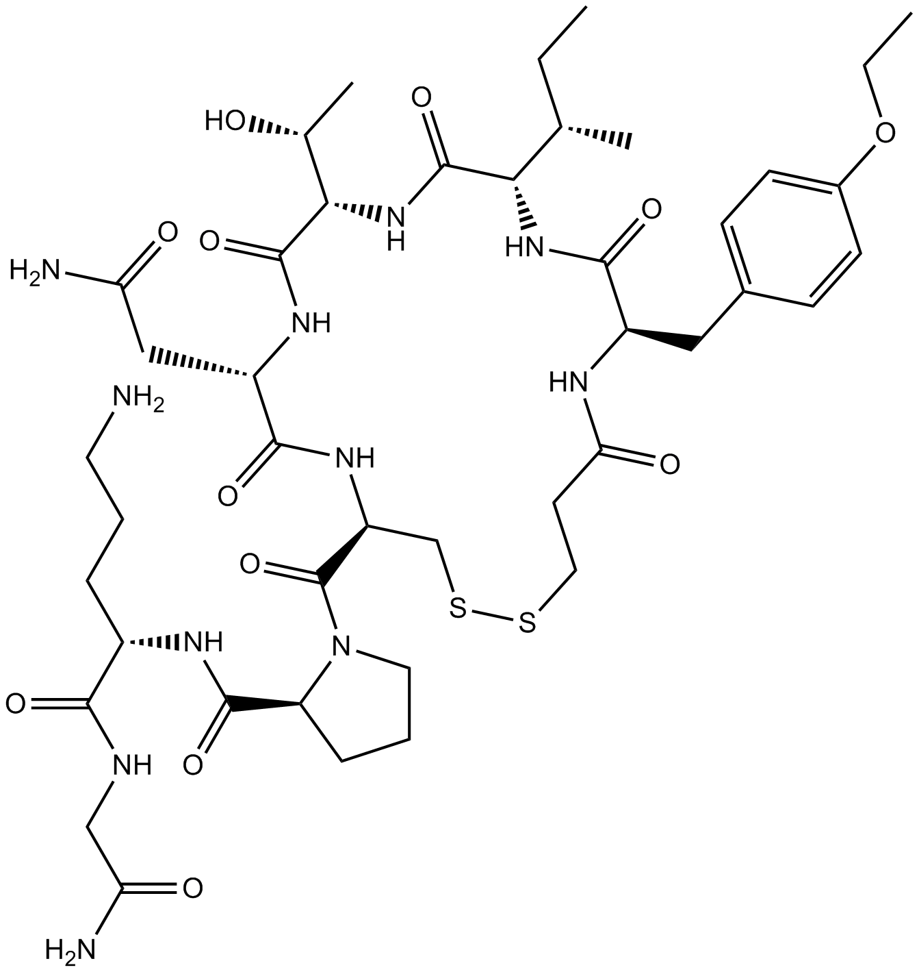
Cas No.: 90779-69-4
Sample solution is provided at 25 µL, 10mM.
_1-3.$$$39978408@51.jpg');)
_1-9.$$$38538795@65.jpg');)
_1-9.$$$39112701@51.jpg');)
_1-7.$$$37316672@70.jpg');)
_366-372.$$$36198801@70.jpg');)
.$$$38565142@65.jpg');)
_1867-1883.$$$33248023@67.jpg');)
_eads6055.$$$39977546@45.jpg');)
_eadm9805.$$$40080571@45.jpg');)
_eadj3020.$$$39666821@45.jpg');)
_1-20.$$$39757301@28.jpg');)
_1-24.$$$38898113@28.jpg');)
_273-287.$$$36806353@46.jpg');)
_1-16.$$$37156877@44.jpg');)
_1-19.$$$37460805@44.jpg');)
_1291-1307.$$$34518654@46.jpg');)
Atosibanはオキシトシンとバソプレシン受容体の混合拮抗剤である。子宮平滑筋でオキシトシン受容体を競合的に遮断することで、子宮収縮を効果的に阻害し、妊娠の時間を延ばす[1, 2]。Atosibanは合成のペプチド子宮収縮抑制剤であり、早産の治療に使われる[3]。
体外で、Atosiban (10μM)でヒト子宮筋細胞を処理した結果、オキシトシン(OT)誘発の細胞内p38キナーゼの活性化とCOX-2の上方制御を著しく減らした[4]。Atosiban (10μM)でヒト臍帯静脈内皮細胞(HUVECs)を処理した結果、HUVECsで、低酸素誘導性因子-1 α (HIF-1α)のプロモーター活性を著しく阻害し、HIF-1αのmRNAとタンパク質の発見を減らした[5]。
体内で、実験性子宮内膜症ラットにAtosiban (0.5mg/kg/日)を21日間腹腔内注射した結果、子宮内膜症インプラントの体積を著しく減らし、移植組織で、増殖細胞核抗原の発見レベルを著しく減らした[6]。Atosiban (6mg/kg/日)を妊娠したラットに21日間腹腔内注射した結果、新生ラットの血漿と心臓組織で、酸化ストレスのレベルを著しく増やした[7]。
References:
[1] McCafferty G P, Pullen M A, Wu C, et al. Use of a novel and highly selective oxytocin receptor antagonist to characterize uterine contractions in the rat[J]. American Journal of Physiology-Regulatory, Integrative and Comparative Physiology, 2007, 293(1): R299-R305.
[2] Kuć P, Laudański P, Pierzyński P, et al. The effect of combined tocolysis on in vitro uterine contractility in preterm labour[J]. Advances in medical sciences, 2011, 56(1): 88-94.
[3] Di Renzo G C. Safety and efficacy of new drugs in preterm labor[J]. Expert Review of Obstetrics & Gynecology, 2007, 2(1): 19-24.
[4] Kim S H, Pohl O, Chollet A, et al. Differential effects of oxytocin receptor antagonists, Atosiban and Nolasiban, on oxytocin receptor–mediated signaling in human amnion and myometrium[J]. Molecular Pharmacology, 2017, 91(4): 403-415.
[5] Zhu J, Wang H, Zhang X, et al. Regulation of angiogenic behaviors by oxytocin receptor through Gli1-indcued transcription of HIF-1α in human umbilical vein endothelial cells[J]. Biomedicine & Pharmacotherapy, 2017, 90: 928-934.
[6] Simsek Y, Celik O, Karaer A, et al. Therapeutic efficiency of Atosiban, an oxytocin receptor blocking agent in the treatment of experimental endometriosis[J]. Archives of gynecology and obstetrics, 2012, 286(3): 777-783.
[7] Simsek Y, Celik O, Karaer A, et al. Elevated cardiac oxidative stress in newborn rats from mothers treated with atosiban[J]. Archives of gynecology and obstetrics, 2012, 285(3): 655-661.
| 細胞実験[1]: | |
細胞株 | 出生前の子宮筋層平滑筋細胞 |
準備方法 | Atosiban (10μM)のある/ない条件下で、オキシトシン(OT) (10nM)で出生前の子宮筋層平滑筋細胞を5分間、15分間、30分間、2時間、4時間と6時間刺激した。リン酸化されたNF-κB p65サブユニット、ERK1/2、p38 MAPK、並びにCOX-2とp-cPLA2の発見をウエスタンブロットで検出した。 |
反応条件 | 10μM;5分間、15分間、30分間、2時間、4時間と6時間 |
アプリケーション | Atosibanの処理は、ヒト子宮筋層細胞で、p38キナーゼのOT誘発の活性化と、COX-2の上方制御を著しく減らした。 |
| 動物実験 [2]: | |
動物モデル | 雌のWistarラット |
準備方法 | 発情期にある35匹の雌ラットに、外科手術で子宮内膜症を誘発した。この手続きの4週間後に、再開腹を行った。子宮内膜症病巣の生存能力と大きさを記録した。その後、ラットをランダムに三つのグループに分けた。最初のグループ(n=8)で、毎日の用量が0.2mL 0.9% NaClを腹腔内注射(i.p.)した(対照)。第二と第三のグループ(n=8とn=8)で、0.5mg/kg/日のAtosibanと1mg/日のdiltiazemをそれぞれ腹腔内注射した。毎日ほぼ同じ時間で、処理を21日間連続で行った。処理の終わりに、開腹手術を行い、子宮内膜症病巣の大きさを記録した。組織学と免疫組織化学的な実験で、子宮内膜インプラントを処理した。子宮内膜インプラントの体積を測定し、免疫組織学的な分析を行い、グループの間で比較した。 |
投与形態 | 0.5mg/kg/日;21日間;腹腔内注射 |
アプリケーション | Atosibanの処理後、子宮内膜インプラントの体積は著しく減少した。対照群と比べて、増殖細胞核抗原の発見レベルは、Atosibanとdiltiazem群で、著しく減少した。 |
References: | |
| Cas No. | 90779-69-4 | SDF | |
| 同義語 | RW22164; RWJ22164 | ||
| Formula | C43H67N11O12S2 | M.Wt | 994.19 |
| 溶解度 | DMF: 30 mg/ml,DMSO: 14 mg/ml,Ethanol: 5 mg/ml,PBS (pH 7.2): 5 mg/ml | Storage | Store at -20°C |
| General tips | Please select the appropriate solvent to prepare the stock solution according to the
solubility of the product in different solvents; once the solution is prepared, please store it in
separate packages to avoid product failure caused by repeated freezing and thawing.Storage method
and period of the stock solution: When stored at -80°C, please use it within 6 months; when stored
at -20°C, please use it within 1 month. To increase solubility, heat the tube to 37°C and then oscillate in an ultrasonic bath for some time. |
||
| Shipping Condition | Evaluation sample solution: shipped with blue ice. All other sizes available: with RT, or with Blue Ice upon request. | ||
| Prepare stock solution | |||

|
1 mg | 5 mg | 10 mg |
| 1 mM | 1.0058 mL | 5.0292 mL | 10.0584 mL |
| 5 mM | 201.2 μL | 1.0058 mL | 2.0117 mL |
| 10 mM | 100.6 μL | 502.9 μL | 1.0058 mL |
Step 1: Enter information below (Recommended: An additional animal making an allowance for loss during the experiment)
 g
μL
g
μL
Step 2: Enter the in vivo formulation (This is only the calculator, not formulation. Please contact us first if there is no in vivo formulation at the solubility Section.)
Calculation results:
Working concentration: mg/ml;
Method for preparing DMSO master liquid: mg drug pre-dissolved in μL DMSO ( Master liquid concentration mg/mL, Please contact us first if the concentration exceeds the DMSO solubility of the batch of drug. )
Method for preparing in vivo formulation: Take μL DMSO master liquid, next addμL PEG300, mix and clarify, next addμL Tween 80, mix and clarify, next add μL saline, mix and clarify.
Method for preparing in vivo formulation: Take μL DMSO master liquid, next add μL Corn oil, mix and clarify.
Note: 1. Please make sure the liquid is clear before adding the next solvent.
2. Be sure to add the solvent(s) in order. You must ensure that the solution obtained, in the previous addition, is a clear solution before proceeding to add the next solvent. Physical methods such as vortex, ultrasound or hot water bath can be used to aid dissolving.
3. All of the above co-solvents are available for purchase on the GlpBio website.
Quality Control & SDS
- View current batch:
- Purity: >99.00% Appearance: A solid
- COA (Certificate of Analysis)
- SDS (Safety Data Sheet)
- Datasheet
Average Rating: 5 (Based on Reviews and 9 reference(s) in Google Scholar.)