

DPCPX |
カタログ番号GC16099
 One-Click Copy Product Info
|

DPCPXは有力で選択性の高親和性A1アデノシン受容体拮抗剤であり、ラット脂肪細胞膜で、アデニル酸シクラーゼに対するKi値は0.45 nMである。
Products are for research use only. Not for human use. We do not sell to patients.
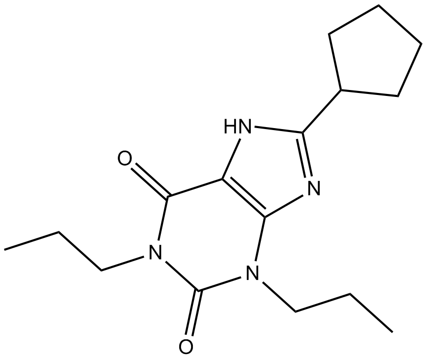
Cas No.: 102146-07-6
Sample solution is provided at 25 µL, 10mM.
_1-3.$$$39978408@51.jpg');)
_1-9.$$$38538795@65.jpg');)
_1-9.$$$39112701@51.jpg');)
_1-7.$$$37316672@70.jpg');)
_366-372.$$$36198801@70.jpg');)
.$$$38565142@65.jpg');)
_1867-1883.$$$33248023@67.jpg');)
_eads6055.$$$39977546@45.jpg');)
_eadm9805.$$$40080571@45.jpg');)
_eadj3020.$$$39666821@45.jpg');)
_1-20.$$$39757301@28.jpg');)
_1-24.$$$38898113@28.jpg');)
_273-287.$$$36806353@46.jpg');)
_1-16.$$$37156877@44.jpg');)
_1-19.$$$37460805@44.jpg');)
_1291-1307.$$$34518654@46.jpg');)
DPCPXは有力で選択性の高親和性A1アデノシン受容体拮抗剤であり、ラット脂肪細胞膜で、アデニル酸シクラーゼに対するKi値は0.45 nMである[1]。A1アデノシン受容体は重要なGタンパク質共役の受容体であり、その活性は、アデニリルシクラーゼ活性を阻害し、カルシウムイオンチャネルを調節し、カリウムイオンチャネルを阻害し、よって心臓機能、ニューロントランスミッションと様々な生理プロセスを調節する[2]。DPCPXはキサンチンの類似物であり、A1アデノシン受容体で、アデノシン結合部位に競合的に結合し、その活性を阻害する。それは、A1アデノシン受容体の機能と、心血管、神経、代謝システムと一部の癌における役割に関する研究に広く使われている[1,3]。
様々な起源のA1アデノシン受容体における薬理特性の差をDPCPXで評価する。[3H]-DPCPXを使い、ヒト、ギニアピッグ、ラットとマウスの脳膜を処理した(処理の濃度と時間は:ヒト: 0.7nM, 120分間;ギニアピッグ: 0.2nM, 60分間;ラットとマウス:0.1nM, 20分間)。DPCPXは異なる種類の膜の結合特性には著しい差を持つが、同種では、異なる脳の領域では、その結合親和性は比較的に一致した[4]。DPCPX (87nM)でMCF-7細胞を24、48と72時間処理した結果、p53、カスパーゼ3、8と9の発見を増やし、細胞の生存率を減らし、DPCPXがMCF-7細胞でアポトーシスを促進することが示唆された[5]。
DPCPX (1mg/kg)をマウスに腹腔内投与した結果、造影剤iohexolによる腎障害を著しく減らした[6]。DPCPX (0.25mg/kg)をラットに皮下投与した結果、中大脳動脈閉塞(MCAO)ラットで、死亡率、神経学的欠損スコア、脳梗塞の体積を著しく増やしたが、tetramethylpyrazine (TSG)の神経保護効果を減らした[7]。
References:
[1] Lohse M, Klotz K N, Lindenborn-Fotinos J, et al., 8-Cyclopentyl-1,3-dipropylxanthine (DPCPX) — a selective high affinity antagonist radioligand for A1 adenosine receptors, Naunyn-Schmiedeberg's archives of pharmacology[J]. 1987, 336: 204-210.
[2] Linden J, Structure and function of A1 adenosine receptors, FASEB journal : official publication of the Federation of American Societies for Experimental Biology[J]. 1991,12: 2668-76.
[3] Moro S, Gao Z G, Jacobson K A, et al., Progress in the pursuit of therapeutic adenosine receptor antagonists, Medicinal Research Reviews[J]. 2005, 26: 131-159.
[4] Maemoto T, Finlayson K, Olverman H J, Akahane A, et al., Species differences in brain adenosine A1 receptor pharmacology revealed by use of xanthine and pyrazolopyridine based antagonists, British Journal of Pharmacology[J]. 1997, 122: 1202-1208.
[5] Dastjerdi M N, Rarani M Z, Valiani A, et al., The effect of adenosine A1 receptor agonist and antagonist on p53 and caspase 3, 8, and 9 expression and apoptosis rate in MCF-7 breast cancer cell line, Research in Pharmaceutical Sciences[J]. 2016, 11: 303-310.
[6] Lee H T, Jan M, Bae S C, et al., A1 adenosine receptor knockout mice are protected against acute radiocontrast nephropathy in vivo, American Journal of Physiology-Renal Physiology[J]. 2006, 290: F1367-F1375.
[7] Ruan L, Li G, Zhao W, et al., Activation of Adenosine A1 Receptor in Ischemic Stroke: Neuroprotection by Tetrahydroxy Stilbene Glycoside as an Agonist, Antioxidants[J]. 2021, 10: 1112.
| 細胞実験[1]: | |
細胞株 | MCF-7細胞 |
準備方法 | 1ウェルあたり104細胞で、MCF-7細胞を24ウェルのプレートに播種し、24時間培養した。様々な濃度(0, 87nM)のDPCPXで処理した。DMSOを24、48、72と96時間で添加し、100μlの可溶性画分アリコートを96ウェルのプレートに移し、光学密度(OD)をプレートリーダーで測定した。 |
反応条件 | 0, 87nM;24, 48, 72時間 |
アプリケーション | DPCPXはMCF-7細胞の生存率を時間依存的に著しく阻害した。 |
| 動物実験 [2]: | |
動物モデル | 腎障害モデルマウス |
準備方法 | マウスに一酸化窒素合成酵素阻害剤(NG-nitro-L-arginine methyl ester, 10mg/kg)とプロスタグランジン合成の阻害剤(indomethacin, 10mg/kg)を腹腔内(ip)投与し、腎臓における保護メカニズムを阻害する。一部を1 mg/kg DPCPXで腹腔内処理した。15分後、マウスにiohexol (350mg iodine/ml)を腹腔内注射した。造影剤注入後24時間で比色法で血漿Crを測定し、腎臓の機能を評価した。 |
投与形態 | 1mg/kg、15分間;腹腔内注射 |
アプリケーション | DPCPXの処理は、造影剤腎症から著しく保護した。 |
References: | |
| Cas No. | 102146-07-6 | SDF | |
| Chemical Name | 8-cyclopentyl-1,3-dipropyl-1H-purine-2,6(3H,7H)-dione | ||
| Canonical SMILES | O=C1N(CCC)C(N(CCC)C2=C1NC(C3CCCC3)=N2)=O | ||
| Formula | C16H24N4O2 | M.Wt | 304.39 |
| 溶解度 | <1.52mg/ml in DMSO; <3.04mg/ml in ethanol | Storage | Store at -20°C |
| General tips | Please select the appropriate solvent to prepare the stock solution according to the
solubility of the product in different solvents; once the solution is prepared, please store it in
separate packages to avoid product failure caused by repeated freezing and thawing.Storage method
and period of the stock solution: When stored at -80°C, please use it within 6 months; when stored
at -20°C, please use it within 1 month. To increase solubility, heat the tube to 37°C and then oscillate in an ultrasonic bath for some time. |
||
| Shipping Condition | Evaluation sample solution: shipped with blue ice. All other sizes available: with RT, or with Blue Ice upon request. | ||
| Prepare stock solution | |||

|
1 mg | 5 mg | 10 mg |
| 1 mM | 3.2853 mL | 16.4263 mL | 32.8526 mL |
| 5 mM | 657.1 μL | 3.2853 mL | 6.5705 mL |
| 10 mM | 328.5 μL | 1.6426 mL | 3.2853 mL |
Step 1: Enter information below (Recommended: An additional animal making an allowance for loss during the experiment)
 g
μL
g
μL
Step 2: Enter the in vivo formulation (This is only the calculator, not formulation. Please contact us first if there is no in vivo formulation at the solubility Section.)
Calculation results:
Working concentration: mg/ml;
Method for preparing DMSO master liquid: mg drug pre-dissolved in μL DMSO ( Master liquid concentration mg/mL, Please contact us first if the concentration exceeds the DMSO solubility of the batch of drug. )
Method for preparing in vivo formulation: Take μL DMSO master liquid, next addμL PEG300, mix and clarify, next addμL Tween 80, mix and clarify, next add μL saline, mix and clarify.
Method for preparing in vivo formulation: Take μL DMSO master liquid, next add μL Corn oil, mix and clarify.
Note: 1. Please make sure the liquid is clear before adding the next solvent.
2. Be sure to add the solvent(s) in order. You must ensure that the solution obtained, in the previous addition, is a clear solution before proceeding to add the next solvent. Physical methods such as vortex, ultrasound or hot water bath can be used to aid dissolving.
3. All of the above co-solvents are available for purchase on the GlpBio website.
Quality Control & SDS
- View current batch:
- Purity: >98.00% Appearance: A solid
- COA (Certificate of Analysis)
- SDS (Safety Data Sheet)
- Datasheet
Average Rating: 5 (Based on Reviews and 23 reference(s) in Google Scholar.)