

Sunitinib |
カタログ番号GC17651
 One-Click Copy Product Info
|

スニチニブ(Sunitinib (SU 11248))は経口活性の多標的受容体チロシンキナーゼ阻害剤であり、血管内皮増殖因子受容体(VEGFR2)と血小板由来増殖因子受容体(PDGFRβ)に対するIC50値はそれぞれ80nMと2nMである。
Products are for research use only. Not for human use. We do not sell to patients.
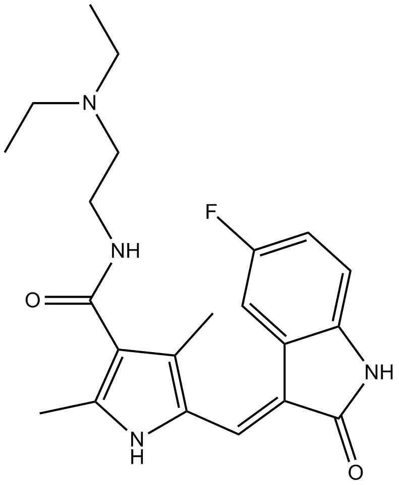
Cas No.: 557795-19-4
Sample solution is provided at 25 µL, 10mM.
スニチニブ(Sunitinib (SU 11248))は経口活性の多標的受容体チロシンキナーゼ阻害剤であり、血管内皮増殖因子受容体(VEGFR2)と血小板由来増殖因子受容体(PDGFRβ)に対するIC50値はそれぞれ80nMと2nMである[1]。スニチニブはATP競合的な阻害剤で、自己リン酸化とその後のRNase活性化を阻害することで、Ire1αのリン酸化を効果的に阻害する[2]。スニチニブは進行性腎細胞癌、消化管間質癌と非小細胞肺癌の治療によく使われている[3]。
試験管内実験では、腎細胞癌細胞786-Oをスニチニブ(2, 5 μM)で処理した結果、細胞内リン酸化プロテオームで変化を誘発し、180のリン酸化ペプチドが1.5倍の変化を示し、86のリン酸化ペプチドが下方制御され、94のリン酸化ペプチドが上方制御された[4]。髄芽腫VC312細胞をスニチニブ(0-5μM)で24あるいは48時間処理した結果、分断されたカスパーゼ3とPARPのレベルを用量依存的に増強させ、細胞のアポトーシスを誘発し、STAT3とAKT活性を阻害した[5]。
生体内実験では、4T1-lucとRENCA-luc腫瘍細胞を接種されたマウスにスニチニブ(30, 60, 120mg/kg)を経口投与した結果、マウス肺移植の後の4T1腫瘍の成長を阻害できなかったものの、RENCA腫瘍の成長を著しく阻害した[6]。糖尿病ラットにスニチニブ(1mg/kg)を4週間投与した結果、卵巣間質変性、間質線維症、卵胞変性、そしてNF-kappaBの免疫発現を著しく減らした[7]。
References:
[1] Sun L, Liang C, Shirazian S, et al. Discovery of 5-[5-fluoro-2-oxo-1, 2-dihydroindol-(3 Z)-ylidenemethyl]-2, 4-dimethyl-1 H-pyrrole-3-carboxylic acid (2-diethylaminoethyl) amide, a novel tyrosine kinase inhibitor targeting vascular endothelial and platelet-derived growth factor receptor tyrosine kinase[J]. Journal of medicinal chemistry, 2003, 46(7): 1116-1119.
[2] Ali M M U, Bagratuni T, Davenport E L, et al. Structure of the Ire1 autophosphorylation complex and implications for the unfolded protein response[J]. The EMBO journal, 2011, 30(5): 894-905.
[3] Gridelli C, Maione P, Del Gaizo F, et al. Sorafenib and sunitinib in the treatment of advanced non-small cell lung cancer[J]. The oncologist, 2007, 12(2): 191-200.
[4] Van Der Mijn J C, Broxterman H J, Knol J C, et al. Sunitinib activates Axl signaling in renal cell cancer[J]. International journal of cancer, 2016, 138(12): 3002-3010.
[5] Yang F, Jove V, Xin H, et al. Sunitinib induces apoptosis and growth arrest of medulloblastoma tumor cells by inhibiting STAT3 and AKT signaling pathways[J]. Molecular Cancer Research, 2010, 8(1): 35-45.
[6] Welti J C, Powles T, Foo S, et al. Contrasting effects of sunitinib within in vivo models of metastasis[J]. Angiogenesis, 2012, 15: 623-641.
[7] Erbas O, Pala H G, Pala E E, et al. Therapeutic effect of sunitinib on diabetes mellitus related ovarian injury: an experimental rat model study[J]. Gynecological Endocrinology, 2015, 31(5): 388-391.
| 細胞実験[1]: | |
細胞株 | 786-O細胞 |
準備方法 | リン酸化プロテオーム分析は未処理の、あるいは2と5μMのスニチニブに2時間暴露した786-O細胞のライセートで行われた。1.0 μMのスタウロスポリンの処理は、チロシンキナーゼの阻害のための陽性対照として使われた。 |
反応条件 | 2、5μM; 2時間 |
アプリケーション | 二つのスニチニブ濃度で、129のタンパク質からの180のリン酸化ペプチドは三回の執権で二回以上の実験で、1.5倍以上の変化を示した。これらの中で、86のリン酸化ペプチドが下方制御され、94のリン酸化ペプチドが上方制御された。 |
| 動物実験 [2]: | |
動物モデル | 雌のBalb/cマウス |
準備方法 | 4T1-lucまたはRENCA-luc腫瘍細胞を雌のBalb/cマウスに静脈内注射した。薬物(30, 60,または120 mg/kg/日のスニチニブ)は7日間経口投与した。 |
投与形態 | 30, 60, 120 mg/kg/日; p.o.(経口) |
アプリケーション | スニチニブは、マウス肺移植の後の4T1腫瘍の成長を阻害できなかったものの、RENCA腫瘍の成長を著しく阻害した。 |
References: | |
| Cas No. | 557795-19-4 | SDF | |
| Chemical Name | N-[2-(diethylamino)ethyl]-5-[(Z)-(5-fluoro-2-oxo-1H-indol-3-ylidene)methyl]-2,4-dimethyl-1H-pyrrole-3-carboxamide | ||
| Canonical SMILES | CCN(CC)CCNC(=O)C1=C(NC(=C1C)C=C2C3=C(C=CC(=C3)F)NC2=O)C | ||
| Formula | C22H27FN4O2 | M.Wt | 398.47 |
| 溶解度 | 19.9mg/mL in DMSO with gentle warming | Storage | Store at -20°C |
| General tips | Please select the appropriate solvent to prepare the stock solution according to the
solubility of the product in different solvents; once the solution is prepared, please store it in
separate packages to avoid product failure caused by repeated freezing and thawing.Storage method
and period of the stock solution: When stored at -80°C, please use it within 6 months; when stored
at -20°C, please use it within 1 month. To increase solubility, heat the tube to 37°C and then oscillate in an ultrasonic bath for some time. |
||
| Shipping Condition | Evaluation sample solution: shipped with blue ice. All other sizes available: with RT, or with Blue Ice upon request. | ||
| Prepare stock solution | |||

|
1 mg | 5 mg | 10 mg |
| 1 mM | 2.5096 mL | 12.548 mL | 25.096 mL |
| 5 mM | 501.9 μL | 2.5096 mL | 5.0192 mL |
| 10 mM | 251 μL | 1.2548 mL | 2.5096 mL |
Step 1: Enter information below (Recommended: An additional animal making an allowance for loss during the experiment)
 g
μL
g
μL
Step 2: Enter the in vivo formulation (This is only the calculator, not formulation. Please contact us first if there is no in vivo formulation at the solubility Section.)
Calculation results:
Working concentration: mg/ml;
Method for preparing DMSO master liquid: mg drug pre-dissolved in μL DMSO ( Master liquid concentration mg/mL, Please contact us first if the concentration exceeds the DMSO solubility of the batch of drug. )
Method for preparing in vivo formulation: Take μL DMSO master liquid, next addμL PEG300, mix and clarify, next addμL Tween 80, mix and clarify, next add μL saline, mix and clarify.
Method for preparing in vivo formulation: Take μL DMSO master liquid, next add μL Corn oil, mix and clarify.
Note: 1. Please make sure the liquid is clear before adding the next solvent.
2. Be sure to add the solvent(s) in order. You must ensure that the solution obtained, in the previous addition, is a clear solution before proceeding to add the next solvent. Physical methods such as vortex, ultrasound or hot water bath can be used to aid dissolving.
3. All of the above co-solvents are available for purchase on the GlpBio website.
Quality Control & SDS
- View current batch:
- Purity: >98.00% Appearance: A solid
- COA (Certificate of Analysis)
- SDS (Safety Data Sheet)
- Datasheet
Average Rating: 5 (Based on Reviews and 19 reference(s) in Google Scholar.)