

Sunitinib |
Catalog No.GC17651
 One-Click Copy Product Info
|

Sunitinib (SU 11248)은 구복으로 활성화의 다목적 수용체 틸로신 키나제 이펙터로 혈관 내피 성장 인자 수용체 (VEGFR2)와 혈소판 유래 성장 인자 수용체 (PDGFRβ)의 IC50 값이 각각 80 nM과 2 nM이다.
Products are for research use only. Not for human use. We do not sell to patients.
Cas No.: 557795-19-4
Sample solution is provided at 25 µL, 10mM.
Sunitinib (SU 11248)은 구복으로 활성화의 다목적 수용체 틸로신 키나제 이펙터로 혈관 내피 성장 인자 수용체 (VEGFR2)와 혈소판 유래 성장 인자 수용체 (PDGFRβ)의 IC50 값이 각각 80 nM과 2 nM이다. Sunitinib는 ATP 경쟁성 억제제로 자인산화와 후속 RNase 활성화를 억제하고 Ire1α의 인산화를 효과적으로 억제한다[2]. Sunitinib는 일반적으로 후기 신세포암과 위장도 기질 종양, 비소세포 폐암 치료에 사용된다[3].
체외 실험에서 신장암 세포 786-O에 Sunitinib (2, 5 μM)를 처리하고 세포 내인산화 단백질군에 변화를 일으켰다. 이 중 180개의 인산화 펩타이드에서 1.5배 이상의 변화를 보였고86개의 인산화 펩타이드가 감소하고 94개의 인산화 펩타이드가 증가했다[4]. 수골모세포괴VC312 세포에 Sunitinib (0-5μM)를 24시간이나 48시간 동안 처리한 결과는 용량 의존적으로 분리된 caspase-3와 PARP의 수준이 증가하고 세포 조각을 유도하고 STAT3과 AKT의 활동을 억제했다[5].
체내 실험에서 4T1-luc와 RENCA-luc 종양 세포를 이식한 쥐에 대해 Sunitinib (30, 60, 120mg/kg)를 구강 투여한 결과는 쥐 폐 이식 후 4T1 종양의 성장을 억제하지 못했고RENCA 종양의 성장을 현저히 억제했다[6]. 다이어벳 쥐에 대해 Sunitinib (1mg/kg)를 4주간 치료한 결과는 자궁 기질 퇴행성 변화, 기질적 섬유증, 포식구 퇴행성 변화, NF-카파B의 면역 표현을 현저히 줄였다[7].
References:
[1] Sun L, Liang C, Shirazian S, et al. Discovery of 5-[5-fluoro-2-oxo-1, 2-dihydroindol-(3 Z)-ylidenemethyl]-2, 4-dimethyl-1 H-pyrrole-3-carboxylic acid (2-diethylaminoethyl) amide, a novel tyrosine kinase inhibitor targeting vascular endothelial and platelet-derived growth factor receptor tyrosine kinase[J]. Journal of medicinal chemistry, 2003, 46(7): 1116-1119.
[2] Ali M M U, Bagratuni T, Davenport E L, et al. Structure of the Ire1 autophosphorylation complex and implications for the unfolded protein response[J]. The EMBO journal, 2011, 30(5): 894-905.
[3] Gridelli C, Maione P, Del Gaizo F, et al. Sorafenib and sunitinib in the treatment of advanced non-small cell lung cancer[J]. The oncologist, 2007, 12(2): 191-200.
[4] Van Der Mijn J C, Broxterman H J, Knol J C, et al. Sunitinib activates Axl signaling in renal cell cancer[J]. International journal of cancer, 2016, 138(12): 3002-3010.
[5] Yang F, Jove V, Xin H, et al. Sunitinib induces apoptosis and growth arrest of medulloblastoma tumor cells by inhibiting STAT3 and AKT signaling pathways[J]. Molecular Cancer Research, 2010, 8(1): 35-45.
[6] Welti J C, Powles T, Foo S, et al. Contrasting effects of sunitinib within in vivo models of metastasis[J]. Angiogenesis, 2012, 15: 623-641.
[7] Erbas O, Pala H G, Pala E E, et al. Therapeutic effect of sunitinib on diabetes mellitus related ovarian injury: an experimental rat model study[J]. Gynecological Endocrinology, 2015, 31(5): 388-391.
| 세포 실험 [1]: | |
세포 라인 | 786-O세포 |
제조 방법 | 처리되지 않은 786-O 세포 라이사테이트나 2와 5 μM Sunitinib에 2시간 동안 노출된 것에 대한 인산화 단백질군 분석을 실시했다. 1.0 μM 스타우로스포린 처리를 티로신 키나제 억제의 양성 대조으로 사용했다. |
반응 조건 | 2、5μM; 2시간 동안 |
응용 분야 | 두 종류의 Sunitinib 농도에서 129개 단백질로부터 나온 180개의 인산화 펩타이드이 3회의 실험 중 2회 이상에서 1.5배 이상의 변화를 보였습니다. 그 중 86개의 인산화 펩타이드가 감소되고 94개는 증가했습니다. |
| 동물 실험 [2]: | |
동물 모형 | 암컷 Balb/c 쥐 |
제조 방법 | 암컷 Balb/c 쥐는 4T1-luc나 RENCA-luc 종양 세포를 정맥 주입했습니다. 약물 (30, 60또는 120 mg/kg/일 Sunitinib)은 구강 투여로 7일간 행해졌습니다. |
제형 | 30, 60, 120 mg/kg/day; p.o. |
응용 분야 | Sunitinib는 쥐의 폐에 심어진 후 4T1 종양의 성장을 억제하지 못합니다. 그런데 Sunitinib는 RENCA 종양의 성장을 현저히 억제할 수 있습니다. |
References: | |
| Cas No. | 557795-19-4 | SDF | |
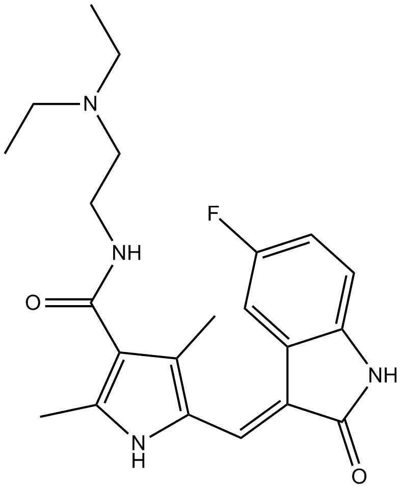
| Chemical Name | N-[2-(diethylamino)ethyl]-5-[(Z)-(5-fluoro-2-oxo-1H-indol-3-ylidene)methyl]-2,4-dimethyl-1H-pyrrole-3-carboxamide | ||
| Canonical SMILES | CCN(CC)CCNC(=O)C1=C(NC(=C1C)C=C2C3=C(C=CC(=C3)F)NC2=O)C | ||
| Formula | C22H27FN4O2 | M.Wt | 398.47 |
| Solubility | 19.9mg/mL in DMSO with gentle warming | Storage | Store at -20°C |
| General tips | Please select the appropriate solvent to prepare the stock solution according to the
solubility of the product in different solvents; once the solution is prepared, please store it in
separate packages to avoid product failure caused by repeated freezing and thawing.Storage method
and period of the stock solution: When stored at -80°C, please use it within 6 months; when stored
at -20°C, please use it within 1 month. To increase solubility, heat the tube to 37°C and then oscillate in an ultrasonic bath for some time. |
||
| Shipping Condition | Evaluation sample solution: shipped with blue ice. All other sizes available: with RT, or with Blue Ice upon request. | ||
| Prepare stock solution | |||

|
1 mg | 5 mg | 10 mg |
| 1 mM | 2.5096 mL | 12.548 mL | 25.096 mL |
| 5 mM | 501.9 μL | 2.5096 mL | 5.0192 mL |
| 10 mM | 251 μL | 1.2548 mL | 2.5096 mL |
Step 1: Enter information below (Recommended: An additional animal making an allowance for loss during the experiment)
 g
μL
g
μL
Step 2: Enter the in vivo formulation (This is only the calculator, not formulation. Please contact us first if there is no in vivo formulation at the solubility Section.)
Calculation results:
Working concentration: mg/ml;
Method for preparing DMSO master liquid: mg drug pre-dissolved in μL DMSO ( Master liquid concentration mg/mL, Please contact us first if the concentration exceeds the DMSO solubility of the batch of drug. )
Method for preparing in vivo formulation: Take μL DMSO master liquid, next addμL PEG300, mix and clarify, next addμL Tween 80, mix and clarify, next add μL saline, mix and clarify.
Method for preparing in vivo formulation: Take μL DMSO master liquid, next add μL Corn oil, mix and clarify.
Note: 1. Please make sure the liquid is clear before adding the next solvent.
2. Be sure to add the solvent(s) in order. You must ensure that the solution obtained, in the previous addition, is a clear solution before proceeding to add the next solvent. Physical methods such as vortex, ultrasound or hot water bath can be used to aid dissolving.
3. All of the above co-solvents are available for purchase on the GlpBio website.
Quality Control & SDS
- View current batch:
- Purity: >98.00% Appearance: A solid
- COA (Certificate of Analysis)
- SDS (Safety Data Sheet)
- Datasheet
Average Rating: 5 (Based on Reviews and 19 reference(s) in Google Scholar.)