

Verteporfin (Synonyms: CL 318952;Visudyne) |
カタログ番号GC14482
 One-Click Copy Product Info
|

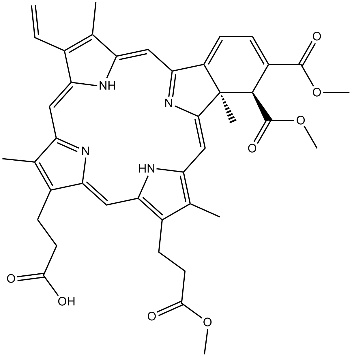
Verteporfinは、ベンゾポルフィリン誘導モノ酸環Aであり、光線力学的療法(PDT)の光感受性物質である。
Products are for research use only. Not for human use. We do not sell to patients.
Cas No.: 129497-78-5
Sample solution is provided at 25 µL, 10mM.
Verteporfinは、ベンゾポルフィリン誘導モノ酸環Aであり、光線力学的療法(PDT)の光感受性物質である[1]。690nmのレーザーで活性化された場合、ROS蓄積を通じて、Verteporfinは光化学的な損傷をミトコンドリアに起こした[2]。Verteporfinは、光活性化なしでYes関連タンパク質(YAP)を抑制し、同時にオートファジーを抑制し、ジャーミナルセンターキナーゼ様のキナーゼ(GLK)とSTE20/SPS1関連のプロリン/アラニンリッチキナーゼ(SPAK)を下方制御する[2]。Verteporfinは、内皮細胞への光線力学的な損傷、後続する血小板の活性化を通じて、血栓を誘発し、血管閉塞を起こす[3]。Verteporfinは、オートファゴーソムの形成を阻害するために、オートファジー阻害剤として広く使われている[4]。
In vitro(体外)実験で、Verteporfinで72時間処理したところ、PANC-1とSW1990細胞の増殖が著しく阻害され、IC50値はそれぞれ1.4µMと1.7µMであった[5]。16µM Verteporfinで48時間処理したところ、MCF-7、BT-474とBT-549細胞の増殖が低下し、同時にYAP、AXLとCYR61 mRNAの発現レベルが低下した[6]。5µg/ml Verteporfinで24時間処理したところ、BxPC3細胞で、脂質過酸化とフェロトーシスが誘発され、ミトコンドリアの機能が破壊された[7]。
In vivo(体内)実験で、20mg/kg/日のVerteporfinを3週間腹腔内注射したところ、マウスの腱切除術/熱傷モデルで、異所性骨化の活性と血管新生が軽減した[8]。75mg/kg/日のVerteporfinを2日間連続的に、腹腔内注射したところ、マウスで、尿酸一ナトリウム誘発の痛風性関節炎と炎症反応が軽減された[9]。
| 細胞実験 [1]: | |
|
細胞株 |
PANC-1細胞 |
|
調製方法 |
5% CO2、37°Cの加湿インキュベーターで、10%のウシ胎児血清を含有するDMEM培地でPANC-1細胞を培養した。1×10⁴個/ウェルの密度で、細胞を96ウェルのプレートに24時間播種培養し、各実験に対して三回繰り返した。細胞を様々な濃度のVerteporfin(0、2、4、6、8と10μM)で処理した。異なる時間の処理(24、48と72時間)後、細胞の生存率を測定した。 |
|
反応条件 |
0、2、4、6、8と10μM;24、48と72時間 |
|
応用 |
Verteporfinの処理により、PANC-1細胞の生存率が用量と時間依存的に阻害された。 |
| 動物実験 [2]: | |
|
動物モデル |
雄のC57BL/6 Jマウス |
|
調製方法 |
21±1°C、60%の相対湿度、12時間の明暗周期というコントロールされた環境で雄のC57BL/6 Jマウス(10週齢)を培養し、食物と水を自由に摂取させた。マウスに、Verteporfin(75mg/kg)(n=8)または殺菌PBS(200µl)(n=8)を2日間連続で腹腔内(i.p.)注射した。二回目の注射の1時間後、2mgの尿酸一ナトリウム結晶を右後肢の足底腱膜に皮下投与した。殺菌PBS(50μl)を左後肢の足底腱膜に皮下投与した。尿酸一ナトリウム注射の0時間後(ベースライン)と24時間後に、マウスの足の浮腫をデジタルノギスで測定した。 |
|
剤形 |
75mg/kg/日;2日間;腹腔内注射 |
|
応用 |
Verteporfinの処理により、マウスで尿酸一ナトリウム誘発性足の浮腫が抑制された。 |
| Cas No. | 129497-78-5 | SDF | |
| 同義語 | CL 318952;Visudyne | ||
| Canonical SMILES | O=C([C@H]1[C@](/C(N=C2/C=C(C(C)=C/3C=C)\NC3=C/C4=N/C(C(CCC(O)=O)=C4C)=C\5)=C/C6=C(C)C(CCC(OC)=O)=C5N6)(C)C2=CC=C1C(OC)=O)OC | ||
| Formula | C41H42N4O8 | M.Wt | 718.79 |
| 溶解度 | ≥ 18.3mg/mL in DMSO | Storage | 4°C, protect from light |
| Shipping Condition | Evaluation sample solution: shipped with blue ice. All other sizes available: with RT, or with Blue Ice upon request. | ||
| Prepare stock solution | |||

|
1 mg | 5 mg | 10 mg |
| 1 mM | 1.3912 mL | 6.9561 mL | 13.9123 mL |
| 5 mM | 278.2 μL | 1.3912 mL | 2.7825 mL |
| 10 mM | 139.1 μL | 695.6 μL | 1.3912 mL |
Step 1: Enter information below (Recommended: An additional animal making an allowance for loss during the experiment)
 g
μL
g
μL
Step 2: Enter the in vivo formulation (This is only the calculator, not formulation. Please contact us first if there is no in vivo formulation at the solubility Section.)
Calculation results:
Working concentration: mg/ml;
Method for preparing DMSO master liquid: mg drug pre-dissolved in μL DMSO ( Master liquid concentration mg/mL, Please contact us first if the concentration exceeds the DMSO solubility of the batch of drug. )
Method for preparing in vivo formulation: Take μL DMSO master liquid, next addμL PEG300, mix and clarify, next addμL Tween 80, mix and clarify, next add μL saline, mix and clarify.
Method for preparing in vivo formulation: Take μL DMSO master liquid, next add μL Corn oil, mix and clarify.
Note: 1. Please make sure the liquid is clear before adding the next solvent.
2. Be sure to add the solvent(s) in order. You must ensure that the solution obtained, in the previous addition, is a clear solution before proceeding to add the next solvent. Physical methods such as vortex, ultrasound or hot water bath can be used to aid dissolving.
3. All of the above co-solvents are available for purchase on the GlpBio website.
Quality Control & SDS
- View current batch:
- Purity: >98.00% Appearance: A solid
- COA (Certificate of Analysis)
- SDS (Safety Data Sheet)
- Datasheet
Average Rating: 5 (Based on Reviews and 24 reference(s) in Google Scholar.)