

Verteporfin (Synonyms: CL 318952;Visudyne) |
Catalog No.GC14482
 One-Click Copy Product Info
|

Verteporfin,a benzoporphyrin derivative monoacid ring A, is a photo-sensitizing agent for photodynamic therapy (PDT).
Products are for research use only. Not for human use. We do not sell to patients.
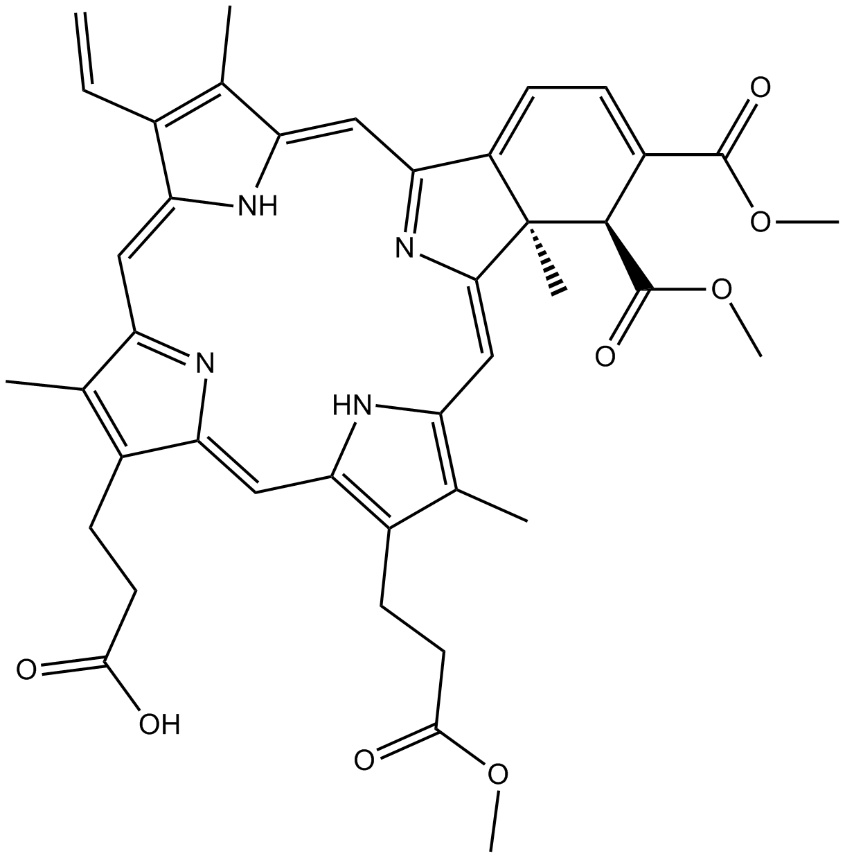
Cas No.: 129497-78-5
Sample solution is provided at 25 µL, 10mM.
Verteporfin, a benzoporphyrin derivative monoacid ring A, is a photo-sensitizing agent for photodynamic therapy (PDT)[1]. Verteporfin causes photochemical damage to the mitochondria via ROS accumulation when activated by a 690nm laser[2]. Verteporfin restrains Yes-associated protein (YAP) without photoactivation, along with suppressing autophagy, and downregulating germinal center kinase-like kinase (GLK) and STE20/SPS1-related proline/alanine-rich kinase (SPAK)[2]. Verteporfin induces thrombosis through photodynamic damage to endothelial cells and subsequent platelet activation, leading to vascular occlusion[3]. Verteporfin has been widely used as an autophagy inhibitor for blocking autophagosome formation[4].
In vitro, Verteporfin treatment for 72 hours significantly inhibited the proliferation of PANC-1 and SW1990 cells, with IC50 values of 1.4µM and 1.7µM, respectively[5]. Treatment with 16µM Verteporfin for 48 hours reduced the growth of MCF-7, BT-474, and BT-549 cells, accompanied by a decrease in the expression levels of YAP, AXL, and CYR61 mRNA[6]. Treatment with 5µg/ml Verteporfin for 24 hours induced lipid peroxidation and ferroptosis in BxPC3 cells, and disrupted mitochondrial function[7].
In vivo, Verteporfin treatment via intraperitoneal injection at a dose of 20mg/kg/day for 3 weeks reduced the heterotopic ossification formation and angiogenesis in the tenotomy/burn model of mice[8]. Intraperitoneal injection of Verteporfin at a dose of 75mg/kg/day for 2 consecutive days alleviated monosodium urate-induced gout arthritis and inflammatory responses in mice[9].
References:
[1] Scott L J, Goa K L. Verteporfin[J]. Drugs & aging, 2000, 16(2): 139-146.
[2] Wei C, Li X. The role of photoactivated and non-photoactivated verteporfin on tumor[J]. Frontiers in pharmacology, 2020, 11: 557429.
[3] Schmidt-Erfurth U, Hasan T. Mechanisms of action of photodynamic therapy with verteporfin for the treatment of age-related macular degeneration[J]. Survey of ophthalmology, 2000, 45(3): 195-214.
[4] Donohue E, Tovey A, Vogl A W, et al. Inhibition of autophagosome formation by the benzoporphyrin derivative verteporfin[J]. Journal of Biological Chemistry, 2011, 286(9): 7290-7300.
[5] Wei H, Wang F, Wang Y, et al. Verteporfin suppresses cell survival, angiogenesis and vasculogenic mimicry of pancreatic ductal adenocarcinoma via disrupting the YAP‐TEAD complex[J]. Cancer science, 2017, 108(3): 478-487.
[6] Wei C, Li X. Verteporfin inhibits cell proliferation and induces apoptosis in different subtypes of breast cancer cell lines without light activation[J]. BMC cancer, 2020, 20(1): 1042.
[7] Zhou W, Lim A, Elmadbouh O H M, et al. Verteporfin induces lipid peroxidation and ferroptosis in pancreatic cancer cells[J]. Free Radical Biology and Medicine, 2024, 212: 493-504.
[8] Luo G, Sun Z, Liu H, et al. Verteporfin attenuates trauma‐induced heterotopic ossification of Achilles tendon by inhibiting osteogenesis and angiogenesis involving YAP/β‐catenin signaling[J]. The FASEB Journal, 2023, 37(7): e23057.
[9] Shippy D C, Ulland T K. Verteporfin attenuates NLRP3 inflammasome activation to alleviate gout arthritis flares[J]. Journal of Inflammation, 2025, 22(1): 28.
| Cell experiment [1]: | |
|
Cell lines |
PANC-1 cells |
|
Preparation Method |
PANC-1 cells were cultured in DMEM medium containing 10% fetal bovine serum in a humidified incubator containing 5% CO2 at 37°C. Cells were seeded in a 96-well plate at a density of 1×10⁴ cells/well for 24h, with three replicates for each treatment. Cells were treated with different concentrations of Verteporfin (0, 2, 4, 6, 8, and 10μM). After a different period of treatment (24, 48, and 72h), the cell viability was determined. |
|
Reaction Conditions |
0, 2, 4, 6, 8, and 10μM; 24, 48, and 72h |
|
Applications |
Verteporfin treatment inhibited the cell viability of PANC-1 cells in a dose and time-dependent manner. |
| Animal experiment [2]: | |
|
Animal models |
Male C57BL/6 J mice |
|
Preparation Method |
Male C57BL/6 J mice (10 weeks old) were maintained in a controlled environment at 21±1°C and 60% relative humidity under a 12h light/dark cycle, and were given free access to food and water. Mice were administered Verteporfin (75mg/kg) (n=8) or sterile PBS (200µl) (n=8) by intraperitoneal (i.p.) injection for two consecutive days. One hour following the second injection, 2mg of monosodium urate crystals were subcutaneously injected into the plantar aponeurosis of the right hind paw. Sterile PBS (50µl) was subcutaneously injected into the plantar aponeurosis of the left hind paw. Mouse paw edema was measured with a digital caliper at 0 (baseline) and 24h post-monosodium urate injection. |
|
Dosage form |
75mg/kg/day; 2 days; i.p. |
|
Applications |
Verteporfin treatment suppressed monosodium urate-induced paw swelling in mice. |
References: [1] Wei H, Wang F, Wang Y, et al. Verteporfin suppresses cell survival, angiogenesis and vasculogenic mimicry of pancreatic ductal adenocarcinoma via disrupting the YAP‐TEAD complex[J]. Cancer science, 2017, 108(3): 478-487. [2] Shippy D C, Ulland T K. Verteporfin attenuates NLRP3 inflammasome activation to alleviate gout arthritis flares[J]. Journal of Inflammation, 2025, 22(1): 28. | |
| Cas No. | 129497-78-5 | SDF | |
| Synonyms | CL 318952;Visudyne | ||
| Canonical SMILES | O=C([C@H]1[C@](/C(N=C2/C=C(C(C)=C/3C=C)\NC3=C/C4=N/C(C(CCC(O)=O)=C4C)=C\5)=C/C6=C(C)C(CCC(OC)=O)=C5N6)(C)C2=CC=C1C(OC)=O)OC | ||
| Formula | C41H42N4O8 | M.Wt | 718.79 |
| Solubility | ≥ 18.3mg/mL in DMSO | Storage | 4°C, protect from light |
| Shipping Condition | Evaluation sample solution: shipped with blue ice. All other sizes available: with RT, or with Blue Ice upon request. | ||
| Prepare stock solution | |||

|
1 mg | 5 mg | 10 mg |
| 1 mM | 1.3912 mL | 6.9561 mL | 13.9123 mL |
| 5 mM | 278.2 μL | 1.3912 mL | 2.7825 mL |
| 10 mM | 139.1 μL | 695.6 μL | 1.3912 mL |
Step 1: Enter information below (Recommended: An additional animal making an allowance for loss during the experiment)
 g
μL
g
μL
Step 2: Enter the in vivo formulation (This is only the calculator, not formulation. Please contact us first if there is no in vivo formulation at the solubility Section.)
Calculation results:
Working concentration: mg/ml;
Method for preparing DMSO master liquid: mg drug pre-dissolved in μL DMSO ( Master liquid concentration mg/mL, Please contact us first if the concentration exceeds the DMSO solubility of the batch of drug. )
Method for preparing in vivo formulation: Take μL DMSO master liquid, next addμL PEG300, mix and clarify, next addμL Tween 80, mix and clarify, next add μL saline, mix and clarify.
Method for preparing in vivo formulation: Take μL DMSO master liquid, next add μL Corn oil, mix and clarify.
Note: 1. Please make sure the liquid is clear before adding the next solvent.
2. Be sure to add the solvent(s) in order. You must ensure that the solution obtained, in the previous addition, is a clear solution before proceeding to add the next solvent. Physical methods such as vortex, ultrasound or hot water bath can be used to aid dissolving.
3. All of the above co-solvents are available for purchase on the GlpBio website.
Quality Control & SDS
- View current batch:
- Purity: >98.00% Appearance: A solid
- COA (Certificate of Analysis)
- SDS (Safety Data Sheet)
- Datasheet
Average Rating: 5 (Based on Reviews and 24 reference(s) in Google Scholar.)