

HADA (Synonyms: HCC-Amino-D-alanine hydrochloride) |
Catalog No.GC50533
 One-Click Copy Product Info
|

HADA(HCC-아미노-D-알라닌 염산염)는 파란색 형광 D-아미노산(FDAA)으로 살아있는 박테리아의 펩타이드에 적합하고 λ ~ 405/460 nm를 자극/발산한다.
Products are for research use only. Not for human use. We do not sell to patients.
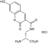
Cas No.: 2253733-10-5
Sample solution is provided at 25 µL, 10mM.
_1-3.$$$39978408@51.jpg');)
_1-9.$$$38538795@65.jpg');)
_1-9.$$$39112701@51.jpg');)
_1-7.$$$37316672@70.jpg');)
_366-372.$$$36198801@70.jpg');)
.$$$38565142@65.jpg');)
_1867-1883.$$$33248023@67.jpg');)
_eads6055.$$$39977546@45.jpg');)
_eadm9805.$$$40080571@45.jpg');)
_eadj3020.$$$39666821@45.jpg');)
_1-20.$$$39757301@28.jpg');)
_1-24.$$$38898113@28.jpg');)
_273-287.$$$36806353@46.jpg');)
_1-16.$$$37156877@44.jpg');)
_1-19.$$$37460805@44.jpg');)
_1291-1307.$$$34518654@46.jpg');)
HADA(HCC-아미노-D-알라닌 염산염)는 파란색 형광 D-아미노산(FDAA)으로 살아있는 박테리아의 펩타이드에 적합하고 λ ~ 405/460 nm를 자극/발산한다. 성장률에 영향을 미치지 않고 FDAA는 효과적으로 다양한 박테리아 세포 그룹의 외부 주변 및 격막 표시를 실시하여 미토콘드리아 투과성을 보여준다[1-2].
D-FDAA 조사는 다양한 세균에서PG 성장 지점을 표시할 수 있다. FDAA는 DD 트랜스펩티데이즈(페니실린 결합 단백질, PBP)나 LD 트랜스펩티데이즈(LDT)(존재할 경우)를 통해 PG와 혼합된다. FDAA는 E. coli와 같은 빠른 성장 과정에서 단 30초 만에 종을 표시할 수 있다. HADA는 대부분의 박테리아 종에 대한 PG에 대해 반복 가능하고 안정적인 표시를 할 수 있고 보통은 광범위한 최적화 없이도 가능한다[1-3].
References:
[1] Botella H, Yang G, Ouerfelli O, et al. Distinct spatiotemporal dynamics of peptidoglycan synthesis between Mycobacterium smegmatis and Mycobacterium tuberculosis[J]. MBio, 2017, 8(5): 10.1128/mbio. 01183-17.
[2] Peters K, Pazos M, Edoo Z, Hugonnet JE, Martorana AM, Polissi A, VanNieuwenhze MS, Arthur M, Vollmer W. Copper inhibits peptidoglycan LD-transpeptidases suppressing β-lactam resistance due to bypass of penicillin-binding proteins. Proc Natl Acad Sci U S A. 2018 Oct 16;115(42):10786-10791.
[3] Mendes S S, Marques J, Mesterházy E, et al. Synergetic antimicrobial activity and mechanism of clotrimazole-linked CO-releasing molecules[J]. ACS bio & med Chem Au, 2022, 2(4): 419-436.
이 프로토콜은 지침을 제공하고 구체적인 요구에 따라 수정되어야 합니다.
1.다음 용액을 미리 준비하십시오: TSB (또는 LB), TSB (LB) 아가르 판, 50 mM HADA 원액(DMSO에 녹여), 10× 및 1× 구연산 나트륨 버퍼, 1배 PBS 및 3% 파라포름알데히드.
2. 얼음 위에서 박테리아 균주의 기뢰 저장액을 녹입니다. 재배양 루프를 사용하여 TSB 아가르 판에 줄무늬로 그은 37°C에서 통기하여야 합니다. 그 후 TSB (또는 LB) 매체에 하나의 박테리아 균주을 접중하고 37°C에서 통기하여야 합니다.
3.TSB (또는 LB) 매체에서 통기하여야 OD578을 0.1로 희석하고 37°C에서 배양하고 지수 성장기의 OD578이 0.4에 도달할 때까지 성장시킵니다.
4. TSB (또는 LB)를 37°C로 예열하고 세포를 OD578 0.1까지 희석하고 500μl를 멸균된 1.5 ml 마이크로 튜브에 넣습니다.
5.샘플에 50 mM HADA 원액 2.5 µl를 추가하여 최종 농도를 250 μM로 하고 37°C에서 흔들며 30분 동안 재배합니다.
6.성장 중인 배양물에 미리 냉각된 10배 나트륨 버퍼의 최종량의 열분의 일 (50 µl, pH 2.25)을 추가합니다.
7. 4°C에서 16,200 xg로 샘플을 2분간 원심분리합니다. 초산액을 버리고 얼음 차가운 1× 구연산나트륨 버퍼 (pH 3.0) 1.5 ml로 침전물을 재수정합니다.
8.4°C에서 16,200 xg로 샘플을 2분간 원심 분리합니다. 상청을 버리고 얼음 차가운 1× PBS (pH 7.4) 1.5 ml로 침전물을 재수정한 후 샘플을 다시 원심분리합니다. 얼음 차가운 1×PBS (pH 7.4)로 씻기 단계를 한 번 더 반복합니다. 세포 침전물을 건드리지 않고 상청을 조심해서 버리세요.
9. 세포를 12.5 µl 1×PBS와 12.5 μl 3% 패라포맄을 재수정하여 고정합니다. 피펫으로 잘 혼합하고 얼음 위나 4°C에서 마이크로스코피 분석할 때까지 보관합니다.
10. 현미경 분석: 고정된 세포 3 μl을 준비된 현미경에 아가르 슬라이드의 웰에 피펫하고 사각 조심 스럽게 올려 살짝 누르면서 놓습니다. 레이블된 세포를 Eclipse Ti 현미경 (Plan Fluor×100/1.30 Oil Ph3 DLL 목록)에 Cool SNAP HQ2CCD 카메라로 위상대조와 DAPI 채널 (발광파장 350/50 nm, 방출파장 460/50 nm)을 사용하여 분석합니다. 위상대조 분석은 100 ms의 노출 시간을 사용하고 형광 이미지 분석은 1 s의 노출 시간을 사용합니다.
노트:
1.사용하기 전에 0.22 μm 필터 막을 사용하여 필터하고 10×및 1× 구연산 나트륨 버퍼, 1× PBS와 3% 패라포맄 용액을 얼음 위에서 냉각시킵니다.
2.HADA는 산성 pH나 무버퍼드 물에서 거의 형광을 띄지 않고 pH 7.0 이상에서의 세포 이미지에서 HADA 표시의 최대 밝기를 보입니다. 마지막 두 씻기 단계는 매우 중요합니다.
3. 레이블된 세포의 현미경 분석은 고정 직후에 수행되어야 합니다.
4.이 프로토콜을 사용하면 모든 분석된 세포가 완전히 레이블링되어 수축하는 세포의 septa에서 더 강한 신호를 보입니다. 성장 중인 세포에 10× 소다시트레이트 버퍼를 추가하고 pH 3.0의 1× 구연산 나트륨 버퍼로 최초 씻기를 진행하고 얼음 위에서 샘플을 빠르게 처리하면 미결합된 HADA의 제거율을 높이고 배경 형광 신호를 줄이고 신호 검출을 향상시킬 수 있습니다.
References:
[1] Peters K, Pazos M, VanNieuwenhze M S, et al. Optimized Protocol for the Incorporation of FDAA (HADA Labeling) for in situ Labeling of Peptidoglycan[J]. Bio-protocol, 2019, 9(15): e3316-e3316.
| Cas No. | 2253733-10-5 | SDF | |
| Synonyms | HCC-Amino-D-alanine hydrochloride | ||
| Canonical SMILES | N[C@@H](C(O)=O)CNC(C1=CC(C=CC(O)=C2)=C2OC1=O)=O.Cl | ||
| Formula | C13H12N2O6.HCl | M.Wt | 328.71 |
| Solubility | DMSO : 125 mg/mL (380.27 mM; Need ultrasonic) | Storage | Store at -20°C |
| General tips | Please select the appropriate solvent to prepare the stock solution according to the
solubility of the product in different solvents; once the solution is prepared, please store it in
separate packages to avoid product failure caused by repeated freezing and thawing.Storage method
and period of the stock solution: When stored at -80°C, please use it within 6 months; when stored
at -20°C, please use it within 1 month. To increase solubility, heat the tube to 37°C and then oscillate in an ultrasonic bath for some time. |
||
| Shipping Condition | Evaluation sample solution: shipped with blue ice. All other sizes available: with RT, or with Blue Ice upon request. | ||
| Prepare stock solution | |||

|
1 mg | 5 mg | 10 mg |
| 1 mM | 3.0422 mL | 15.211 mL | 30.422 mL |
| 5 mM | 608.4 μL | 3.0422 mL | 6.0844 mL |
| 10 mM | 304.2 μL | 1.5211 mL | 3.0422 mL |
Step 1: Enter information below (Recommended: An additional animal making an allowance for loss during the experiment)
 g
μL
g
μL
Step 2: Enter the in vivo formulation (This is only the calculator, not formulation. Please contact us first if there is no in vivo formulation at the solubility Section.)
Calculation results:
Working concentration: mg/ml;
Method for preparing DMSO master liquid: mg drug pre-dissolved in μL DMSO ( Master liquid concentration mg/mL, Please contact us first if the concentration exceeds the DMSO solubility of the batch of drug. )
Method for preparing in vivo formulation: Take μL DMSO master liquid, next addμL PEG300, mix and clarify, next addμL Tween 80, mix and clarify, next add μL saline, mix and clarify.
Method for preparing in vivo formulation: Take μL DMSO master liquid, next add μL Corn oil, mix and clarify.
Note: 1. Please make sure the liquid is clear before adding the next solvent.
2. Be sure to add the solvent(s) in order. You must ensure that the solution obtained, in the previous addition, is a clear solution before proceeding to add the next solvent. Physical methods such as vortex, ultrasound or hot water bath can be used to aid dissolving.
3. All of the above co-solvents are available for purchase on the GlpBio website.
Quality Control & SDS
- View current batch:
- Purity: >98.00% Appearance: A solid
- COA (Certificate of Analysis)
- SDS (Safety Data Sheet)
- Datasheet
Average Rating: 5 (Based on Reviews and 3 reference(s) in Google Scholar.)