

HADA (Synonyms: HCC-Amino-D-alanine hydrochloride) |
カタログ番号GC50533
 One-Click Copy Product Info
|

HADA(HCC-アミノ-D-アラニン塩酸塩)は青色の蛍光Dアミノ酸(FDAA)であり、生きている細菌のペプチドに適切であり、λ ~ 405/460 nmを刺激/発光する。
Products are for research use only. Not for human use. We do not sell to patients.
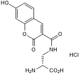
Cas No.: 2253733-10-5
Sample solution is provided at 25 µL, 10mM.
_1-3.$$$39978408@51.jpg');)
_1-9.$$$38538795@65.jpg');)
_1-9.$$$39112701@51.jpg');)
_1-7.$$$37316672@70.jpg');)
_366-372.$$$36198801@70.jpg');)
.$$$38565142@65.jpg');)
_1867-1883.$$$33248023@67.jpg');)
_eads6055.$$$39977546@45.jpg');)
_eadm9805.$$$40080571@45.jpg');)
_eadj3020.$$$39666821@45.jpg');)
_1-20.$$$39757301@28.jpg');)
_1-24.$$$38898113@28.jpg');)
_273-287.$$$36806353@46.jpg');)
_1-16.$$$37156877@44.jpg');)
_1-19.$$$37460805@44.jpg');)
_1291-1307.$$$34518654@46.jpg');)
HADA(HCC-アミノ-D-アラニン塩酸塩)は青色の蛍光Dアミノ酸(FDAA)であり、生きている細菌のペプチドに適切であり、λ ~ 405/460 nmを刺激/発光する。成長速度を影響せずに、FDAAはミトコンドリア透過性を示すために、様々な細菌細胞群の外部末梢と横隔膜に効率よくマーキングする[1-2]。
D-FDAAプローブは様々な細菌の中でPG成長部位をマークできる。FDAAはDDトランスぺプチダーゼ(ペニシリン結合タンパク質、PBP)またはLDトランスぺプチダーゼ(LDT)(存在するのであれば)を通じてPGと混合する。FDAAは大腸菌などの急速に増殖する種でただの30秒以内でマークされる。HADAはほとんどの細菌種に対して、徹底的な最適化をせずに、PGの再現と安定した標識をすることができる[1-3]。
References:
[1] Botella H, Yang G, Ouerfelli O, et al. Distinct spatiotemporal dynamics of peptidoglycan synthesis between Mycobacterium smegmatis and Mycobacterium tuberculosis[J]. MBio, 2017, 8(5): 10.1128/mbio. 01183-17.
[2] Peters K, Pazos M, Edoo Z, Hugonnet JE, Martorana AM, Polissi A, VanNieuwenhze MS, Arthur M, Vollmer W. Copper inhibits peptidoglycan LD-transpeptidases suppressing β-lactam resistance due to bypass of penicillin-binding proteins. Proc Natl Acad Sci U S A. 2018 Oct 16;115(42):10786-10791.
[3] Mendes S S, Marques J, Mesterházy E, et al. Synergetic antimicrobial activity and mechanism of clotrimazole-linked CO-releasing molecules[J]. ACS bio & med Chem Au, 2022, 2(4): 419-436.
このプロトコールはあくまでもガイドラインであり、お客様にはご自分のニーズに合わせてご調整ください。
1.下記の溶液を調製する:TSB (またはLB), TSB (LB)寒天培地, 50 mMのHADA貯蔵液(DMSOの中に), 10×と1×のクエン酸ナトリウムの緩衝液、1×PBSと3%のパラホルムアルデヒド。
2.氷の上で細菌株のグリセロールストックを解凍する。接種ループを使って菌株をTSB寒天培地に塗布し、37℃で一晩中培養する。その後、シングル細胞コローニで25mlのTSB(またはLB)培地を接種して、37℃で一晩中培養する。
3.一夜間培養された細胞をTSB (またはLB)培地で0.1のOD578になるまで薄めて、指数関数的OD578が0.4になるまで、37度で成長させる。
4.37度に事前加熱されたTSB(またはLB)で細胞を0.1のOD578になるまで薄めて、500μlは無菌1.5mlのマイクロチューブに入れた。
5.2.5μlの50mMのHADA貯蔵液をサンプルに入れて、最終濃度を250μMに達成させ、37度で30分間振りながら培養した。
6.予め冷却した10×クエン酸ナトリウム緩衝液 (50 µl、pH 2.25) の最終量の 10分の1を増殖中の培養物に加える。
7.サンプルを16,200 xg、4℃で二分間遠心分離する。上清を廃棄し、沈殿物を1.5 mlの氷冷した1×クエン酸ナトリウム緩衝液(pH 3.0)で再懸濁した。
8.サンプルを16,200 xg、4℃で二分間遠心分離する。上清を廃棄し、沈殿物を1.5 mlの氷冷した1×PBS (pH 7.4)で再懸濁した。その後サンプルをもう一度遠心分離した。氷冷した1×PBS(pH 7.4)で洗浄ステップをもう1回繰り返す。細胞沈殿物を触らずに注意を払いながら上清を廃棄する。
9.細胞ペレットを12.5 µlの1×PBSと12.5 µlの3%のパラホルムアルデヒドに再懸濁して細胞を固定する。ピペットでよく混ぜ、顕微鏡で分析するまで氷上または4°Cで保存する。
10.顕微鏡の分析:3μlの固定した細胞を、準備された顕微鏡用のアガローススライドのウェルにピペットで移し、正方形のカバーガラスを注意を払いながら上に置き、軽く押し付けた。標識された細胞は、Cool SNAP HQ2CCD カメラを備えたEclipse Ti顕微鏡 (Plan Fluor×100/1.30 Oil Ph3 DLLの対物レンズ) を使って、位相差とDAPIチャネル(励起波長は350/50 nm、発光波長は460/50 nm)を使用して分析した。位相差分析には100 msの露出時間を使用し、蛍光画像には1 sの露出時間を使用した。
注:
1.使用の前に、0.22μmのフィルター膜で濾過し、10×と1×のクエン酸ナトリウムの緩衝液、1× PBSと3%のパラホルムアルデヒド溶液を氷の上で予冷する。
2.HADAは酸性pHまたは緩衝液のない水の中ではほとんど蛍光を発さないが、7.0以上のpHで撮られた細胞はHADA標識の最大輝度を示した。最後の二つの洗浄の手続きはとても重要である。
3.標識された細胞の顕微鏡の分析は固定の直後に実行するべきである。
4.このプロトコールで、分析された細胞はすべて完全に標識され、収縮細胞の隔壁でより強いシグナルを示す。成長中の細胞に10×のクエン酸ナトリウムの緩衝液を追加すること、pH 3.0、1×のクエン酸ナトリウムの緩衝液で最初に洗浄すること、氷の上にサンプルを迅速に処理することは、取り込まれなかったHADAの除去を改善し、バックグラウンドの蛍光シグナルを減らし、シグナル検出を改善する。
References:
[1] Peters K, Pazos M, VanNieuwenhze M S, et al. Optimized Protocol for the Incorporation of FDAA (HADA Labeling) for in situ Labeling of Peptidoglycan[J]. Bio-protocol, 2019, 9(15): e3316-e3316.
| Cas No. | 2253733-10-5 | SDF | |
| 同義語 | HCC-Amino-D-alanine hydrochloride | ||
| Canonical SMILES | N[C@@H](C(O)=O)CNC(C1=CC(C=CC(O)=C2)=C2OC1=O)=O.Cl | ||
| Formula | C13H12N2O6.HCl | M.Wt | 328.71 |
| 溶解度 | DMSO : 125 mg/mL (380.27 mM; Need ultrasonic) | Storage | Store at -20°C |
| General tips | Please select the appropriate solvent to prepare the stock solution according to the
solubility of the product in different solvents; once the solution is prepared, please store it in
separate packages to avoid product failure caused by repeated freezing and thawing.Storage method
and period of the stock solution: When stored at -80°C, please use it within 6 months; when stored
at -20°C, please use it within 1 month. To increase solubility, heat the tube to 37°C and then oscillate in an ultrasonic bath for some time. |
||
| Shipping Condition | Evaluation sample solution: shipped with blue ice. All other sizes available: with RT, or with Blue Ice upon request. | ||
| Prepare stock solution | |||

|
1 mg | 5 mg | 10 mg |
| 1 mM | 3.0422 mL | 15.211 mL | 30.422 mL |
| 5 mM | 608.4 μL | 3.0422 mL | 6.0844 mL |
| 10 mM | 304.2 μL | 1.5211 mL | 3.0422 mL |
Step 1: Enter information below (Recommended: An additional animal making an allowance for loss during the experiment)
 g
μL
g
μL
Step 2: Enter the in vivo formulation (This is only the calculator, not formulation. Please contact us first if there is no in vivo formulation at the solubility Section.)
Calculation results:
Working concentration: mg/ml;
Method for preparing DMSO master liquid: mg drug pre-dissolved in μL DMSO ( Master liquid concentration mg/mL, Please contact us first if the concentration exceeds the DMSO solubility of the batch of drug. )
Method for preparing in vivo formulation: Take μL DMSO master liquid, next addμL PEG300, mix and clarify, next addμL Tween 80, mix and clarify, next add μL saline, mix and clarify.
Method for preparing in vivo formulation: Take μL DMSO master liquid, next add μL Corn oil, mix and clarify.
Note: 1. Please make sure the liquid is clear before adding the next solvent.
2. Be sure to add the solvent(s) in order. You must ensure that the solution obtained, in the previous addition, is a clear solution before proceeding to add the next solvent. Physical methods such as vortex, ultrasound or hot water bath can be used to aid dissolving.
3. All of the above co-solvents are available for purchase on the GlpBio website.
Quality Control & SDS
- View current batch:
- Purity: >98.00% Appearance: A solid
- COA (Certificate of Analysis)
- SDS (Safety Data Sheet)
- Datasheet
Average Rating: 5 (Based on Reviews and 3 reference(s) in Google Scholar.)