Apoptosis
As one of the cellular death mechanisms, apoptosis, also known as programmed cell death, can be defined as the process of a proper death of any cell under certain or necessary conditions. Apoptosis is controlled by the interactions between several molecules and responsible for the elimination of unwanted cells from the body.
Many biochemical events and a series of morphological changes occur at the early stage and increasingly continue till the end of apoptosis process. Morphological event cascade including cytoplasmic filament aggregation, nuclear condensation, cellular fragmentation, and plasma membrane blebbing finally results in the formation of apoptotic bodies. Several biochemical changes such as protein modifications/degradations, DNA and chromatin deteriorations, and synthesis of cell surface markers form morphological process during apoptosis.
Apoptosis can be stimulated by two different pathways: (1) intrinsic pathway (or mitochondria pathway) that mainly occurs via release of cytochrome c from the mitochondria and (2) extrinsic pathway when Fas death receptor is activated by a signal coming from the outside of the cell.
Different gene families such as caspases, inhibitor of apoptosis proteins, B cell lymphoma (Bcl)-2 family, tumor necrosis factor (TNF) receptor gene superfamily, or p53 gene are involved and/or collaborate in the process of apoptosis.
Caspase family comprises conserved cysteine aspartic-specific proteases, and members of caspase family are considerably crucial in the regulation of apoptosis. There are 14 different caspases in mammals, and they are basically classified as the initiators including caspase-2, -8, -9, and -10; and the effectors including caspase-3, -6, -7, and -14; and also the cytokine activators including caspase-1, -4, -5, -11, -12, and -13. In vertebrates, caspase-dependent apoptosis occurs through two main interconnected pathways which are intrinsic and extrinsic pathways. The intrinsic or mitochondrial apoptosis pathway can be activated through various cellular stresses that lead to cytochrome c release from the mitochondria and the formation of the apoptosome, comprised of APAF1, cytochrome c, ATP, and caspase-9, resulting in the activation of caspase-9. Active caspase-9 then initiates apoptosis by cleaving and thereby activating executioner caspases. The extrinsic apoptosis pathway is activated through the binding of a ligand to a death receptor, which in turn leads, with the help of the adapter proteins (FADD/TRADD), to recruitment, dimerization, and activation of caspase-8 (or 10). Active caspase-8 (or 10) then either initiates apoptosis directly by cleaving and thereby activating executioner caspase (-3, -6, -7), or activates the intrinsic apoptotic pathway through cleavage of BID to induce efficient cell death. In a heat shock-induced death, caspase-2 induces apoptosis via cleavage of Bid.
Bcl-2 family members are divided into three subfamilies including (i) pro-survival subfamily members (Bcl-2, Bcl-xl, Bcl-W, MCL1, and BFL1/A1), (ii) BH3-only subfamily members (Bad, Bim, Noxa, and Puma9), and (iii) pro-apoptotic mediator subfamily members (Bax and Bak). Following activation of the intrinsic pathway by cellular stress, pro‑apoptotic BCL‑2 homology 3 (BH3)‑only proteins inhibit the anti‑apoptotic proteins Bcl‑2, Bcl-xl, Bcl‑W and MCL1. The subsequent activation and oligomerization of the Bak and Bax result in mitochondrial outer membrane permeabilization (MOMP). This results in the release of cytochrome c and SMAC from the mitochondria. Cytochrome c forms a complex with caspase-9 and APAF1, which leads to the activation of caspase-9. Caspase-9 then activates caspase-3 and caspase-7, resulting in cell death. Inhibition of this process by anti‑apoptotic Bcl‑2 proteins occurs via sequestration of pro‑apoptotic proteins through binding to their BH3 motifs.
One of the most important ways of triggering apoptosis is mediated through death receptors (DRs), which are classified in TNF superfamily. There exist six DRs: DR1 (also called TNFR1); DR2 (also called Fas); DR3, to which VEGI binds; DR4 and DR5, to which TRAIL binds; and DR6, no ligand has yet been identified that binds to DR6. The induction of apoptosis by TNF ligands is initiated by binding to their specific DRs, such as TNFα/TNFR1, FasL /Fas (CD95, DR2), TRAIL (Apo2L)/DR4 (TRAIL-R1) or DR5 (TRAIL-R2). When TNF-α binds to TNFR1, it recruits a protein called TNFR-associated death domain (TRADD) through its death domain (DD). TRADD then recruits a protein called Fas-associated protein with death domain (FADD), which then sequentially activates caspase-8 and caspase-3, and thus apoptosis. Alternatively, TNF-α can activate mitochondria to sequentially release ROS, cytochrome c, and Bax, leading to activation of caspase-9 and caspase-3 and thus apoptosis. Some of the miRNAs can inhibit apoptosis by targeting the death-receptor pathway including miR-21, miR-24, and miR-200c.
p53 has the ability to activate intrinsic and extrinsic pathways of apoptosis by inducing transcription of several proteins like Puma, Bid, Bax, TRAIL-R2, and CD95.
Some inhibitors of apoptosis proteins (IAPs) can inhibit apoptosis indirectly (such as cIAP1/BIRC2, cIAP2/BIRC3) or inhibit caspase directly, such as XIAP/BIRC4 (inhibits caspase-3, -7, -9), and Bruce/BIRC6 (inhibits caspase-3, -6, -7, -8, -9).
Any alterations or abnormalities occurring in apoptotic processes contribute to development of human diseases and malignancies especially cancer.
References:
1.Yağmur Kiraz, Aysun Adan, Melis Kartal Yandim, et al. Major apoptotic mechanisms and genes involved in apoptosis[J]. Tumor Biology, 2016, 37(7):8471.
2.Aggarwal B B, Gupta S C, Kim J H. Historical perspectives on tumor necrosis factor and its superfamily: 25 years later, a golden journey.[J]. Blood, 2012, 119(3):651.
3.Ashkenazi A, Fairbrother W J, Leverson J D, et al. From basic apoptosis discoveries to advanced selective BCL-2 family inhibitors[J]. Nature Reviews Drug Discovery, 2017.
4.McIlwain D R, Berger T, Mak T W. Caspase functions in cell death and disease[J]. Cold Spring Harbor perspectives in biology, 2013, 5(4): a008656.
5.Ola M S, Nawaz M, Ahsan H. Role of Bcl-2 family proteins and caspases in the regulation of apoptosis[J]. Molecular and cellular biochemistry, 2011, 351(1-2): 41-58.
What is Apoptosis? The Apoptotic Pathways and the Caspase Cascade
أهداف لـ نبسب؛ Apoptosis
- Pyroptosis(15)
- Caspase(77)
- 14.3.3 Proteins(3)
- Apoptosis Inducers(71)
- Bax(15)
- Bcl-2 Family(136)
- Bcl-xL(13)
- c-RET(15)
- IAP(32)
- KEAP1-Nrf2(73)
- MDM2(21)
- p53(137)
- PC-PLC(6)
- PKD(8)
- RasGAP (Ras- P21)(2)
- Survivin(8)
- Thymidylate Synthase(12)
- TNF-α(141)
- Other Apoptosis(1145)
- Apoptosis Detection(0)
- Caspase Substrate(0)
- APC(6)
- PD-1/PD-L1 interaction(60)
- ASK1(4)
- PAR4(2)
- RIP kinase(47)
- FKBP(22)
منتجات لـ نبسب؛ Apoptosis
- القط. رقم اسم المنتج بيانات
-
GC15355
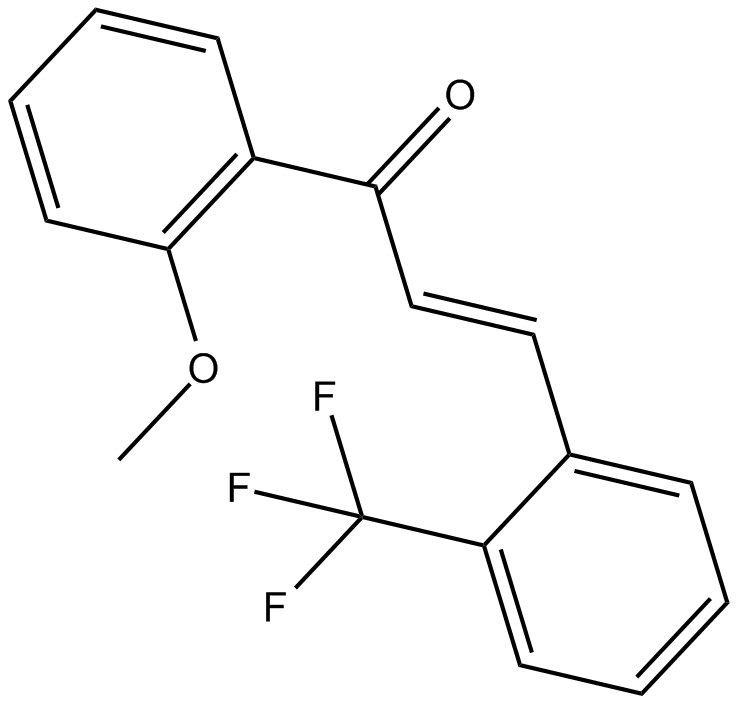
2-Trifluoromethyl-2'-methoxychalcone
Nrf2 activator
-
GN10800
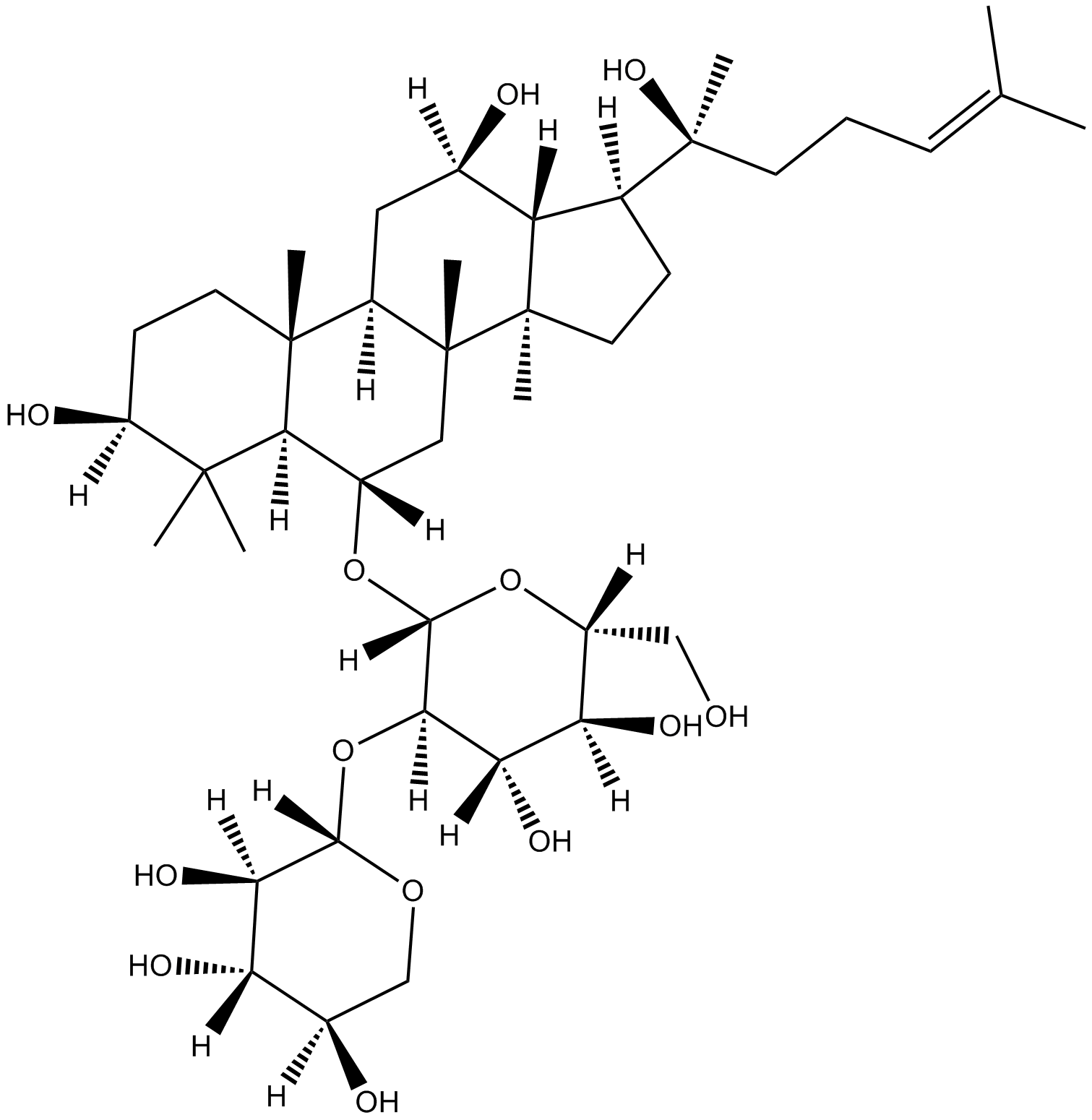
20(S)-NotoginsenosideR2
-
GC46528
25-hydroxy Cholesterol-d6
An internal standard for the quantification of 25hydroxy cholesterol

-
GC48482
28-Acetylbetulin
A lupane triterpenoid with anti-inflammatory and anticancer activities

-
GC35112
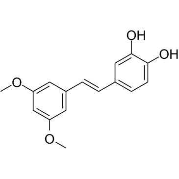
3'-Hydroxypterostilbene
3'-Hydroxypterostilbene هو نظير Pterostilbene3'-Hydroxypterostilbene يثبط نمو خلايا COLO 205 و HCT-116 و HT-29 مع IC50s من 9.0 و 40.2 و 70.9 ميكرومتر على التوالي3'-Hydroxypterostilbene ينظم بشكل كبير مسارات إشارات PI3K / Akt و MAPKs ويمنع بشكل فعال نمو خلايا سرطان القولون البشرية عن طريق إحداث موت الخلايا المبرمج والالتهام الذاتييمكن استخدام 3'-Hydroxypterostilbene في البحث عن السرطان
-
GC12791
3,3'-Diindolylmethane
A phytochemical with antiradiation and chemopreventative effects

-
GC42237
3,5-dimethyl PIT-1
PtdIns-(3,4,5)-P3 (PIP3) serves as an anchor for the binding of signal transduction proteins bearing pleckstrin homology (PH) domains such as phosphatidylinositol 3-kinase (PI3K) or PTEN.

-
GC64762
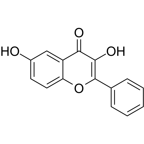
3,6-Dihydroxyflavone
3،6-ديهيدروكسي فلافون هو عامل مضاد للسرطان3،6-Dihydroxyflavone - تقلل الجرعة التي تعتمد على الوقت من قابلية الخلية للحياة وتؤدي إلى موت الخلايا المبرمج عن طريق تنشيط سلسلة caspase ، وشق بوليميريز poly (ADP-ribose) (PARP)3،6-ديهيدروكسي فلافون يزيد من الإجهاد التأكسدي داخل الخلايا وبيروكسيد الدهون
-
GC46583
3-Amino-2,6-Piperidinedione
An active metabolite of (±)-thalidomide

-
GC49849
3-Aminosalicylic Acid
A salicylic acid derivative

-
GC35106
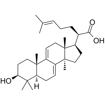
3-Dehydrotrametenolic acid
3- حمض ديهيدروتراميتينوليك ، المعزول من تصلب بوريا كوكوس ، هو مثبط لاكتات ديهيدروجينيز (LDH)3- يعزز حمض ديهيدروتراميتنوليك تمايز الخلايا الشحمية في المختبر ويعمل كمحفز للأنسولين في الجسم الحي3- يحفز حمض ديهيدروتراميتنوليك موت الخلايا المبرمج وله نشاط مضاد للسرطان
-
GC68537
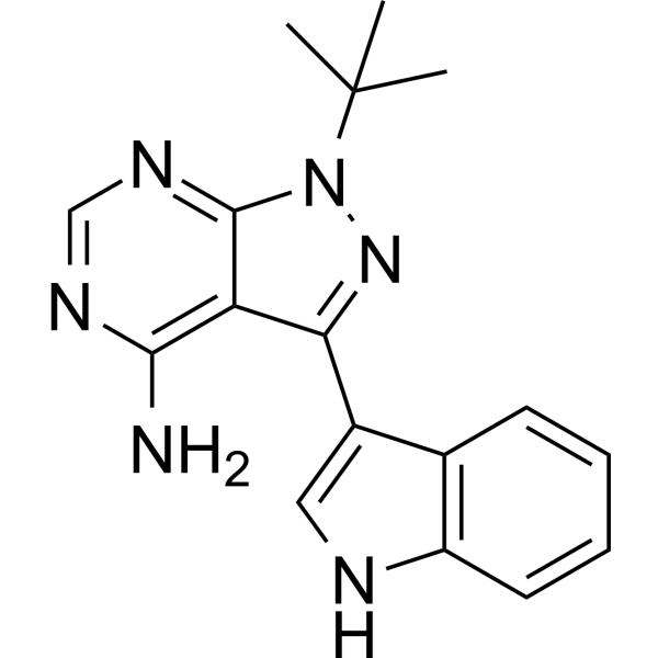
3-IN-PP1
3-IN-PP1 هو مثبط لإنزيم البروتين كيناز د (PKD). يعمل 3-IN-PP1 على تثبيط PKD1 و PKD2 و PKD3 بفعالية، حيث تكون قيمة IC50 لهذه المركبات على التوالي 108 و 94 و 108 نانومتر. كما أن 3-IN-PP1 هو مضاد سرطان شامل، حيث يحد من نمو خلايا الأورام المختلفة. يستخدم 3-IN-PP1 في الأبحاث المتعلقة بسرطان.
-
GC17394
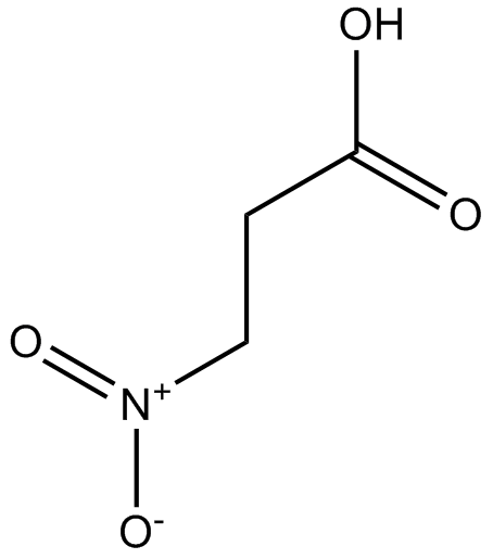
3-Nitropropionic acid
3-حمض النيتروبروبيونيك (β ؛ - حمض النيتروبروبيونيك) هو مثبط لا رجعة فيه لنزعة هيدروجين السكسينات.
-
GC35099
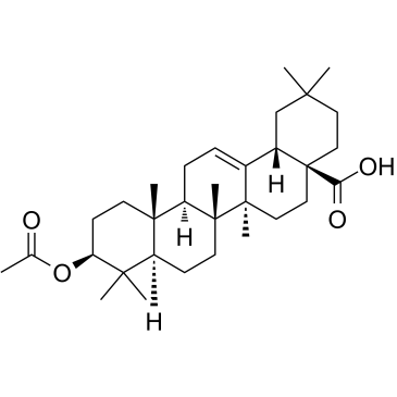
3-O-Acetyloleanolic acid
3-O-Acetyloleanolic acid (3AOA) ، مشتق من حمض الأولينوليك المعزول من بذور Vigna sinensis K. ، يحفز الإصابة بالسرطان ويعرض أيضًا نشاطًا مضادًا لتكوين الأوعية الدموية.
-
GC60507
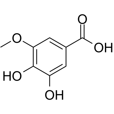
3-O-Methylgallic acid
3-O-Methylgallic acid (3،4-Dihydroxy-5-methoxybenzoic acid) هو مستقلب أنثوسيانين وله قدرة قوية كمضاد للأكسدةيمنع حمض 3-O-methylgallic تكاثر خلايا Caco-2 بقيمة IC50 تبلغ 24.1 ميكرومتريحفز حمض 3-O-methylgallic أيضًا موت الخلايا المبرمج وله تأثيرات مضادة للسرطان
-
GC32767
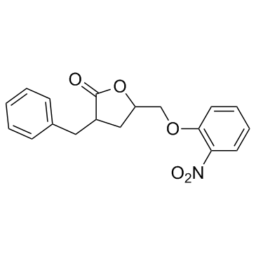
3BDO
3BDO هو منشط جديد لـ mTOR يمكنه أيضًا تثبيط الالتهام الذاتي
-
GC45354
4β-Hydroxywithanolide E
4β؛ -Hydroxywithanolide E ، معزول عن Physalis peruviana L.

-
GC48437
4'-Acetyl Chrysomycin A
A bacterial metabolite with antibacterial and anticancer activities

-
GC42346
4-bromo A23187
4-Bromo A23187 هو نظير مهلجن لحامل أيون الكالسيوم الانتقائي للغاية A-23187

-
GC42401
4-hydroperoxy Cyclophosphamide
نشط تنشيطًا للسيكلوفوسفاميد.

-
GC30896
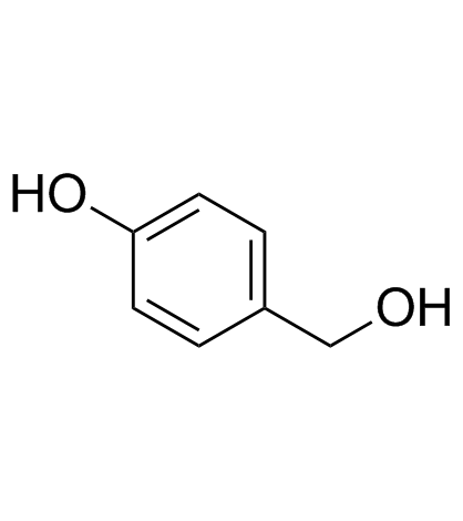
4-Hydroxybenzyl alcohol
4-Hydroxybenzyl alcohol هو مركب فينولي موزع على نطاق واسع في أنواع مختلفة من النباتات
-
GC33815
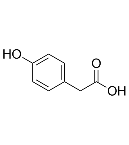
4-Hydroxyphenylacetic acid
حمض 4-هيدروكسي فينيل أسيتيك ، مستقلب رئيسي مشتق من الجراثيم من البوليفينول ، يشارك في العمل المضاد للأكسدة
-
GC35138
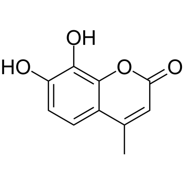
4-Methyldaphnetin
4-ميثيل دافنيتين هو مقدمة في تخليق مشتقات 4-ميثيل الكومارين4-ميثيل دافنيتين له تأثيرات انتقائية قوية مضادة للتكاثر ومحفزة للاستماتة على العديد من خطوط الخلايا السرطانية4-ميثيل دافنيتين يمتلك خاصية الكسح الجذري ويثبط بشدة بيروكسيد الدهون في الغشاء
-
GC68231
4-Methylsalicylic acid

-
GC31648
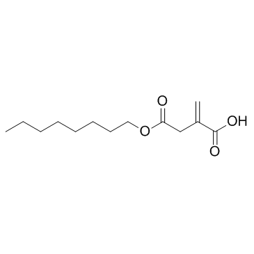
4-Octyl Itaconate
الأوكتيل إيتاكونات (4-OI) هو مشتق إيتاكونات يخترق الخلية. كان لإيتاكونات والأوكتيل إيتاكونات نفس التفاعل مع ثيول، مما يجعل الأوكتيل إيتاكونات بديلاً مناسبًا لدراسة وظائفه الحيوية.
-
GC49127
4-oxo Cyclophosphamide
An inactive metabolite of cyclophosphamide

-
GC45352
4-oxo Withaferin A
4-أوكسو Withaferin A هو نظير withaferin A. Withaferin A هو withanolide معزول من Withania somnifera. 4-oxo Withaferin A لديه القدرة على البحث عن المايلوما المتعددة.

-
GC45353
4-oxo-27-TBDMS Withaferin A
4-oxo-27-TBDMS Withaferin A ، أحد مشتقات withaferin A ، يُظهر تأثيرات قوية مضادة للتكاثر على الخلايا السرطانية .4-oxo-27-TBDMS Withaferin A يستحث موت الخلايا المبرمج للخلايا السرطانية. 4-oxo-27-TBDMS Withaferin A هو عامل مضاد للسرطان.

-
GC60525
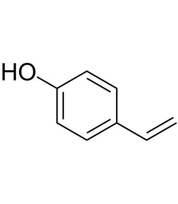
4-Vinylphenol (10%w/w in propylene glycol)
تم العثور على 4-فينيل فينول في الأعشاب الطبية Hedyotis diffusa Willd والأرز البري وهو أيضًا مستقلب حمض p-coumaric وحمض الفيروليك بواسطة بكتيريا حمض اللاكتيك في النبيذ4-فينيل فينول يحث على موت الخلايا المبرمج ويمنع تكوين الأوعية الدموية ويثبط نمو أورام الثدي الغازية في الجسم الحي
-
GC10468
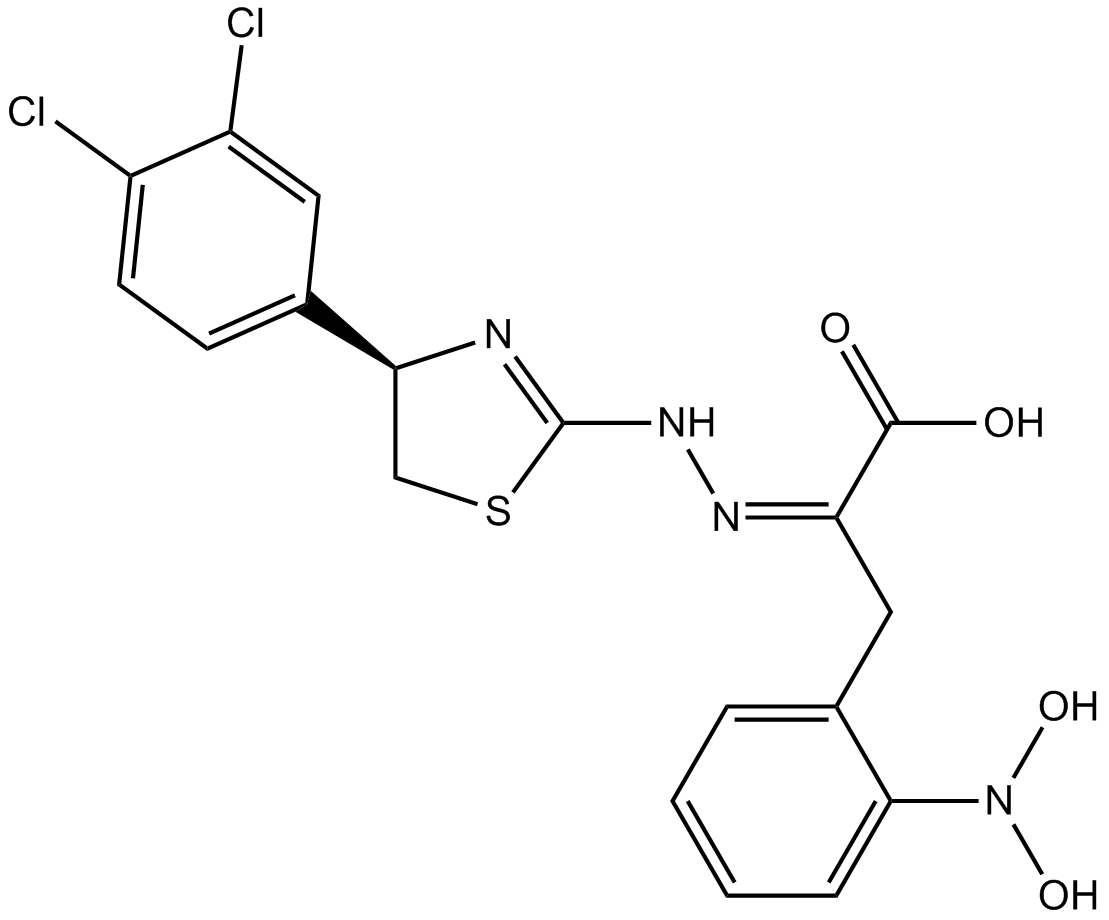
4EGI-1
4EGI-1 هو مثبط لتفاعل eIF4E / eIF4G ، مع Kd 25 ميكرومتر مقابل ربط eIF4E
-
GC35150
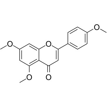
5,7,4'-Trimethoxyflavone
تم عزل 5،7،4'-Trimethoxyflavone من Kaempferia parviflora (KP) وهو نبات طبي مشهور من تايلاند. يحفز 5،7،4'-Trimethoxyflavone موت الخلايا المبرمج ، كما يتضح من الزيادات في المرحلة الفرعية G1 ، وتجزئة الحمض النووي ، وتلطيخ الملحق V / PI ، ونسبة Bax / Bcl-xL ، وتفعيل التحلل البروتيني لـ caspase-3 ، وتدهور بولي بروتين بوليميراز (ADP-ribose) (PARP) 5،7،4'-Trimethoxyflavone فعال بشكل كبير في منع تكاثر خلايا سرطان المعدة البشرية SNU-16 بطريقة تعتمد على التركيز.
-
GN10629
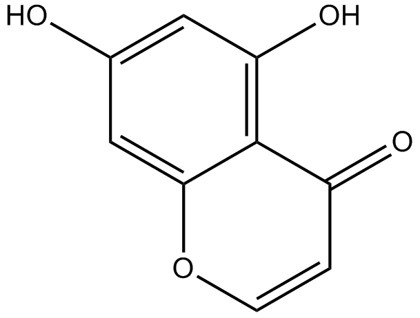
5,7-dihydroxychromone
-
GC63972
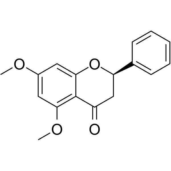
5,7-Dimethoxyflavanone
يظهر 5،7-Dimethoxyflavanone نشاطًا قويًا مضادًا للطفرات ضد طفرات MeIQ في اختبار Ames باستخدام S
-
GC52227
5-(3',4'-Dihydroxyphenyl)-γ-Valerolactone
An active metabolite of various polyphenols

-
GC35147
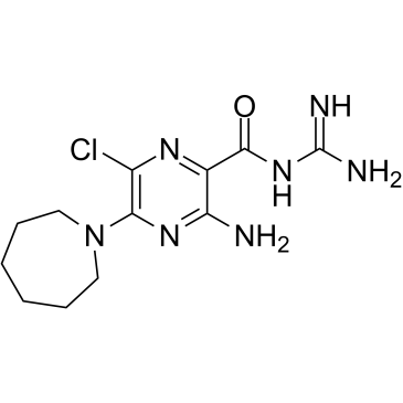
5-(N,N-Hexamethylene)-amiloride
5- (N ، N-Hexamethylene) -amiloride (Hexamethylene amiloride) مشتق من amiloride وهو مثبط قوي لمبادل Na + / H ، مما يقلل من درجة الحموضة داخل الخلايا (pHi) ويحفز موت الخلايا المبرمج في ابيضاض الدم الخلايا
-
GC45356
5-Aminolevulinic Acid (hydrochloride)

-
GC46681
5-Bromouridine
A brominated uridine analog

-
GC42545
5-Fluorouracil-13C,15N2
5-Fluorouracil-13C,15N2 is intended for use as an internal standard for the quantification of 5-flurouracil by GC- or LC-MS.

-
GC46705
5-Methoxycanthinone
5-ميثوكسيكانثينون هو مثبط نشط عن طريق الفم لسلالات الليشمانيا.

-
GC42586
6α-hydroxy Paclitaxel
6α ؛ -هيدروكسي باكليتاكسيل هو مستقلب أساسي للباكليتاكسيل. 6α ؛ - يحتفظ هيدروكسي باكليتاكسيل بتأثير يعتمد على الوقت على بولي ببتيدات نقل الأنيون العضوي 1B1 / SLCO1B1 (OATP1B1) مع فعالية تثبيط مماثلة لباكليتاكسيل ، في حين أنه لم يعد يظهر تثبيطًا معتمدًا على الوقت لـ OATP1B3. 6α ؛ -يمكن استخدام هيدروكسي باكليتاكسيل لأبحاث السرطان.

-
GC45772
6(5H)-Phenanthridinone
An inhibitor of PARP1 and 2

-
GN10093
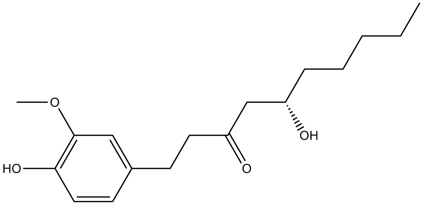
6-gingerol
-
GC49429
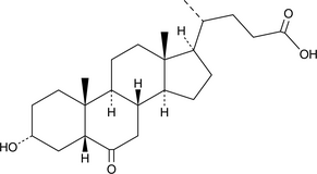
6-keto Lithocholic Acid
A metabolite of lithocholic acid
-
GC35184
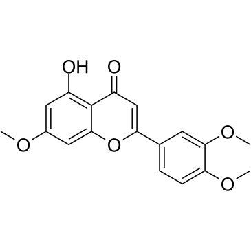
7,3',4'-Tri-O-methylluteolin
7،3 '، 4'-Tri-O-methylluteolin (5-Hydroxy-3'، 4 '، 7-trimethoxyflavone) ، مركب فلافونويد ، له تأثيرات قوية مضادة للالتهابات في خط خلايا البلاعم المستحث بـ LPS بوساطة تثبيط إطلاق وسطاء التهابات ، NO ، PGE2 ، والسيتوكينات المؤيدة للالتهابات
-
GC45673
7,8-Dihydroneopterin
7،8-ديهيدرونوبترين ، علامة الالتهاب ، تحفز موت الخلايا المبرمج الخلوي في الخلايا النجمية والخلايا العصبية من خلال تعزيز تعبير سينثاز أكسيد النيتريك (iNOS)

-
GC16853
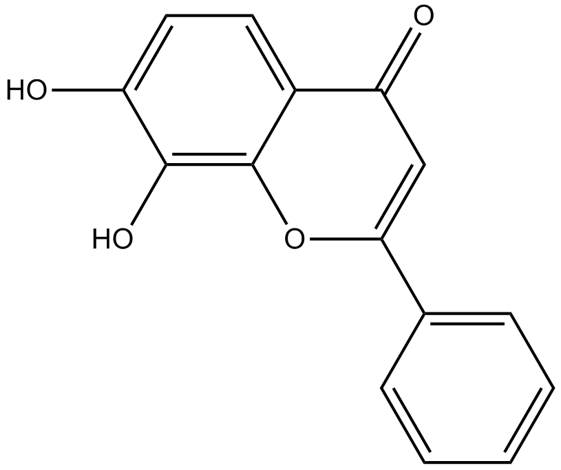
7,8-Dihydroxyflavone
7،8-ديهيدروكسي فلافون هو ناهض TrkB قوي وانتقائي يحاكي الإجراءات الفسيولوجية لعامل التغذية العصبية المشتق من الدماغ (BDNF)
-
GC42616
7-oxo Staurosporine
7-oxo Staurosporine is an antibiotic originally isolated from S.

-
GC16037
7BIO
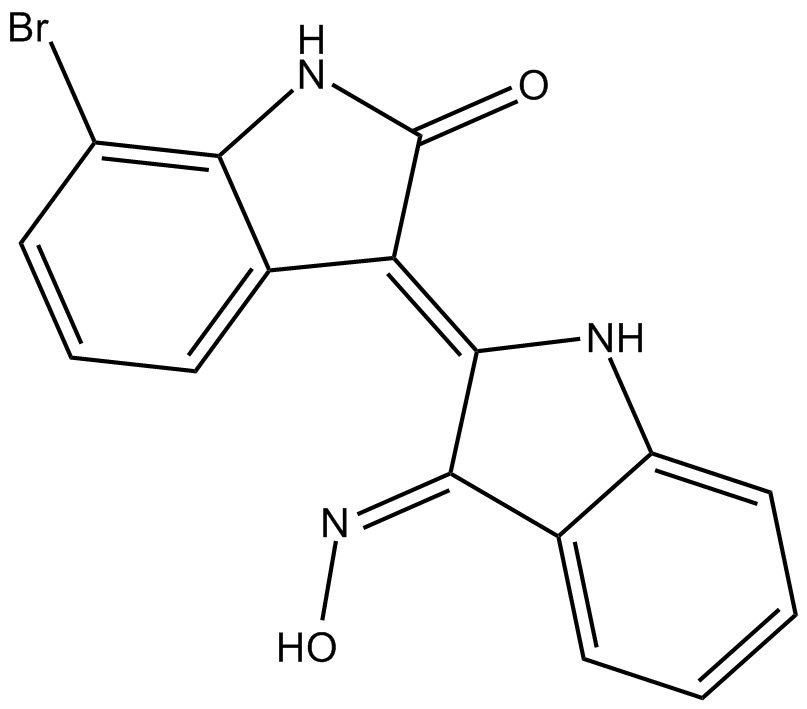
7BIO (7-بروموينديروبين-3-أوكسيم) هو أحد مشتقات الإندروبين
-
GC46741
8(E),10(E),12(Z)-Octadecatrienoic Acid
A conjugated PUFA

-
GC42622
8-bromo-Cyclic AMP
8-bromo-Cyclic AMP is a brominated derivative of cAMP that remains long-acting due to its resistance to degradation by cAMP phosphodiesterase.

-
GC49275
8-Oxycoptisine
8-Oxycoptisine هو قلويد بروتوبيربيرين طبيعي مع نشاط مضاد للسرطان.

-
GC17119
8-Prenylnaringenin
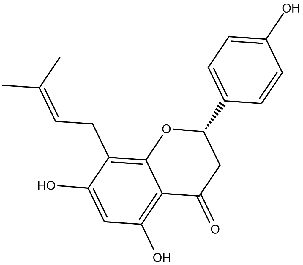
8-برينيلنارينجين هو برينيل فلافونويد معزول من مخاريط القفزة (Humulus lupulus) ، مع السمية الخلوية. 8-prenylnaringenin له نشاط مضاد للتكاثر ضد خلايا سرطان القولون HCT-116 عن طريق تحريض موت الخلايا المبرمج الداخلي والخارجي بوساطة المسار. يعزز 8-Prenylnaringenin أيضًا التعافي من ضمور العضلات الناجم عن عدم الحركة من خلال تنشيط مسار الفسفرة Akt في الفئران.
-
GC41642
9(E),11(E),13(E)-Octadecatrienoic Acid
9(E),11(E),13(E)-Octadecatrienoic acid (β-ESA) is a conjugated polyunsaturated fatty acid that is found in plant seed oils and in mixtures of conjugated linolenic acids synthesized by the alkaline isomerization of linolenic acid.

-
GC41643
9(Z),11(E),13(E)-Octadecatrienoic Acid
9(Z),11(E),13(E)-Octadecatrienoic Acid (α-ESA) is a conjugated polyunsaturated fatty acid commonly found in plant seed oil.

-
GC40785
9(Z),11(E),13(E)-Octadecatrienoic Acid ethyl ester
9(Z),11(E),13(E)-Octadecatrienoic Acid ethyl ester (α-ESA) is a conjugated polyunsaturated fatty acid commonly found in plant seed oil.

-
GC40710
9(Z),11(E),13(E)-Octadecatrienoic Acid methyl ester
9Z,11E,13E-octadecatrienoic acid (α-ESA) is a conjugated polyunsaturated fatty acid commonly found in plant seed oil.

-
GC39152
9-ING-41
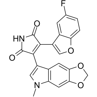
9-ING-41 عبارة عن مثبط انتقائي للجليكوجين سينثيز كيناز 3β (GSK-3β) قائم على ATP منافس لمايليميد مع مثبط IC50 يبلغ 0.71 ميكرومتر9-ING-41 يؤدي بشكل كبير إلى توقف الدورة الخلوية ، الالتهام الذاتي وموت الخلايا المبرمج في الخلايا السرطانية9-ING-41 له نشاط مضاد للسرطان ولديه القدرة على تعزيز التأثيرات المضادة للأورام لأدوية العلاج الكيميائي
-
GN10035
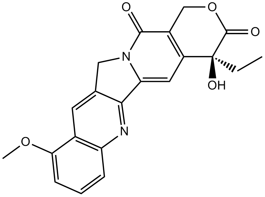
9-Methoxycamptothecin
-
GC45960
9c(i472)
9c (i472) هو مثبط قوي لـ 15-LOX-1 (15-lipoxygenase-1) بقيمة IC50 تبلغ 0.19 μ ؛ M.

-
GC50465
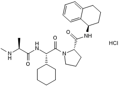
A 410099.1
High affinity XIAP antagonist; active in vivo
-
GC17512
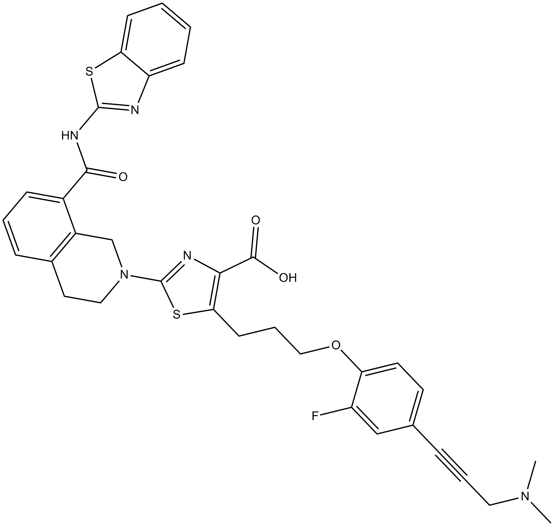
A-1155463
A-1155463 هو مثبط قوي للغاية وانتقائي BCL-XL مع EC50 من 70 نانومتر في خلية Molt-4
-
GC16278
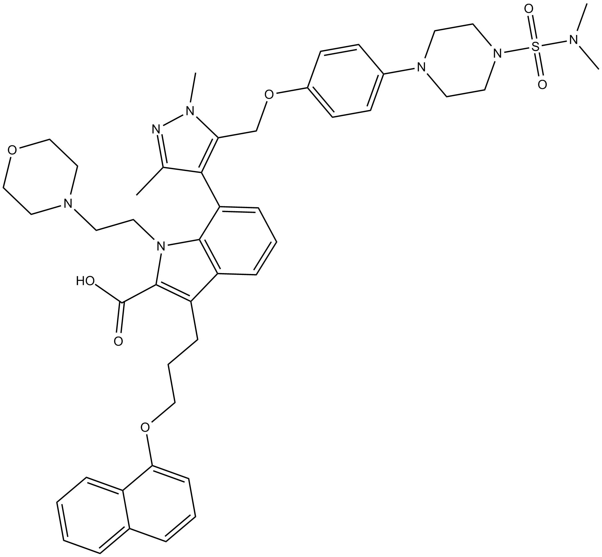
A-1210477
A-1210477 هو مثبط قوي وانتقائي لـ MCL-1 مع Ki يبلغ 0.45 نانومتريربط A-1210477 MCL-1 على وجه التحديد ويعزز موت الخلايا المبرمج للخلايا السرطانية بطريقة تعتمد على MCL-1
-
GC17513
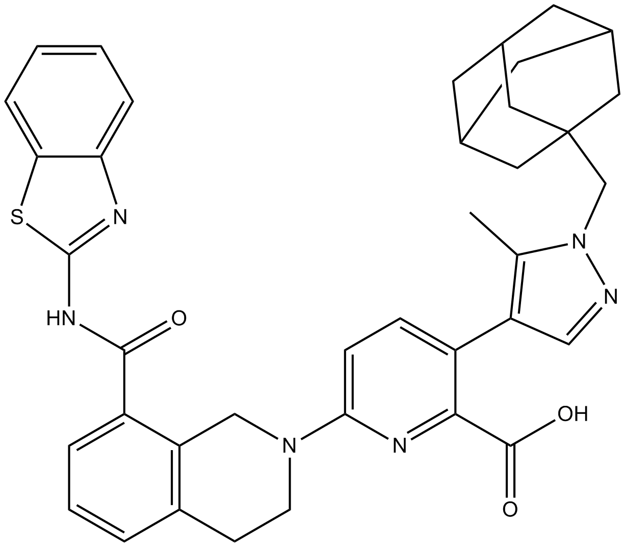
A-1331852
A-1331852 هو مثبط انتقائي BCL-XL متاح عن طريق الفم مع Ki أقل من 10 ميكرومتر
-
GC60544
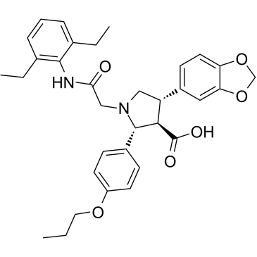
A-192621
A-192621 هو مناهض لمستقبلات الإندوثيلين B (ETB) قوي ، غير ببتيد ، فعال عن طريق الفم وانتقائي مع IC50 من 4.5 نانومتر وكي من 8.8 نانومتر
-
GC32981
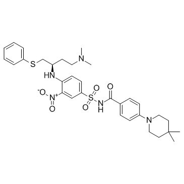
A-385358
A-385358 هو مثبط انتقائي لـ Bcl-XL مع Kis من 0.80 و 67 نانومتر لـ Bcl-XL و Bcl-2 ، على التوالي
-
GC11200
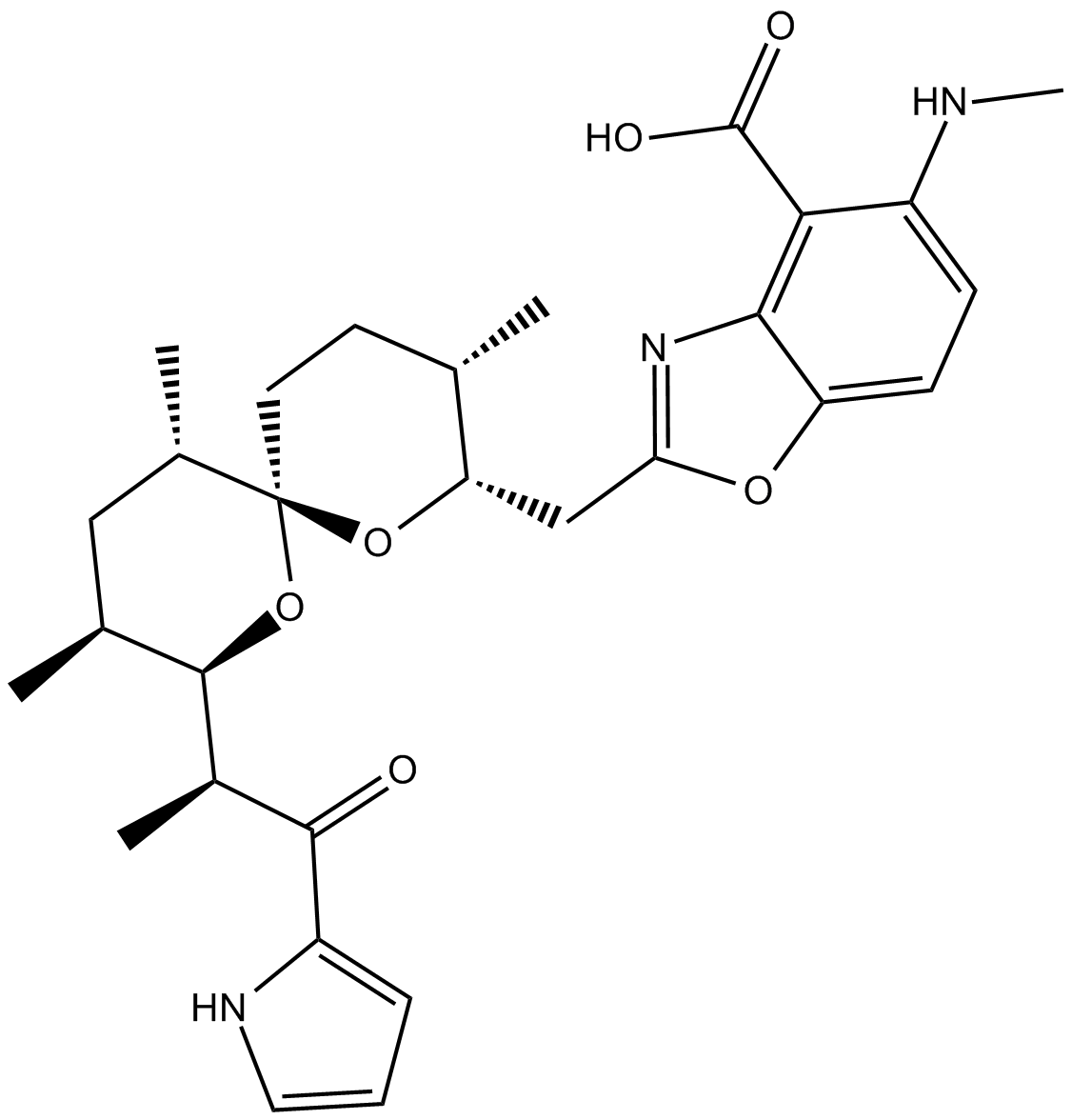
A23187
A23187 (A-23187) هو مضاد حيوي ومنقل الأيونات ثنائية التكافؤ فريدة من نوعها (مثل الكالسيوم والمغنيسيوم).
-
GC42659
A23187 (calcium magnesium salt)
A23187 is a divalent cation ionophore.

-
GC35216
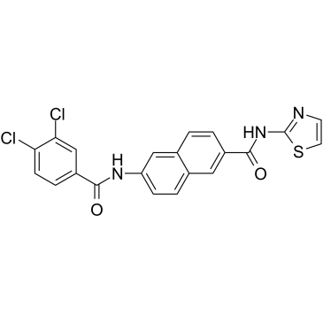
AAPK-25
AAPK-25 هو مثبط مزدوج قوي وانتقائي Aurora / PLK مع نشاط مضاد للورم ، والذي يمكن أن يتسبب في تأخير الانقسام وتوقف الخلايا في مرحلة ما قبل الطور ، مما يعكسه الفسفرة هيستون H3Ser10 للعلامة الحيوية ويتبعها زيادة في موت الخلايا المبرمجتستهدف AAPK-25 Aurora-A و -B و -C بقيم Kd تتراوح من 23-289 نانومتر ، بالإضافة إلى PLK-1 و -2 و -3 بقيم Kd تتراوح من 55-456 نانومتر
-
GC13805
Abacavir
-
GC64674
ABBV-167
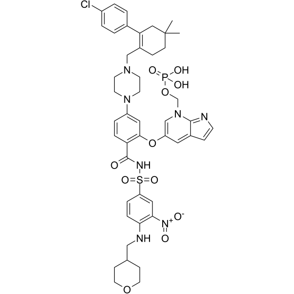
ABBV-167 هو دواء أولي للفوسفات لمثبط BCL-2 venetoclax
-
GC60548
ABT-100
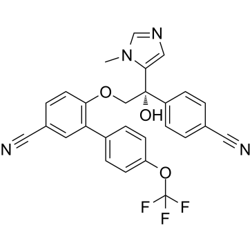
ABT-100 هو مثبط قوي ، انتقائي للغاية وفعال عن طريق الفمABT-100 يمنع تكاثر الخلايا (IC50s من 2.2 نانومتر ، 3.8 نانومتر ، 5.9 نانومتر ، 6.9 نانومتر ، 9.2 نانومتر ، 70 نانومتر و 818 نانومتر لـ EJ-1 ، DLD-1 ، MDA-MB-231 ، HCT-116 ، MiaPaCa- 2 ، PC-3 ، و DU-145 ، على التوالي) ، يزيد من موت الخلايا المبرمج ويقلل من تولد الأوعيةيمتلك ABT-100 نشاطًا مضادًا للورم واسع الطيف
-
GC14069
ABT-199
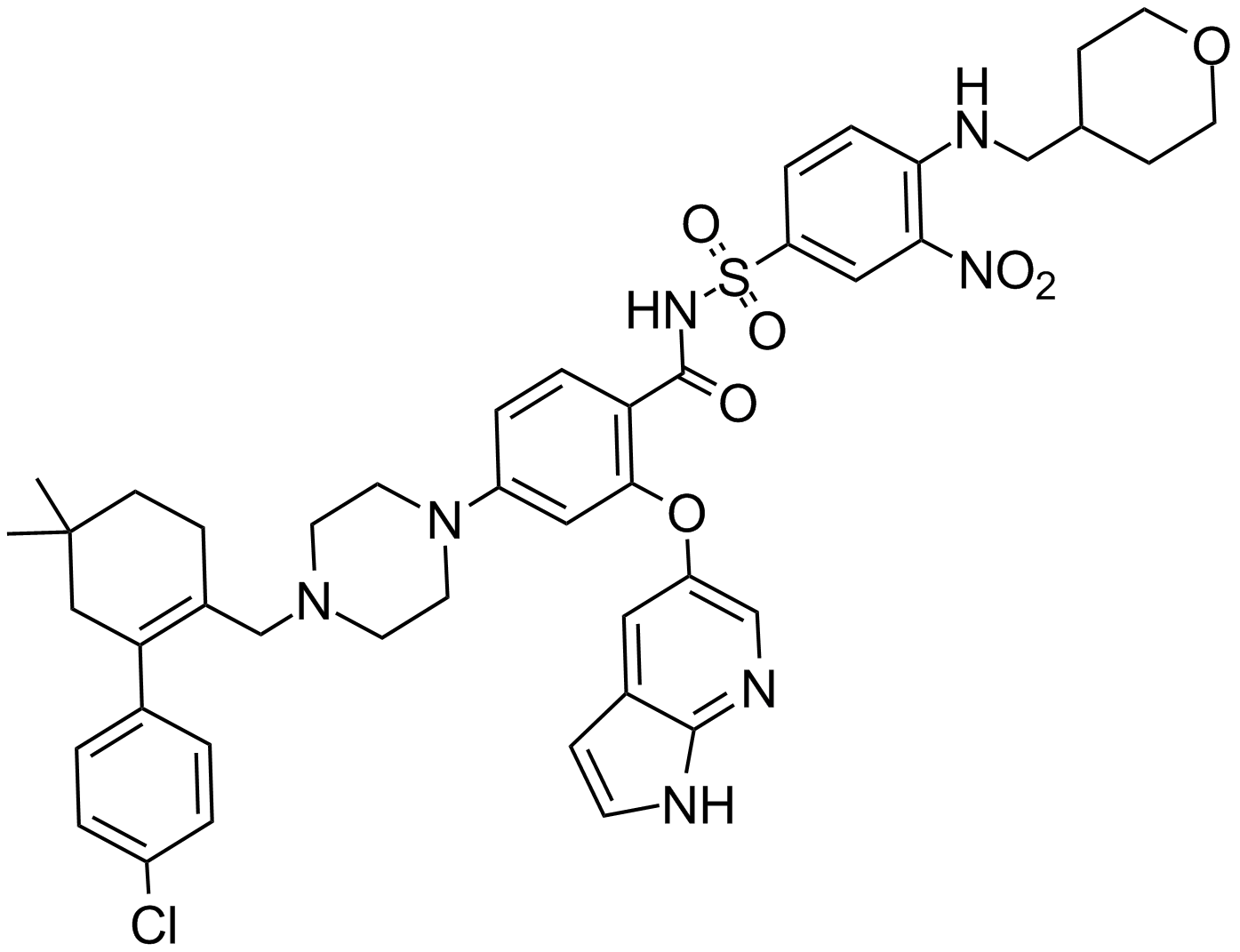
مثبط Bcl-2
-
GC12405
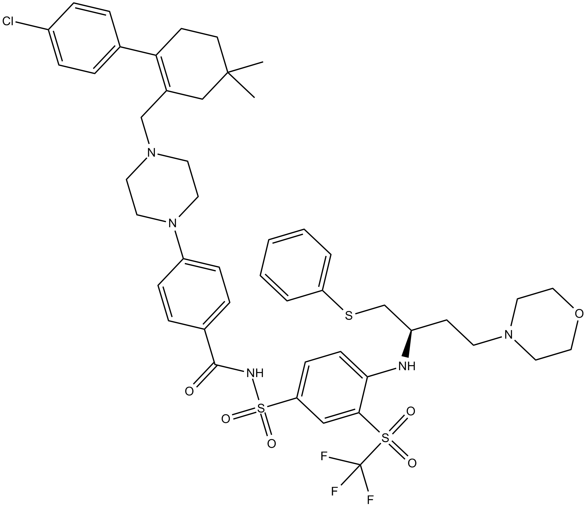
ABT-263 (Navitoclax)
مثبط لبروتينات عائلة Bcl-2
-
GC49745
ABT-263-d8
ABT-263-d8 هو الديوتيريوم المسمى Navitoclax. Navitoclax (ABT-263) هو مثبط قوي وفعال للبروتين من عائلة Bcl-2 والذي يرتبط بالعديد من بروتينات عائلة Bcl-2 المضادة للاستماتة ، مثل Bcl-xL و Bcl-2 و Bcl-w ، مع Ki أقل. من 1 نانومتر.

-
GC17234
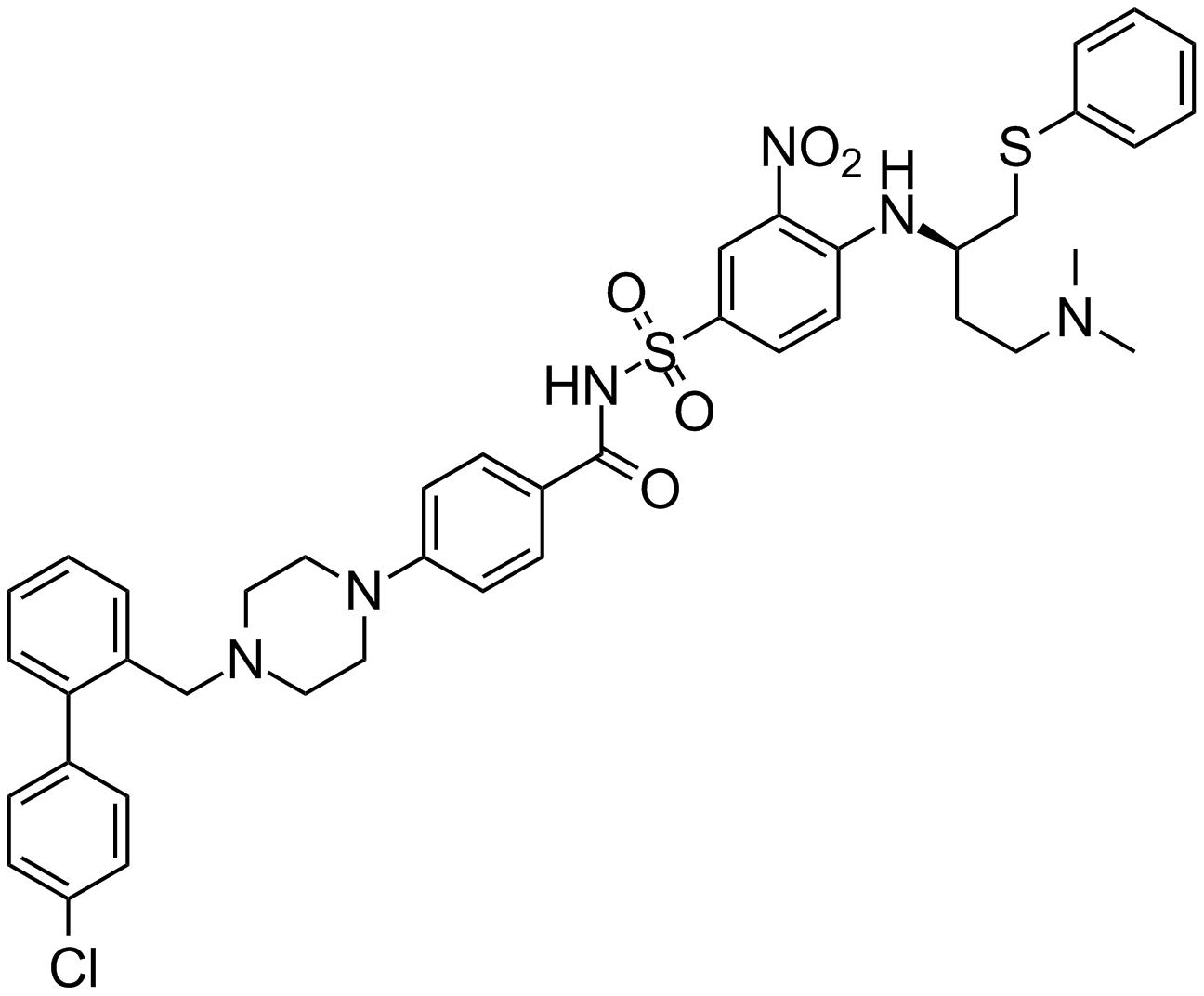
ABT-737
An inhibitor of anti-apoptotic Bcl-2 proteins
-
GA20494
Ac-Asp-Glu-Val-Asp-pNA
The cleavage of the chromogenic caspase-3 substrate Ac-DEVD-pNA can be monitored at 405 nm.
-
GC17602
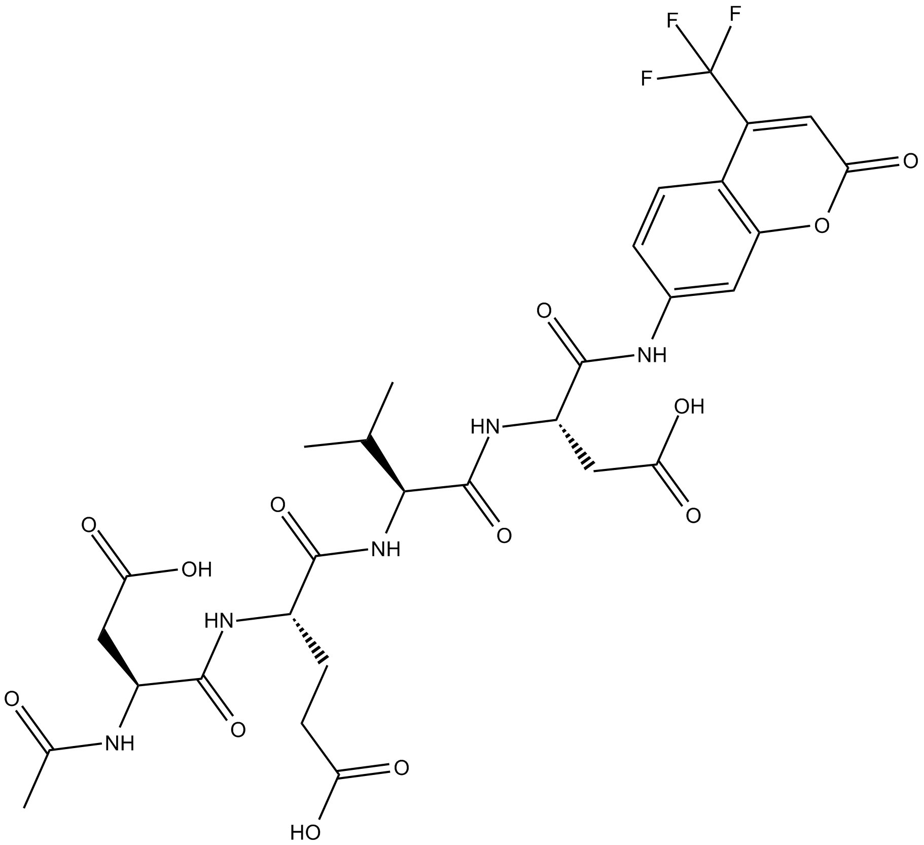
Ac-DEVD-AFC
Ac-DEVD-AFC عبارة عن ركيزة فلورية (Λex = 400 نانومتر ، Λem = 530 نانومتر)
-
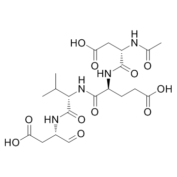
GC32695
Ac-DEVD-CHO
Ac-DEVD-CHO هو مثبط محدد لـ Caspase-3 بقيمة Ki تبلغ 230 pM
-
GC48470
Ac-DEVD-CHO (trifluoroacetate salt)
A dual caspase3/caspase7 inhibitor

-
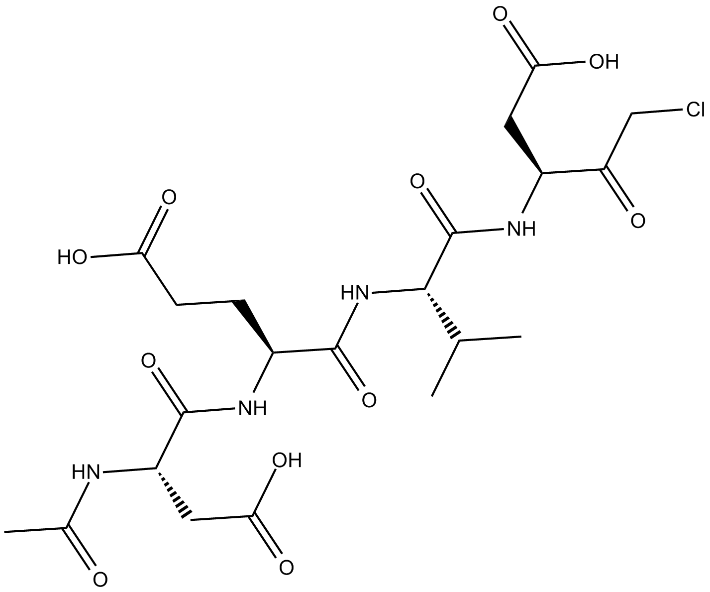
GC10951
Ac-DEVD-CMK
cell-permeable, and irreversible inhibitor of caspase
-
GC42689
Ac-DNLD-AMC
Ac-WLA-AMC عبارة عن ركيزة فلوروجينية من caspase-3.

-
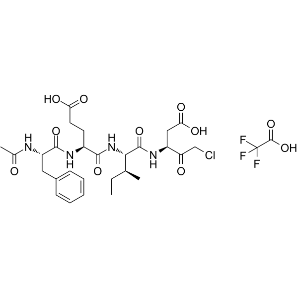
GC65107
Ac-FEID-CMK TFA
Ac-FEID-CMK TFA هو مثبط ببتيد قوي مشتق من GSDMEb
-
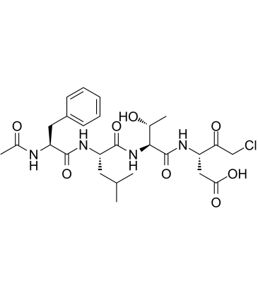
GC60558
Ac-FLTD-CMK
Ac-FLTD-CMK ، مثبط مشتق من غازديرمين D (GSDMD) ، هو مثبط محدد للالتهابات
-
GC49704
Ac-FLTD-CMK (trifluoroacetate salt)
An inhibitor of caspase-1, -4, -5, and -11

-
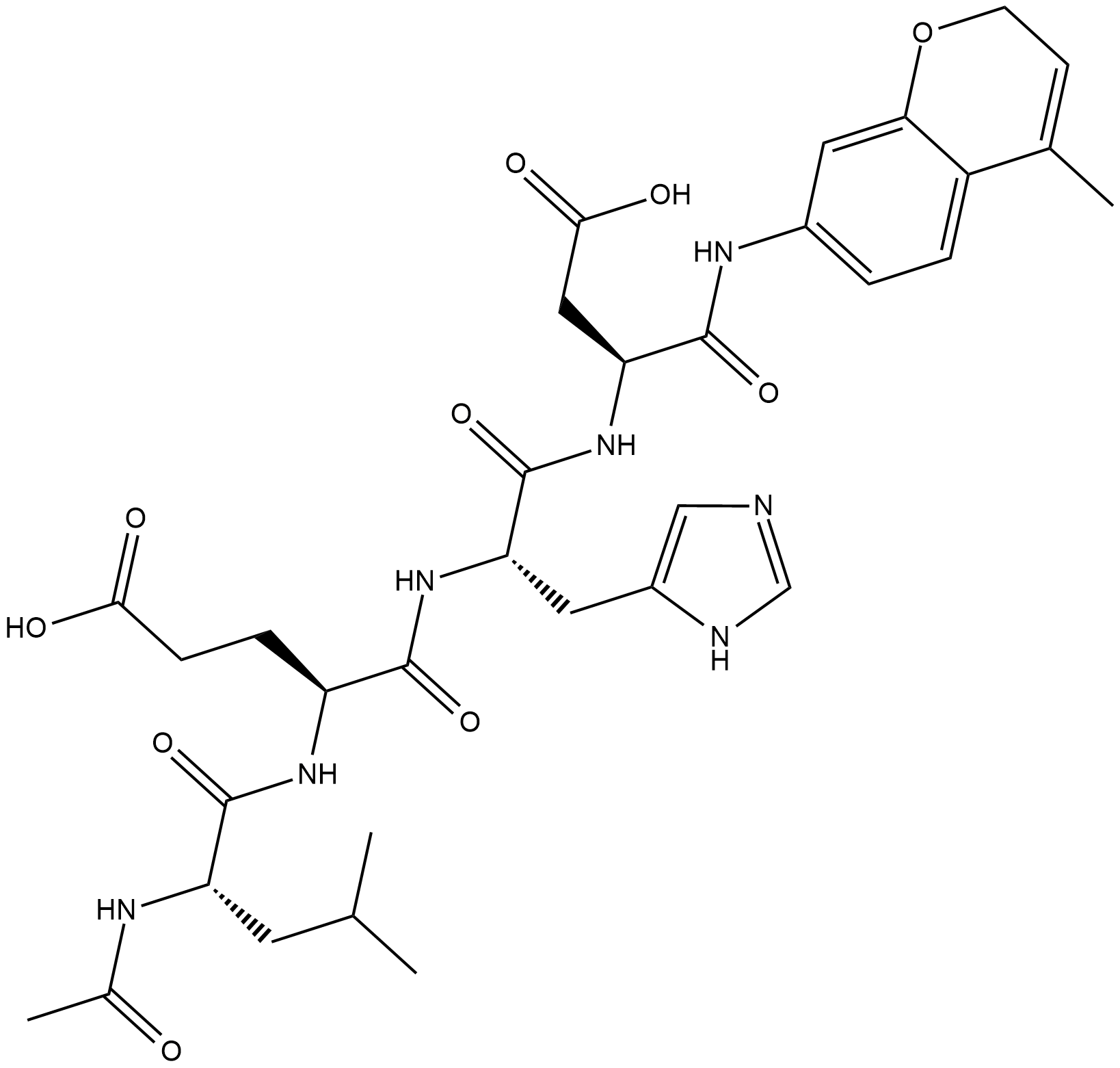
GC18226
Ac-LEHD-AMC (trifluoroacetate salt)
Ac-LEHD-AMC (ملح ثلاثي فلورو أسيتات) هو ركيزة فلورية لكاسباس 9 (الإثارة: 341 نانومتر ؛ الانبعاث: 441 نانومتر).
-
GC40556
Ac-LETD-AFC
Ac-LETD-AFC عبارة عن ركيزة فلورية من كاسباس 8.

-
GC13400
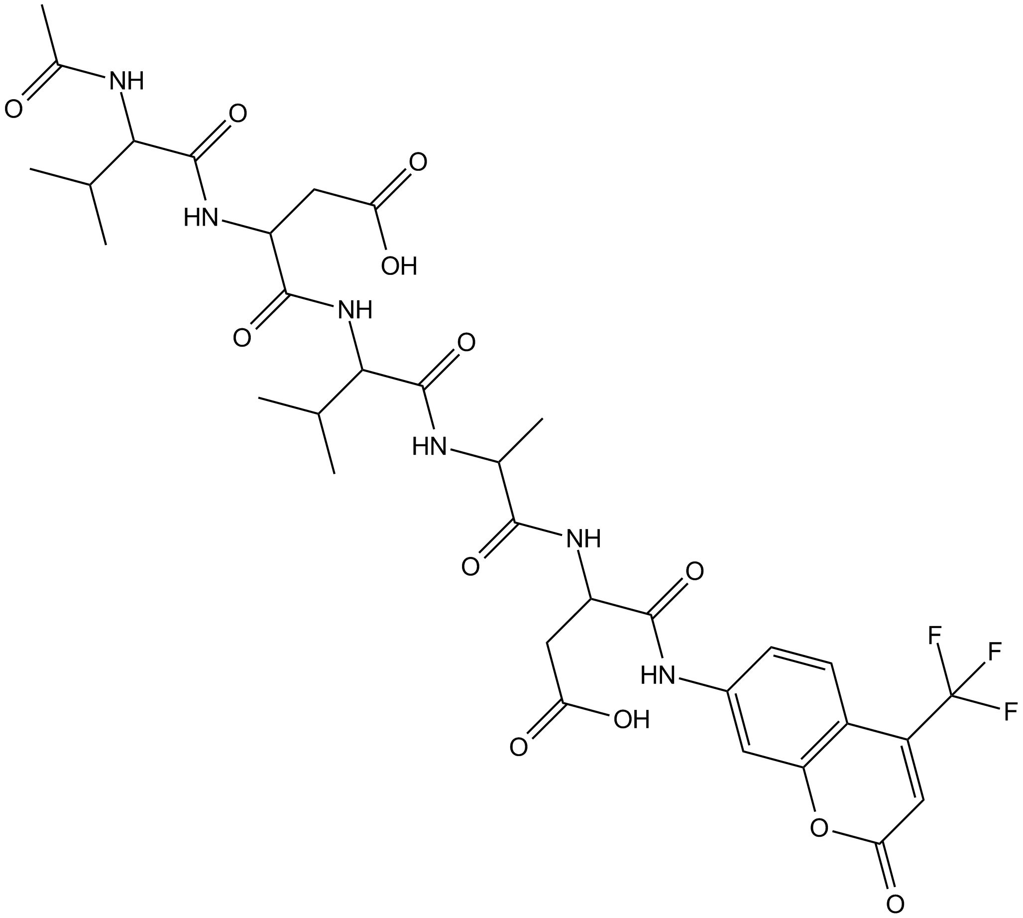
Ac-VDVAD-AFC
Ac-VDVAD-AFC عبارة عن ركيزة فلورية خاصة بكاسبيز. يمكن لـ Ac-VDVAD-AFC قياس النشاط الشبيه بـ caspase-3 ونشاط caspase-2 ويمكن استخدامه في البحث عن الورم والسرطان.
-
GC52372
Ac-VDVAD-AFC (trifluoroacetate salt)
A fluorogenic substrate for caspase-2

-
GC48974
Ac-VEID-AMC (ammonium acetate salt)
A caspase-6 fluorogenic substrate

-
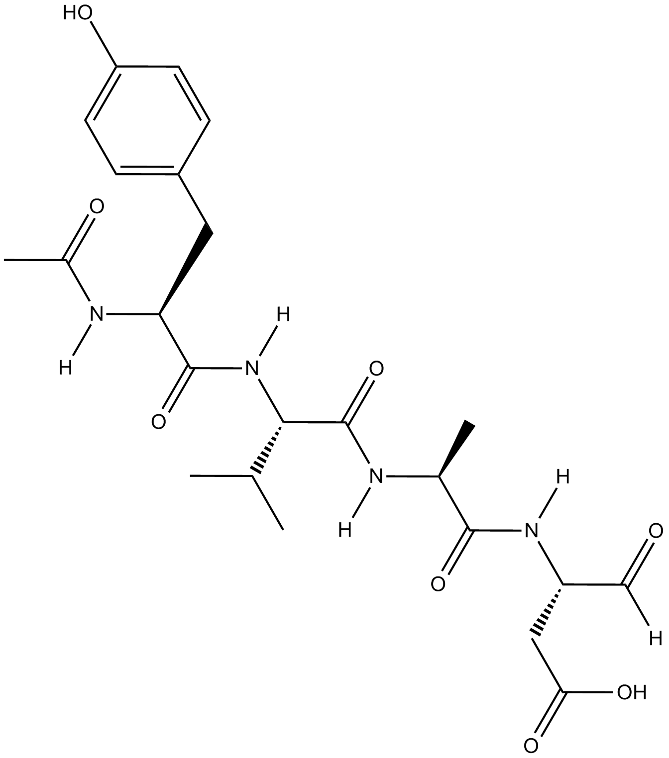
GC18021
Ac-YVAD-CHO
Ac-YVAD-CHO (L-709049) هو مثبط إنزيم (ICE) المحول لرباعي الببتيد (ICE) الفعال والقابل للانعكاس مع قيم Ki للإنسان والفأر 3.0 و 0.76 نانومتر.
-
GC42721
Ac-YVAD-CMK
أسي- YVAD-CMK هو مثبط لا رجعة فيه وانتقائي للكاسبيز-1 (K_i = 0.8 نانومول)، الذي يمكن أن يمنع تنشيط سايتوكين IL-1β المحفز للالتهاب. يمكن لأسي- YVAD-CMK تقليل الاستجابة الالتهابية وإحداث تأثير عصبي واقي طويل الأمد.

-
GC35227
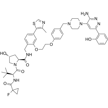
ACBI1
ACBI1 عبارة عن مزيل مواد قوي وتعاوني SMARCA2 و SMARCA4 و PBRM1 مع DC50s من 6 و 11 و 32 نانومتر ، على التواليACBI1 هو محلل بروتاكيظهر ACBI1 نشاطًا مضادًا للتكاثرACBI1 يحث على موت الخلايا المبرمج
-
GN10341
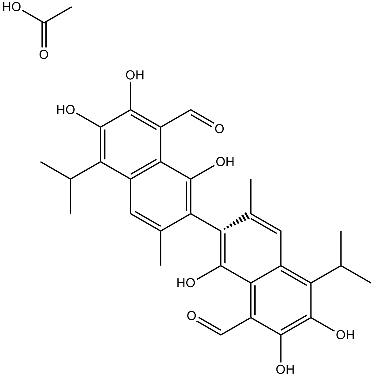
Acetate gossypol
-
GC11786
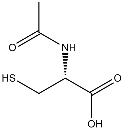
Acetylcysteine
أسيتيل سيستئين هو مشتق N-أسيتيل من السيستئين.
-
GC17094
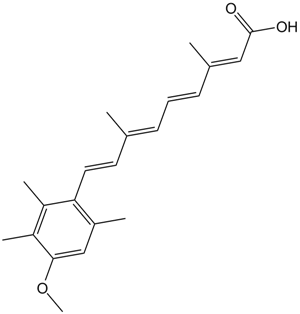
Acitretin
Acitretin (Ro 10-1670) هو الجيل الثاني من الريتينويد الجهازي الذي تم استخدامه في علاج الصدفية
-
GC35242
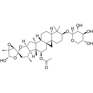
Actein
الأكتين عبارة عن جليكوسيد ترايتيربين معزول من جذور Cimicifuga foetidaيمنع Actein تكاثر الخلايا ، ويحث على الالتهام الذاتي وموت الخلايا المبرمج من خلال تعزيز تنشيط ROS / JNK ، وتثبيط مسار AKT في سرطان المثانة البشريأكتين له سمية قليلة في الجسم الحي
-
GC16866
Actinomycin D
مانع تفاعل الحمض النووي مع المتكلمات الجينية ذو التأثير المضاد للسرطان

-
GC16350
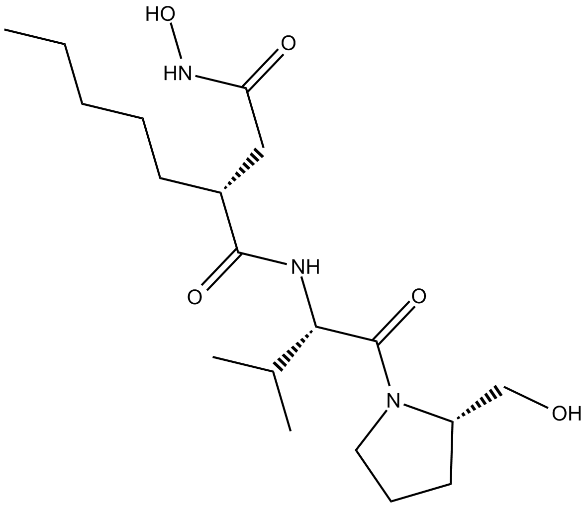
Actinonin
الأكتينونين ((-) - الأكتينونين) هو عامل مضاد للجراثيم يحدث بشكل طبيعي وينتج عن طريق الأكتينوميسيس
-
GC16362
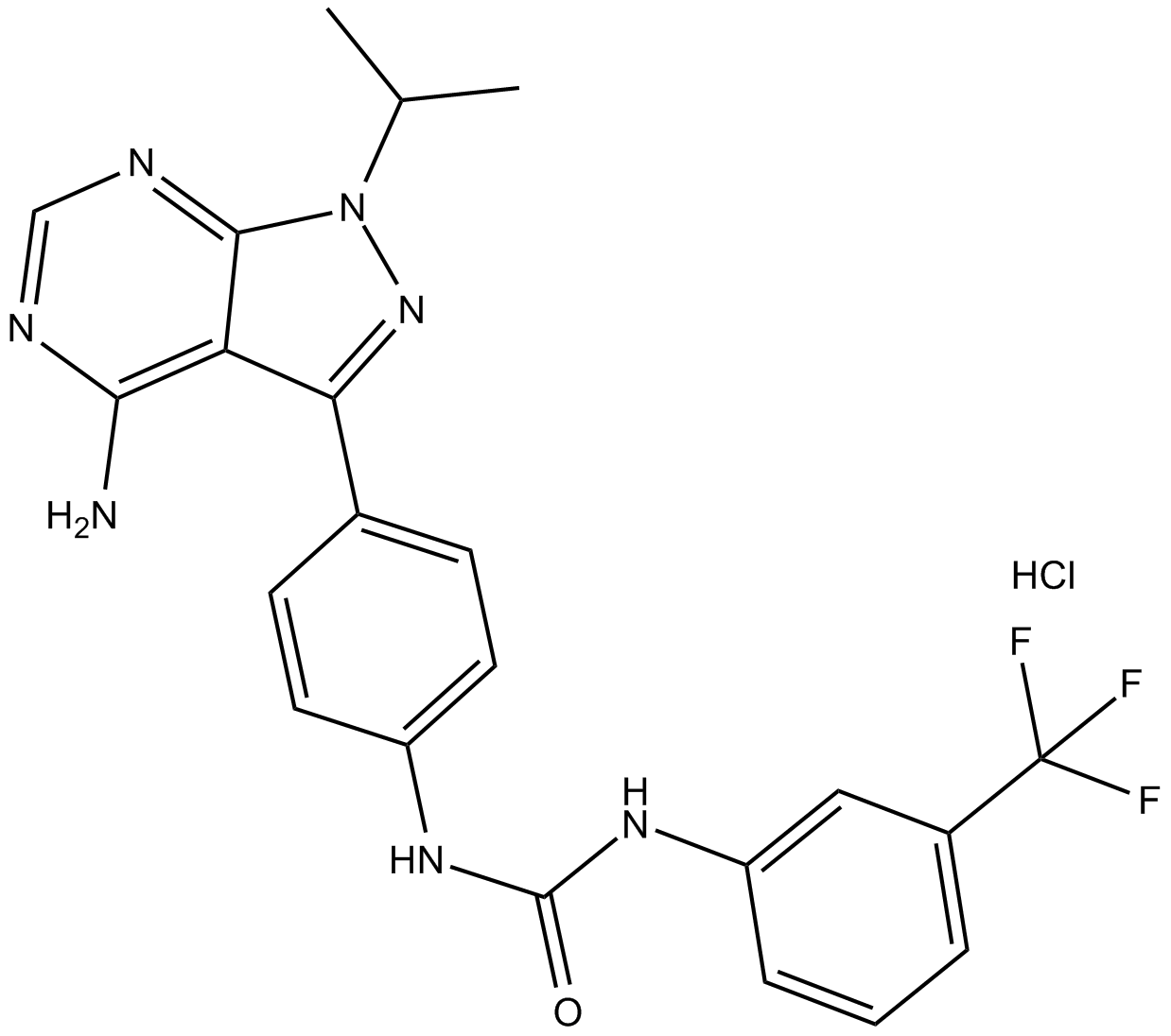
AD57 (hydrochloride)
polypharmacological cancer therapeutic that inhibits RET.
-
GC34214
Adalimumab (Anti-Human TNF-alpha, Human Antibody)
أداليموماب (مضاد لتفاعل عامل نخر الورم-ألفا البشري، أجسام مضادة بشرية) هو أحد أنواع الأجسام المضادة المونوكلونية من فئة IgG1 التي تستهدف عاملاً يُعرف باسم تفاعل نخر الورم-ألفا (TNF-α) في جسم الإنسان.


