Apoptosis
As one of the cellular death mechanisms, apoptosis, also known as programmed cell death, can be defined as the process of a proper death of any cell under certain or necessary conditions. Apoptosis is controlled by the interactions between several molecules and responsible for the elimination of unwanted cells from the body.
Many biochemical events and a series of morphological changes occur at the early stage and increasingly continue till the end of apoptosis process. Morphological event cascade including cytoplasmic filament aggregation, nuclear condensation, cellular fragmentation, and plasma membrane blebbing finally results in the formation of apoptotic bodies. Several biochemical changes such as protein modifications/degradations, DNA and chromatin deteriorations, and synthesis of cell surface markers form morphological process during apoptosis.
Apoptosis can be stimulated by two different pathways: (1) intrinsic pathway (or mitochondria pathway) that mainly occurs via release of cytochrome c from the mitochondria and (2) extrinsic pathway when Fas death receptor is activated by a signal coming from the outside of the cell.
Different gene families such as caspases, inhibitor of apoptosis proteins, B cell lymphoma (Bcl)-2 family, tumor necrosis factor (TNF) receptor gene superfamily, or p53 gene are involved and/or collaborate in the process of apoptosis.
Caspase family comprises conserved cysteine aspartic-specific proteases, and members of caspase family are considerably crucial in the regulation of apoptosis. There are 14 different caspases in mammals, and they are basically classified as the initiators including caspase-2, -8, -9, and -10; and the effectors including caspase-3, -6, -7, and -14; and also the cytokine activators including caspase-1, -4, -5, -11, -12, and -13. In vertebrates, caspase-dependent apoptosis occurs through two main interconnected pathways which are intrinsic and extrinsic pathways. The intrinsic or mitochondrial apoptosis pathway can be activated through various cellular stresses that lead to cytochrome c release from the mitochondria and the formation of the apoptosome, comprised of APAF1, cytochrome c, ATP, and caspase-9, resulting in the activation of caspase-9. Active caspase-9 then initiates apoptosis by cleaving and thereby activating executioner caspases. The extrinsic apoptosis pathway is activated through the binding of a ligand to a death receptor, which in turn leads, with the help of the adapter proteins (FADD/TRADD), to recruitment, dimerization, and activation of caspase-8 (or 10). Active caspase-8 (or 10) then either initiates apoptosis directly by cleaving and thereby activating executioner caspase (-3, -6, -7), or activates the intrinsic apoptotic pathway through cleavage of BID to induce efficient cell death. In a heat shock-induced death, caspase-2 induces apoptosis via cleavage of Bid.
Bcl-2 family members are divided into three subfamilies including (i) pro-survival subfamily members (Bcl-2, Bcl-xl, Bcl-W, MCL1, and BFL1/A1), (ii) BH3-only subfamily members (Bad, Bim, Noxa, and Puma9), and (iii) pro-apoptotic mediator subfamily members (Bax and Bak). Following activation of the intrinsic pathway by cellular stress, pro‑apoptotic BCL‑2 homology 3 (BH3)‑only proteins inhibit the anti‑apoptotic proteins Bcl‑2, Bcl-xl, Bcl‑W and MCL1. The subsequent activation and oligomerization of the Bak and Bax result in mitochondrial outer membrane permeabilization (MOMP). This results in the release of cytochrome c and SMAC from the mitochondria. Cytochrome c forms a complex with caspase-9 and APAF1, which leads to the activation of caspase-9. Caspase-9 then activates caspase-3 and caspase-7, resulting in cell death. Inhibition of this process by anti‑apoptotic Bcl‑2 proteins occurs via sequestration of pro‑apoptotic proteins through binding to their BH3 motifs.
One of the most important ways of triggering apoptosis is mediated through death receptors (DRs), which are classified in TNF superfamily. There exist six DRs: DR1 (also called TNFR1); DR2 (also called Fas); DR3, to which VEGI binds; DR4 and DR5, to which TRAIL binds; and DR6, no ligand has yet been identified that binds to DR6. The induction of apoptosis by TNF ligands is initiated by binding to their specific DRs, such as TNFα/TNFR1, FasL /Fas (CD95, DR2), TRAIL (Apo2L)/DR4 (TRAIL-R1) or DR5 (TRAIL-R2). When TNF-α binds to TNFR1, it recruits a protein called TNFR-associated death domain (TRADD) through its death domain (DD). TRADD then recruits a protein called Fas-associated protein with death domain (FADD), which then sequentially activates caspase-8 and caspase-3, and thus apoptosis. Alternatively, TNF-α can activate mitochondria to sequentially release ROS, cytochrome c, and Bax, leading to activation of caspase-9 and caspase-3 and thus apoptosis. Some of the miRNAs can inhibit apoptosis by targeting the death-receptor pathway including miR-21, miR-24, and miR-200c.
p53 has the ability to activate intrinsic and extrinsic pathways of apoptosis by inducing transcription of several proteins like Puma, Bid, Bax, TRAIL-R2, and CD95.
Some inhibitors of apoptosis proteins (IAPs) can inhibit apoptosis indirectly (such as cIAP1/BIRC2, cIAP2/BIRC3) or inhibit caspase directly, such as XIAP/BIRC4 (inhibits caspase-3, -7, -9), and Bruce/BIRC6 (inhibits caspase-3, -6, -7, -8, -9).
Any alterations or abnormalities occurring in apoptotic processes contribute to development of human diseases and malignancies especially cancer.
References:
1.Yağmur Kiraz, Aysun Adan, Melis Kartal Yandim, et al. Major apoptotic mechanisms and genes involved in apoptosis[J]. Tumor Biology, 2016, 37(7):8471.
2.Aggarwal B B, Gupta S C, Kim J H. Historical perspectives on tumor necrosis factor and its superfamily: 25 years later, a golden journey.[J]. Blood, 2012, 119(3):651.
3.Ashkenazi A, Fairbrother W J, Leverson J D, et al. From basic apoptosis discoveries to advanced selective BCL-2 family inhibitors[J]. Nature Reviews Drug Discovery, 2017.
4.McIlwain D R, Berger T, Mak T W. Caspase functions in cell death and disease[J]. Cold Spring Harbor perspectives in biology, 2013, 5(4): a008656.
5.Ola M S, Nawaz M, Ahsan H. Role of Bcl-2 family proteins and caspases in the regulation of apoptosis[J]. Molecular and cellular biochemistry, 2011, 351(1-2): 41-58.
対象は Apoptosis
- Pyroptosis(15)
- Caspase(77)
- 14.3.3 Proteins(3)
- Apoptosis Inducers(71)
- Bax(15)
- Bcl-2 Family(136)
- Bcl-xL(13)
- c-RET(15)
- IAP(32)
- KEAP1-Nrf2(73)
- MDM2(21)
- p53(137)
- PC-PLC(6)
- PKD(8)
- RasGAP (Ras- P21)(2)
- Survivin(8)
- Thymidylate Synthase(12)
- TNF-α(141)
- Other Apoptosis(1145)
- Apoptosis Detection(0)
- Caspase Substrate(0)
- APC(6)
- PD-1/PD-L1 interaction(60)
- ASK1(4)
- PAR4(2)
- RIP kinase(47)
- FKBP(22)
製品は Apoptosis
- Cat.No. 商品名 インフォメーション
-
GC15355
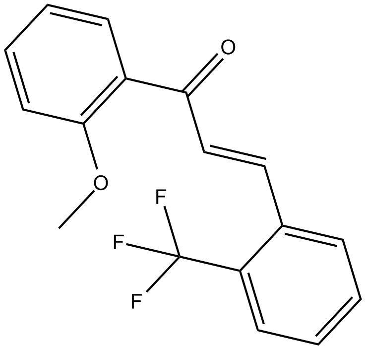
2-Trifluoromethyl-2'-methoxychalcone
Nrf2活性化剤
-
GN10800
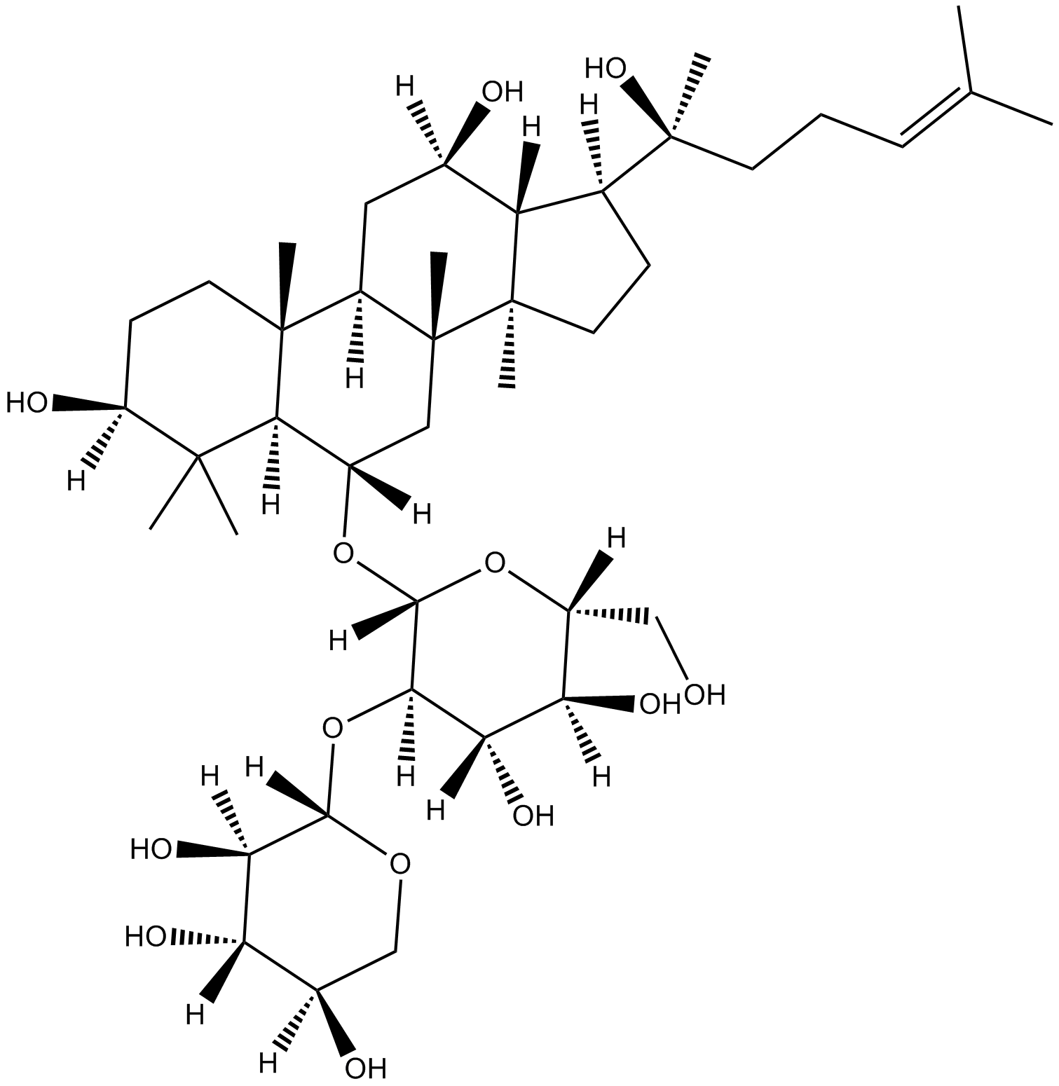
20(S)-NotoginsenosideR2
-
GC46528
25-hydroxy Cholesterol-d6
25ヒドロキシコレステロールの定量化のための内部標準

-
GC48482
28-Acetylbetulin
抗炎症および抗がん作用を持つルパン三萜

-
GC35112
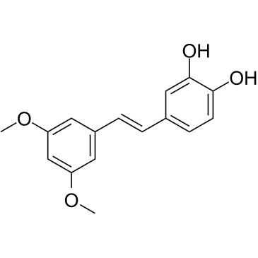
3'-Hydroxypterostilbene
3'-ヒドロキシプテロスチルベンは、プテロスチルベン類似体です。 3'-ヒドロキシプテロスチルベンは、COLO 205、HCT-116、および HT-29 細胞の増殖を、それぞれ 9.0、40.2、および 70.9 μM の IC50 で阻害します。 3'-ヒドロキシプテロスチルベンは、PI3K/Akt および MAPKs シグナル伝達経路を大幅にダウンレギュレートし、アポトーシスとオートファジーを誘導することにより、ヒト結腸癌細胞の増殖を効果的に阻害します。 3'-ヒドロキシプテロスチルベンは、がんの研究に使用できます。
-
GC12791
3,3'-Diindolylmethane
放射線防御および化学予防効果を持つ植物化学物質

-
GC42237
3,5-dimethyl PIT-1
PtdIns-(3,4,5)-P3(PIP3)は、リン脂質3-キナーゼ(PI3K)やPTENなどのpleckstrin homology(PH)ドメインを持つシグナル伝達タンパク質の結合アンカーとして機能します。

-
GC64762
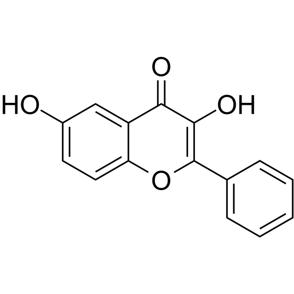
3,6-Dihydroxyflavone
3,6-ジヒドロキシフラボンは抗がん剤です。 3,6-ジヒドロキシフラボンは、用量および時間依存的に細胞生存率を低下させ、カスパーゼカスケードを活性化し、ポリ (ADP-リボース) ポリメラーゼ (PARP) を切断することによってアポトーシスを誘導します。 3,6-ジヒドロキシフラボンは、細胞内の酸化ストレスと脂質過酸化を増加させます。
-
GC46583
3-Amino-2,6-Piperidinedione
(±)-タリドミドの活性代謝物質

-
GC49849
3-Aminosalicylic Acid
サリチル酸誘導体

-
GC35106
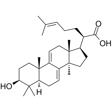
3-Dehydrotrametenolic acid
Poria cocos の菌核から分離された 3- デヒドロトラメテノール酸は、乳酸脱水素酵素 (LDH) 阻害剤です。 3- デヒドロトラメテノール酸は、in vitro で脂肪細胞の分化を促進し、in vivo でインスリン感作物質として作用します。 3- デヒドロトラメテノール酸はアポトーシスを誘導し、抗がん作用があります。
-
GC68537
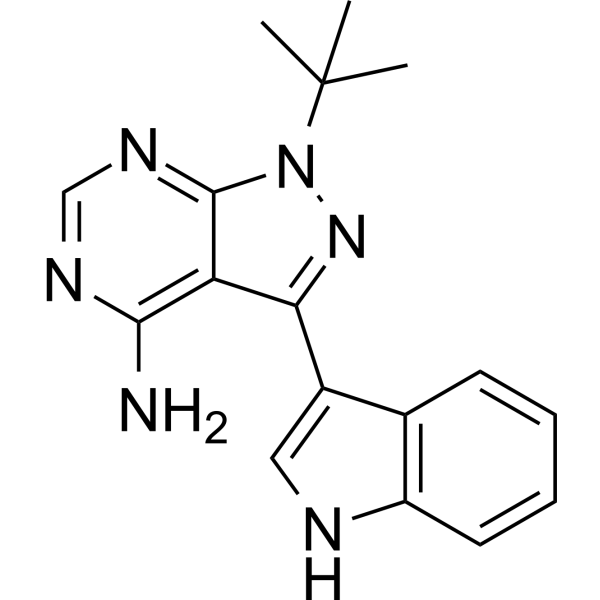
3-IN-PP1
3-IN-PP1は、プロテインキナーゼD(PKD)阻害剤の一種です。 3-IN-PP1は、PKD1、PKD2、およびPKD3に対して広範囲かつ有効なPKD阻害活性を示し、それぞれのIC50値は108、94、および108 nMです。 3-IN-PP1はまた、多くのがん細胞の成長を抑制する広範な抗がん剤でもあります。 3-IN-PP1はがん研究に使用できます。
-
GC17394
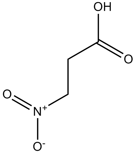
3-Nitropropionic acid
3-ニトロプロピオン酸 (β-ニトロプロピオン酸) は、コハク酸脱水素酵素の不可逆的阻害剤です。
-
GC35099
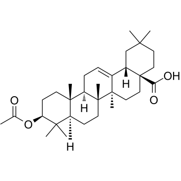
3-O-Acetyloleanolic acid
Vigna sinensis K. の種子から分離されたオレアノール酸誘導体である 3-O-アセチルオレアノール酸 (3AOA) は、癌を誘発し、抗血管新生活性も示します。
-
GC60507
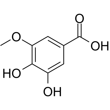
3-O-Methylgallic acid
3-O-メチル没食子酸 (3,4-ジヒドロキシ-5-メトキシ安息香酸) はアントシアニン代謝産物であり、強力な抗酸化能を持っています。 3-O-メチル没食子酸は、24.1 μM の IC50 値で Caco-2 細胞の増殖を阻害します。 3-O-メチル没食子酸も細胞アポトーシスを誘導し、抗がん効果があります。
-
GC32767
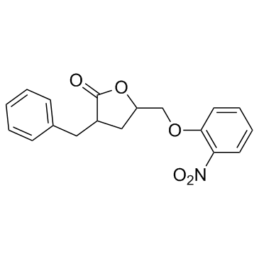
3BDO
3BDO は、オートファジーも阻害できる新しい mTOR アクティベーターです。
-
GC45354
4β-Hydroxywithanolide E
4β-Hydroxywithanolide E、Physalis peruviana L から分離されました。

-
GC48437
4'-Acetyl Chrysomycin A
抗菌および抗がん活性を持つ細菌代謝産物

-
GC42346
4-bromo A23187
4-ブロモ A23187 は、選択性の高いカルシウム イオノフォア A-23187 のハロゲン化類似体です。

-
GC42401
4-hydroperoxy Cyclophosphamide
シクロホスファミドの活性化アナログ

-
GC30896
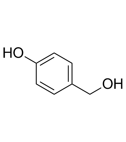
4-Hydroxybenzyl alcohol
4-ヒドロキシベンジルアルコールは、さまざまな種類の植物に広く分布するフェノール化合物です。
-
GC33815
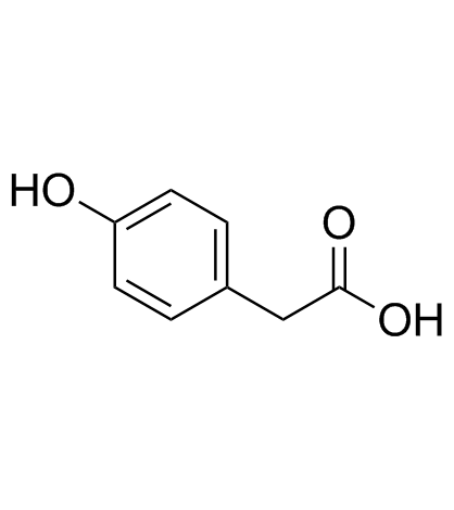
4-Hydroxyphenylacetic acid
ポリフェノールの主要な微生物叢由来の代謝産物である4-ヒドロキシフェニル酢酸は、抗酸化作用に関与しています。
-
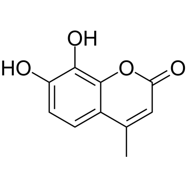
GC35138
4-Methyldaphnetin
4-メチルダフネチンは、4-メチル クマリンの誘導体の合成における前駆体です。 4-メチルダフネチンは、いくつかの癌細胞株に対して、強力で選択的な抗増殖作用とアポトーシス誘導作用を持っています。 4-メチルダフネチンは、ラジカル捕捉特性を有し、膜脂質過酸化を強力に阻害します。
-
GC68231
4-Methylsalicylic acid

-
GC31648
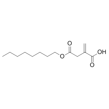
4-Octyl Itaconate
4-オクチルイタコネート(4-OI)は、細胞浸透性のあるイタコン酸誘導体です。イタコン酸と4-オクチルイタコネートは類似したチオール反応性を持っており、生物学的機能を研究するための適切な代替品であると考えられています。
-
GC49127
4-oxo Cyclophosphamide
シクロホスファミドの不活性代謝物

-
GC45352
4-oxo Withaferin A
4-オキソ ウィザフェリン A は、ウィザフェリン A の類似体です。ウィザフェリン A は、ウィタニア ソムニフェラから分離されたウィタノリドです。 4-オキソ ウィザフェリン A は、多発性骨髄腫の研究の可能性を秘めています。

-
GC45353
4-oxo-27-TBDMS Withaferin A
4-オキソ-27-TBDMS ウィザフェリン A、ウィザフェリン A 誘導体は、腫瘍細胞に対して強力な抗増殖効果を示します。4-オキソ-27-TBDMS ウィザフェリン A は、腫瘍細胞のアポトーシスを誘導します。 4-オキソ-27-TBDMS ウィザフェリン A は抗がん剤です。

-
GC60525
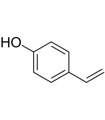
4-Vinylphenol (10%w/w in propylene glycol)
4-ビニルフェノールは、薬草のヘディオティス・ディフサ・ウィルド、ワイルド・ライスに含まれており、ワイン中の乳酸菌によるp-クマル酸とフェルラ酸の代謝産物でもあります。 4-ビニルフェノールは、アポトーシスを誘導し、血管形成を阻害し、in vivo での浸潤性乳房腫瘍の増殖を抑制します。
-
GC10468
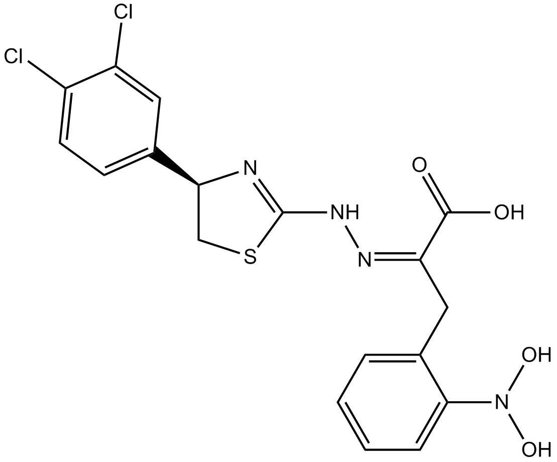
4EGI-1
4EGI-1 は eIF4E/eIF4G 相互作用の阻害剤であり、eIF4E 結合に対する Kd は 25 μM です。
-
GC35150
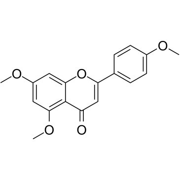
5,7,4'-Trimethoxyflavone
5,7,4'-トリメトキシフラボンは、タイの有名な薬用植物であるケンフェリア パルビフローラ (KP) から分離されました。 5,7,4'-トリメトキシフラボンは、サブ G1 期の増加、DNA 断片化、アネキシン-V/PI 染色、Bax/Bcl-xL 比、カスパーゼ-3 のタンパク質分解活性化、および poly の分解によって証明されるように、アポトーシスを誘導します。 (ADP-リボース) ポリメラーゼ (PARP) タンパク質。5,7,4'-トリメトキシフラボンは、SNU-16 ヒト胃癌細胞の増殖を濃度依存的に阻害するのに非常に効果的です。
-
GN10629
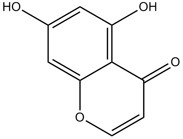
5,7-dihydroxychromone
-
GC63972
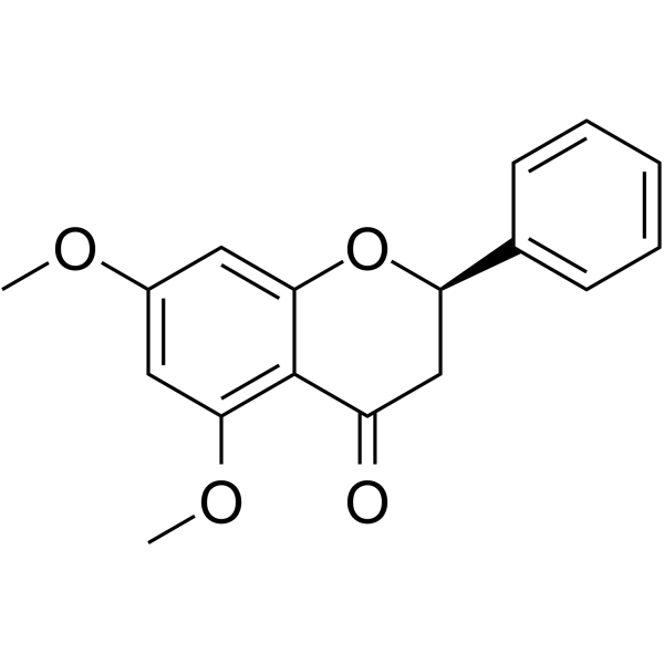
5,7-Dimethoxyflavanone
5,7-ジメトキシフラバノンは、S.
-
GC52227
5-(3',4'-Dihydroxyphenyl)-γ-Valerolactone
様々なポリフェノールの活性代謝物

-
GC35147
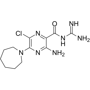
5-(N,N-Hexamethylene)-amiloride
5-(N,N-ヘキサメチレン)-アミロリド (ヘキサメチレンアミロリド) はアミロリドに由来し、強力な Na+/H+ 交換阻害剤であり、細胞内 pH (pHi) を低下させ、白血病患者のアポトーシスを誘導する細胞。
-
GC45356
5-Aminolevulinic Acid (hydrochloride)

-
GC46681
5-Bromouridine
臭素化ウリジンアナログ

-
GC42545
5-Fluorouracil-13C,15N2
5-フルオロウラシル-13C、15N2は、GCまたはLC-MSによる5-フルオロウラシルの定量のための内部標準として使用することを目的としています。

-
GC46705
5-Methoxycanthinone
5-メトキシカンチノンは、リーシュマニア株の経口活性阻害剤です。

-
GC42586
6α-hydroxy Paclitaxel
6α-ヒドロキシ パクリタキセルはパクリタキセルの一次代謝産物です。 6α-ヒドロキシパクリタキセルは、有機陰イオン輸送ポリペプチド 1B1/SLCO1B1 (OATP1B1) に対して、パクリタキセルと同様の阻害効力を持つ時間依存効果を保持しますが、OATP1B3 の時間依存阻害はもはや示しませんでした。 6α-ヒドロキシ パクリタキセルは癌の研究に使用できます。

-
GC45772
6(5H)-Phenanthridinone
PARP1および2の阻害剤

-
GN10093
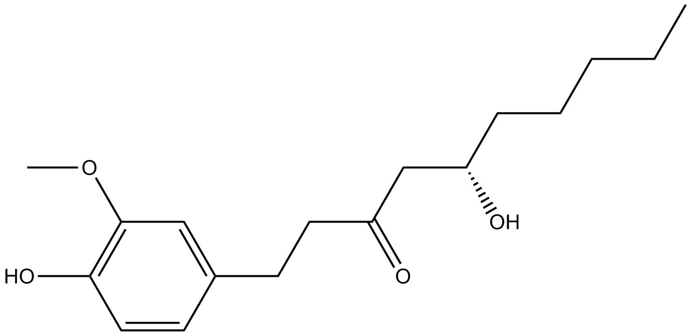
6-gingerol
-
GC49429
6-keto Lithocholic Acid
リトコール酸の代謝産物
-
GC35184
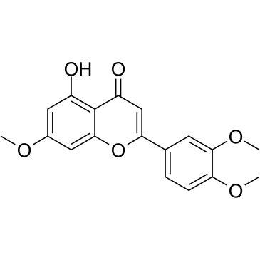
7,3',4'-Tri-O-methylluteolin
7,3',4'-トリ-O-メチルルテオリン (5-ヒドロキシ-3',4',7-トリメトキシフラボン) は、フラボノイド化合物であり、LPS 誘導マクロファージ細胞株において、炎症メディエーター、NO、PGE2、および炎症誘発性サイトカインの放出。
-
GC45673
7,8-Dihydroneopterin
炎症マーカーである 7,8-ジヒドロネオプテリンは、一酸化窒素シンターゼ (iNOS) 発現の増強を介して星状細胞およびニューロンの細胞アポトーシスを誘導します。

-
GC16853
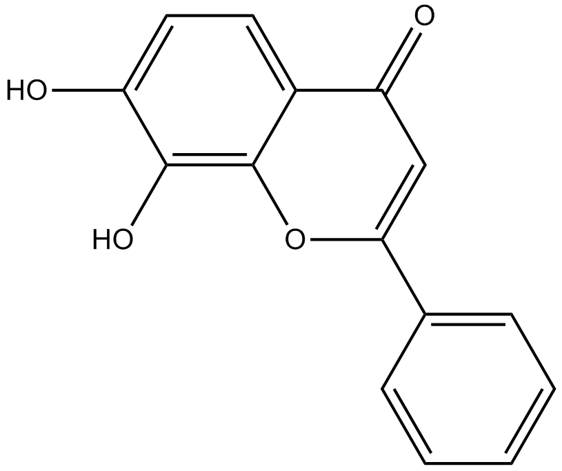
7,8-Dihydroxyflavone
7,8-ジヒドロキシフラボンは、脳由来神経栄養因子 (BDNF) の生理学的作用を模倣する、強力かつ選択的な TrkB アゴニストです。
-
GC42616
7-oxo Staurosporine
7-oxoスタウロスポリンは、元々Sから分離された抗生物質です。

-
GC16037
7BIO
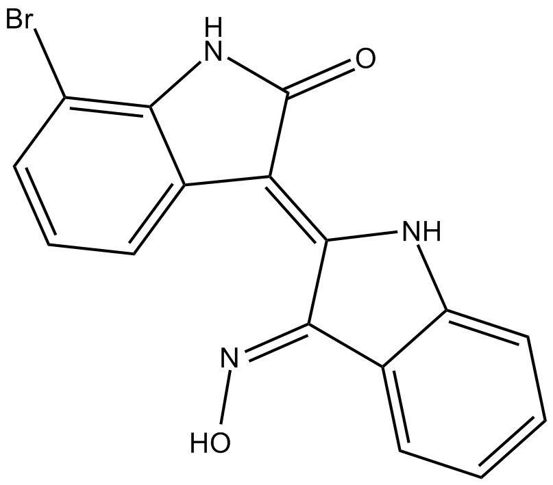
7BIO (7-ブロモインジルビン-3-オキシム) はインジルビンの誘導体です。
-
GC46741
8(E),10(E),12(Z)-Octadecatrienoic Acid
共役PUFA

-
GC42622
8-bromo-Cyclic AMP
8-ブロモシクリックAMPは、cAMPの臭素化誘導体であり、cAMPホスホジエステラーゼによる分解に対する耐性があるため長時間作用します。

-
GC49275
8-Oxycoptisine
8-オキシコプチシンは、抗がん作用を持つ天然のプロトベルベリン アルカロイドです。

-
GC17119
8-Prenylnaringenin
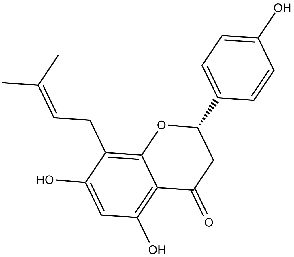
8-プレニルナリンゲニンは、細胞毒性を持つホップ毬花 (Humulus lupulus) から分離されたプレニルフラボノイドです。 8-プレニルナリンゲニンは、内因性および外因性経路を介したアポトーシスの誘導を介して、HCT-116 結腸癌細胞に対して抗増殖活性を持っています。 8-プレニルナリンゲニンは、マウスのAktリン酸化経路の活性化を通じて、固定化による廃用性筋萎縮からの回復も促進します 。
-
GC41642
9(E),11(E),13(E)-Octadecatrienoic Acid
9(E),11(E),13(E)-オクタデカトリエン酸(β-ESA)は、植物の種子油やリノレン酸のアルカリ異性化によって合成された共役リノール酸混合物中に存在する共役多価不飽和脂肪酸です。

-
GC41643
9(Z),11(E),13(E)-Octadecatrienoic Acid
9(Z),11(E),13(E)-オクタデカトリエン酸(α-ESA)は、植物種子油に一般的に存在する共役ポリ不飽和脂肪酸です。

-
GC40785
9(Z),11(E),13(E)-Octadecatrienoic Acid ethyl ester
9(Z),11(E),13(E)-オクタデカトリエン酸エチルエステル(α-ESA)は、植物種子油に一般的に存在する共役ポリ不飽和脂肪酸です。

-
GC40710
9(Z),11(E),13(E)-Octadecatrienoic Acid methyl ester
9Z、11E、13E-オクタデカトリエン酸(α-ESA)は、植物種子油に一般的に存在する共役ポリ不飽和脂肪酸です。

-
GC39152
9-ING-41
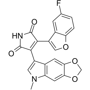
9-ING-41 は、マレイミドベースの ATP 競合的かつ選択的なグリコーゲン合成酵素キナーゼ 3β (GSK-3β) 阻害剤であり、IC50 は 0.71 μM です。 9-ING-41 は、癌細胞の細胞周期停止、オートファジー、およびアポトーシスを有意に引き起こします。 9-ING-41 には抗がん作用があり、化学療法薬の抗腫瘍効果を高める可能性があります。
-
GN10035
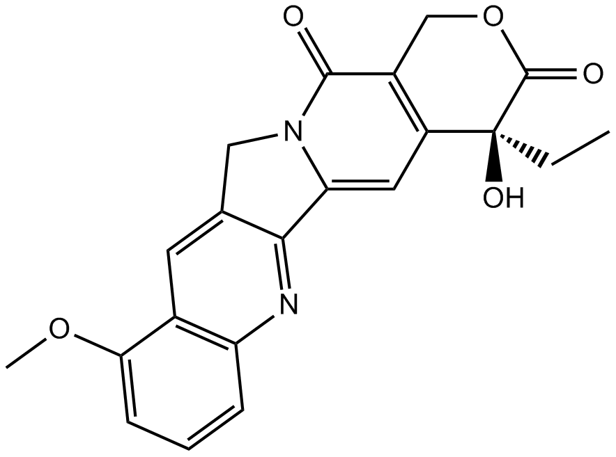
9-Methoxycamptothecin
-
GC45960
9c(i472)
9c(i472) は 15-LOX-1 (15-リポキシゲナーゼ-1) の強力な阻害剤であり、IC50 値は 0.19 μM.

-
GC50465
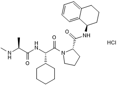
A 410099.1
高親和性のXIAP拮抗剤;in vivoで活性があります
-
GC17512
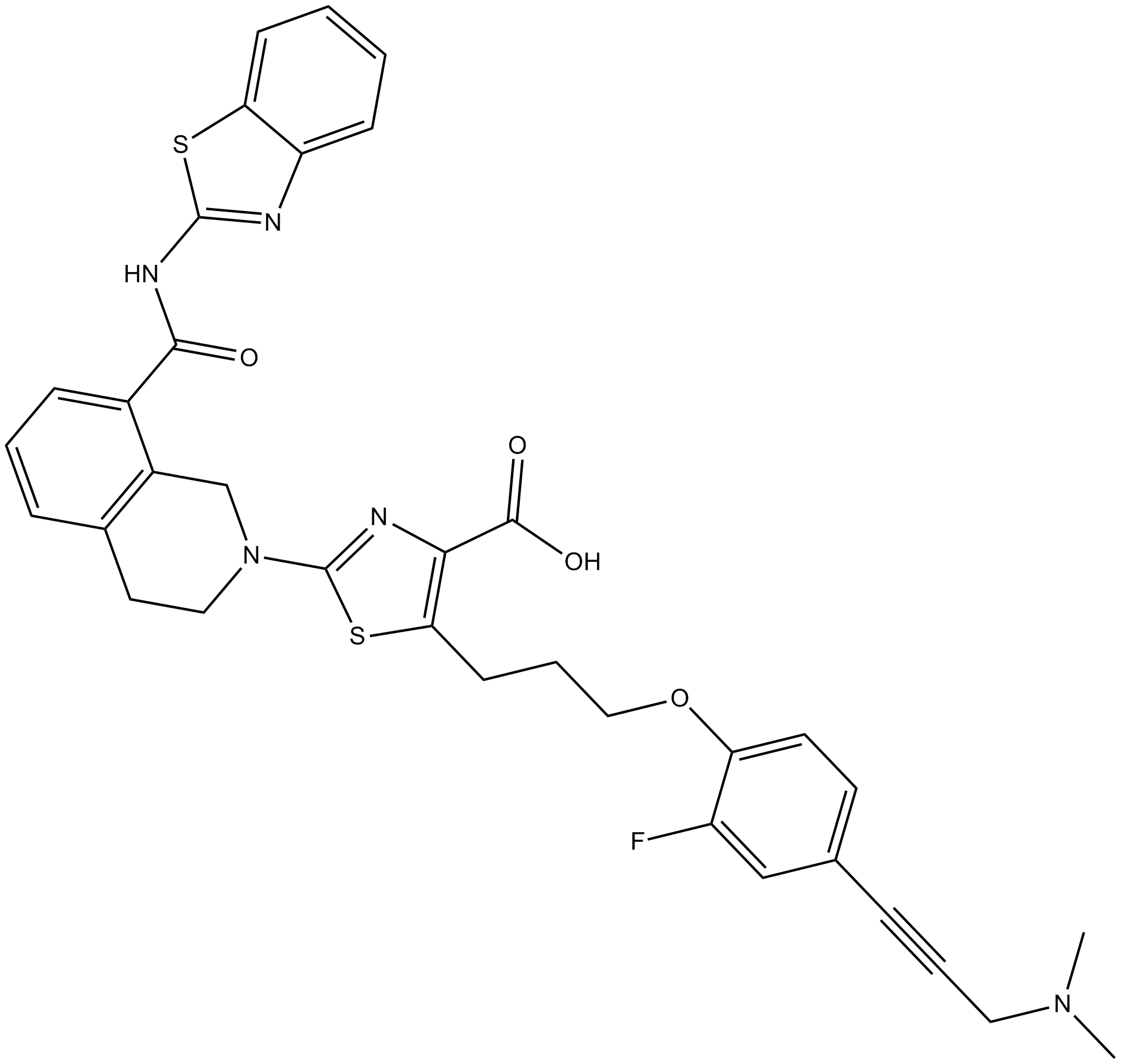
A-1155463
A-1155463 は、Molt-4 細胞で 70 nM の EC50 を持つ非常に強力で選択的な BCL-XL 阻害剤です。
-
GC16278
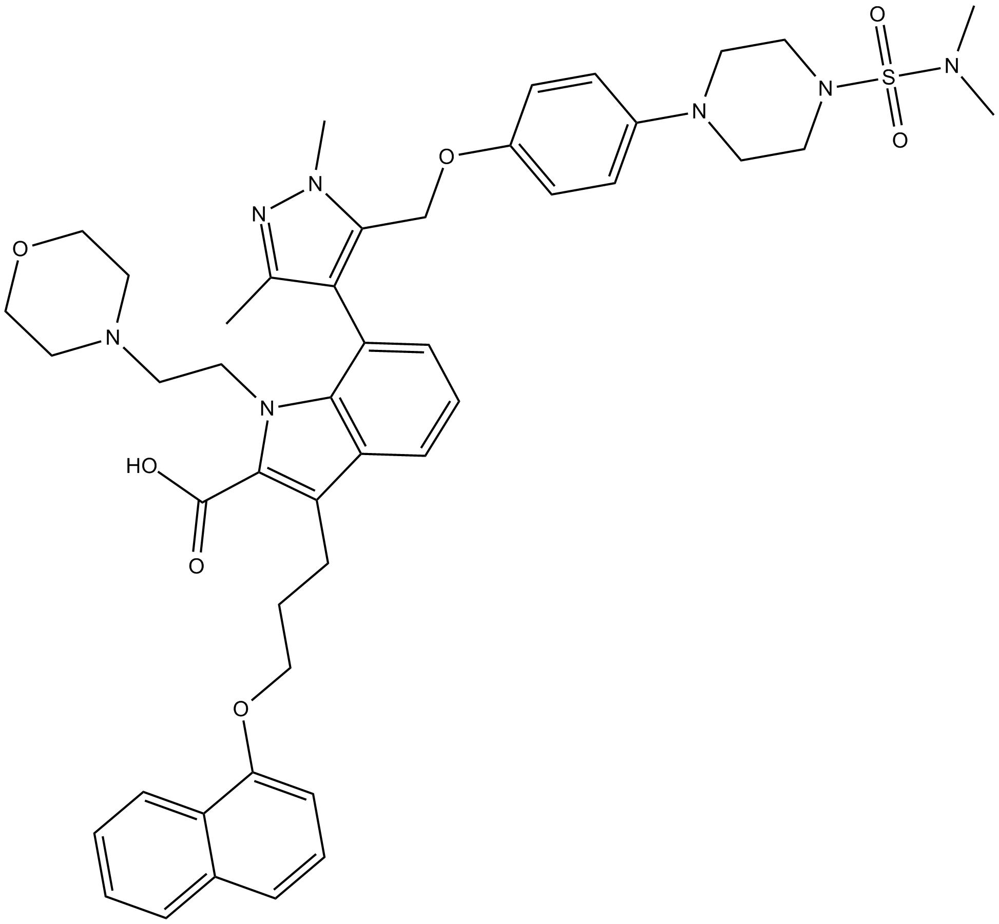
A-1210477
A-1210477 は、MCL-1 の強力かつ選択的な阻害剤で、Ki は 0.45 nM です。 A-1210477 は MCL-1 に特異的に結合し、MCL-1 依存的に癌細胞のアポトーシスを促進します。
-
GC17513
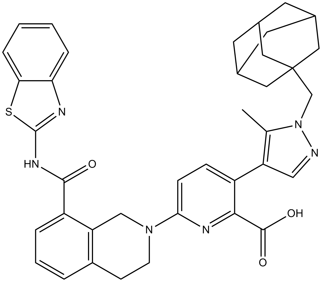
A-1331852
A-1331852 は経口投与可能な BCL-XL 選択的阻害剤で、Ki は 10 pM 未満です。
-
GC60544
A-192621
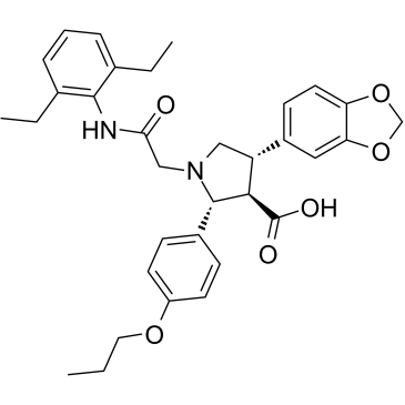
A-192621 は、4.5 nM の IC50 と 8.8 nM の Ki を持つ強力な非ペプチド性経口選択的エンドセリン B (ETB) 受容体アンタゴニストです。
-
GC32981
A-385358
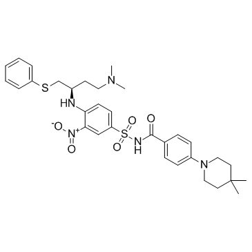
A-385358 は、Bcl-XL と Bcl-2 の Kis がそれぞれ 0.80 と 67 nM の Bcl-XL の選択的阻害剤です。
-
GC11200
A23187
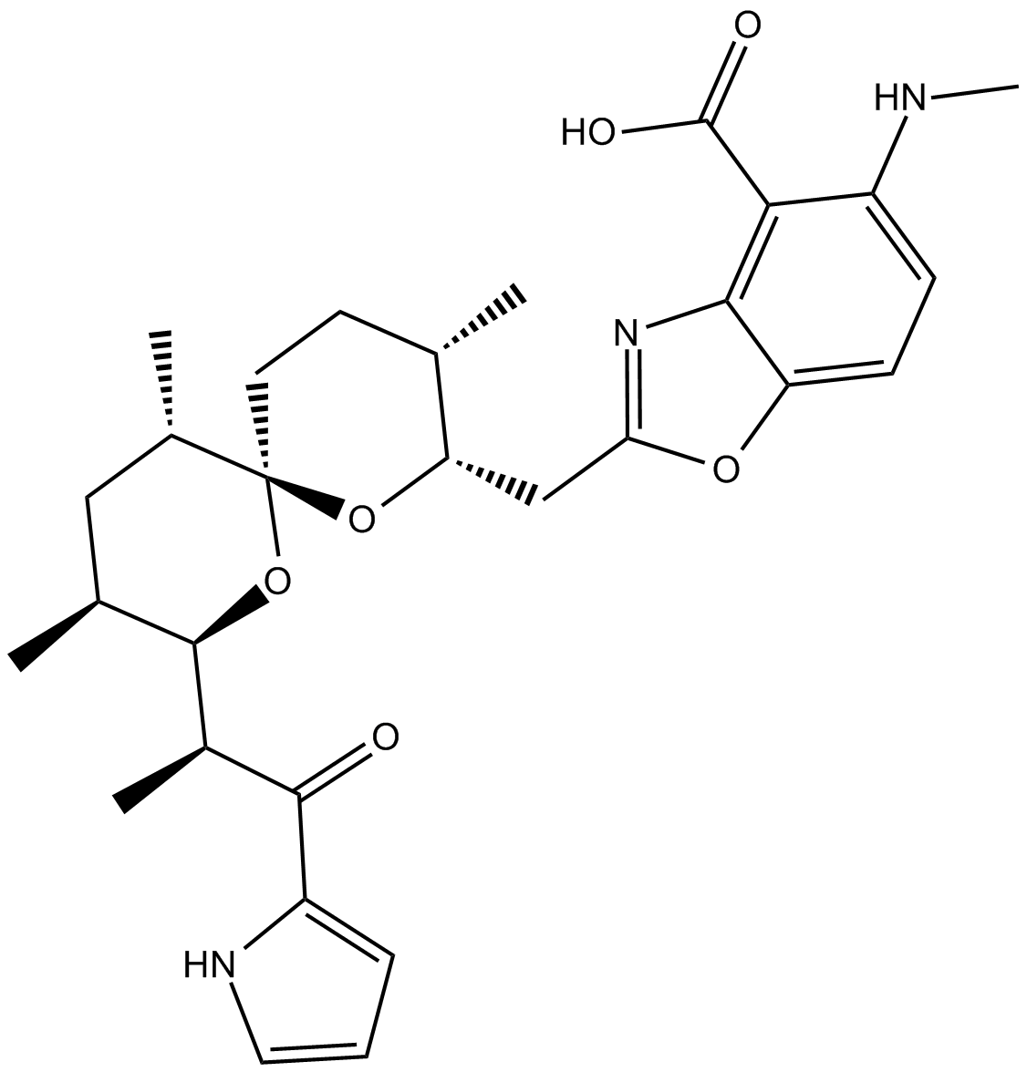
A23187(A-23187)は、抗生物質であり、カルシウムやマグネシウムのような独特の二価陽イオンイオノフォアです。
-
GC42659
A23187 (calcium magnesium salt)
A23187は二価カチオンイオノフォアです。

-
GC35216
AAPK-25
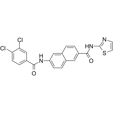
AAPK-25 は、抗腫瘍活性を備えた強力かつ選択的な Aurora/PLK 二重阻害剤であり、有糸分裂の遅延を引き起こし、前中期で細胞を停止させ、バイオマーカーのヒストン H3Ser10 リン酸化を反映し、その後にアポトーシスが急増します。 AAPK-25 は、23 ~ 289 nM の範囲の Kd 値を持つ Aurora-A、-B、および -C と、55 ~ 456 nM の範囲の Kd 値を持つ PLK-1、-2、および -3 をターゲットにしています。
-
GC13805
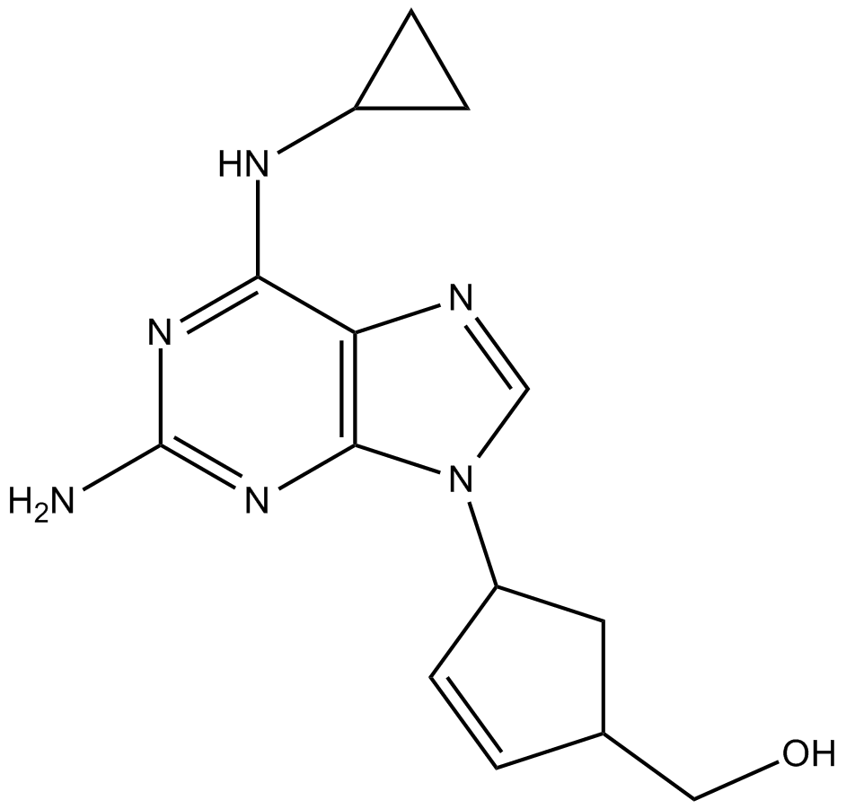
Abacavir
-
GC64674
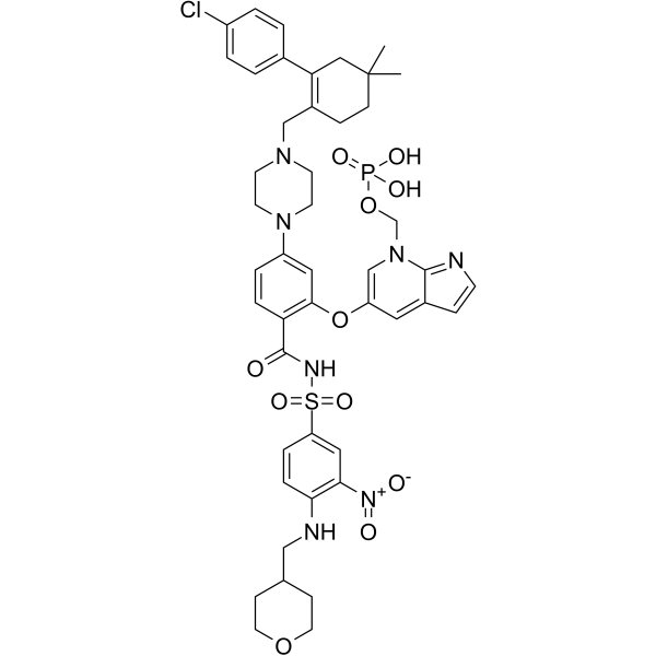
ABBV-167
ABBV-167 は、BCL-2 阻害剤ベネトクラクスのリン酸塩プロドラッグです。
-
GC60548
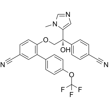
ABT-100
ABT-100 は、強力で選択性の高い経口活性ファルネシルトランスフェラーゼ阻害剤です。 ABT-100 は細胞増殖を阻害します (EJ-1、DLD-1、MDA-MB-231、HCT-116、MiaPaCa- 2、PC-3、および DU-145 細胞) は、アポトーシスを増加させ、血管新生を減少させます。 ABT-100 は、広範囲の抗腫瘍活性を持っています。
-
GC14069
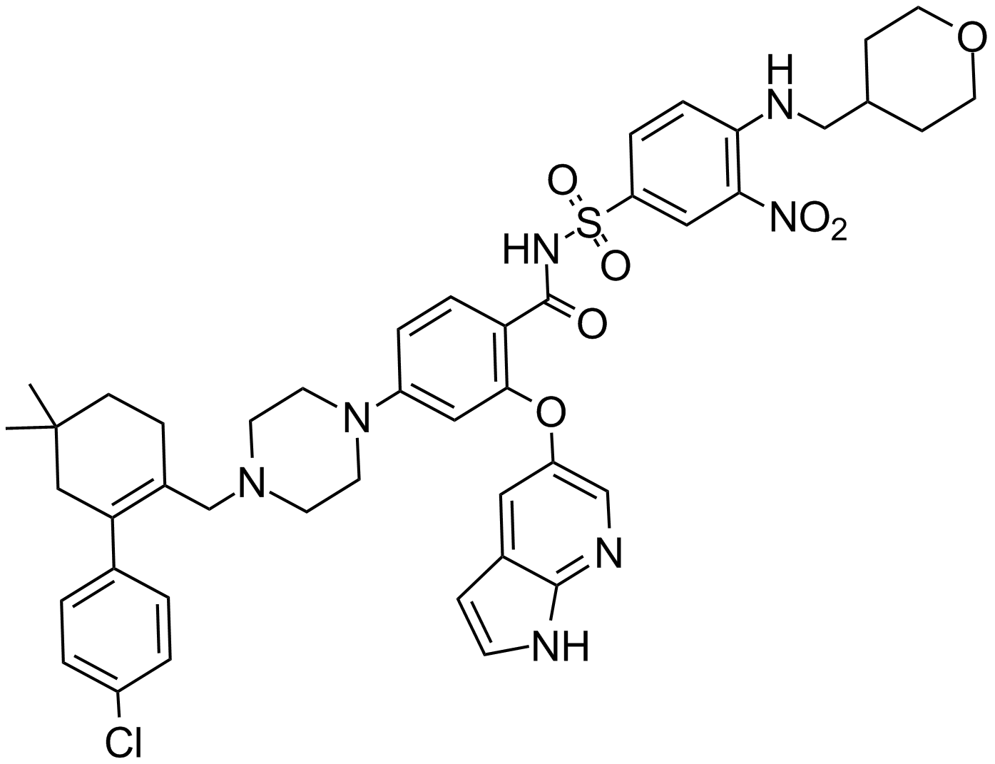
ABT-199
Bcl-2阻害剤
-
GC12405
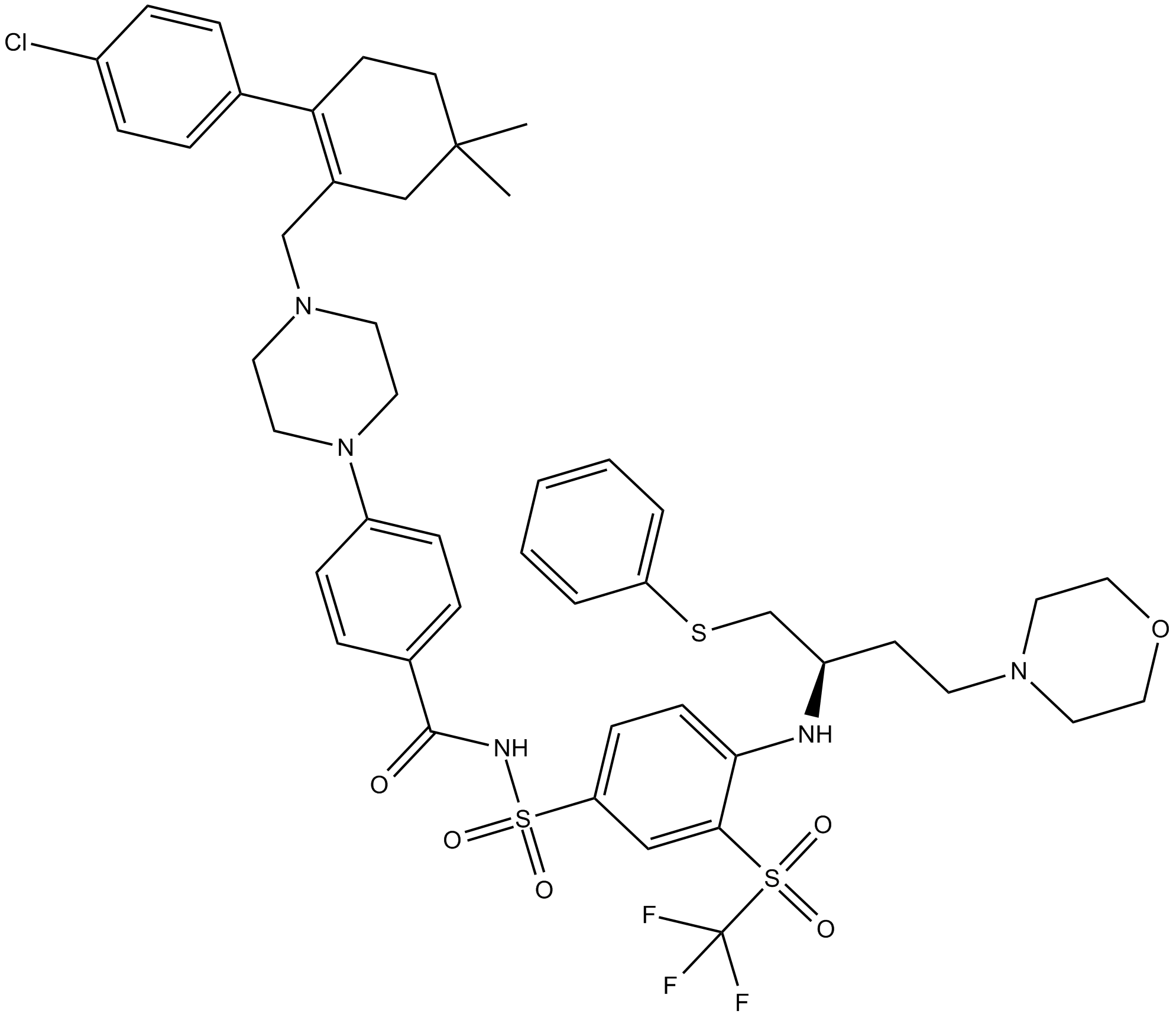
ABT-263 (Navitoclax)
Bcl-2ファミリー蛋白質の阻害剤
-
GC49745
ABT-263-d8
ABT-263-d8 は、Navitoclax とラベル付けされた重水素です。 Navitoclax (ABT-263) は、Bcl-xL、Bcl-2、Bcl-w などの複数の抗アポトーシス Bcl-2 ファミリータンパク質に結合する強力な経口活性 Bcl-2 ファミリータンパク質阻害剤であり、Ki は1 nM 未満。

-
GC17234
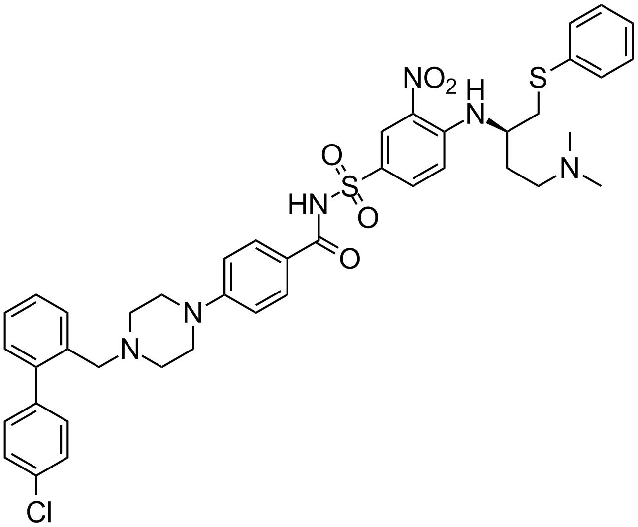
ABT-737
抗アポトーシスBcl-2タンパク質の阻害剤
-
GA20494
Ac-Asp-Glu-Val-Asp-pNA
クロモジェン性カスパーゼ-3基質Ac-DEVD-pNAの切断は、405 nmで監視することができます。
-
GC17602
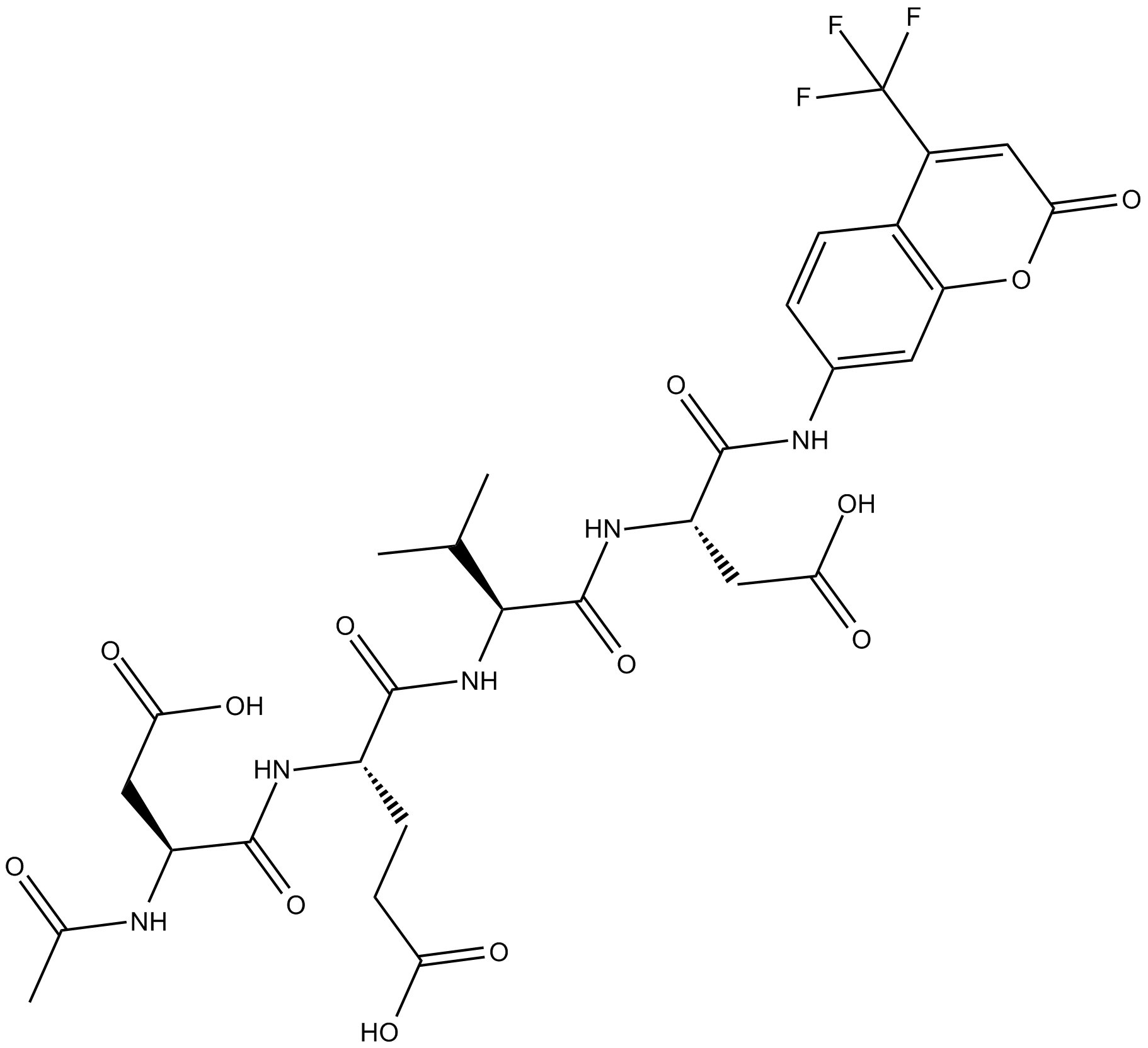
Ac-DEVD-AFC
Ac-DEVD-AFC は蛍光発生基質です (Λex=400 nm、Λem=530 nm)。
-
GC32695
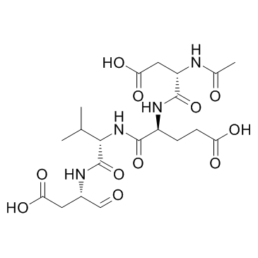
Ac-DEVD-CHO
Ac-DEVD-CHO は、Ki 値が 230 pM の特異的なカスパーゼ 3 阻害剤です。
-
GC48470
Ac-DEVD-CHO (trifluoroacetate salt)
デュアルカスパーゼ3 / カスパーゼ7阻害剤

-
GC10951
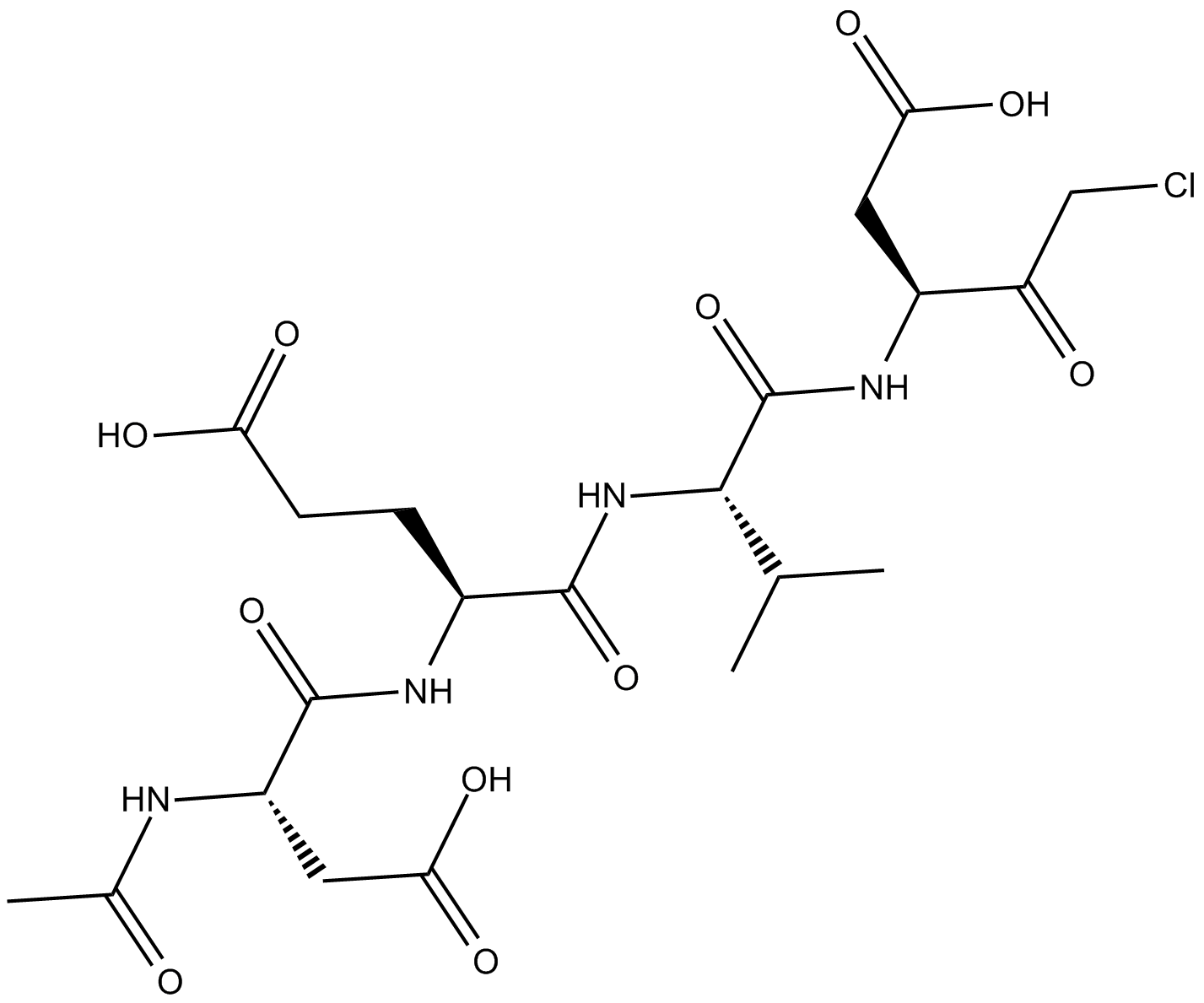
Ac-DEVD-CMK
細胞浸透性で、カスパーゼの不可逆的な阻害剤
-
GC42689
Ac-DNLD-AMC
Ac-WLA-AMC は、カスパーゼ-3 の蛍光基質です。

-
GC65107
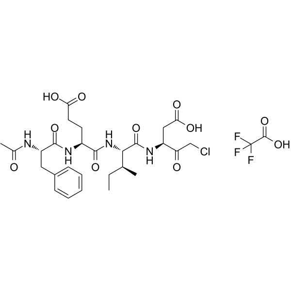
Ac-FEID-CMK TFA
Ac-FEID-CMK TFA は、強力なゼブラフィッシュ固有の GSDMEb 由来のペプチド阻害剤です。
-
GC60558
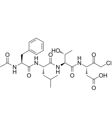
Ac-FLTD-CMK
ガスデルミン D (GSDMD) 由来の阻害剤である Ac-FLTD-CMK は、特定の炎症性カスパーゼ阻害剤です。
-
GC49704
Ac-FLTD-CMK (trifluoroacetate salt)
カスパーゼ-1、-4、-5、および-11の阻害剤

-
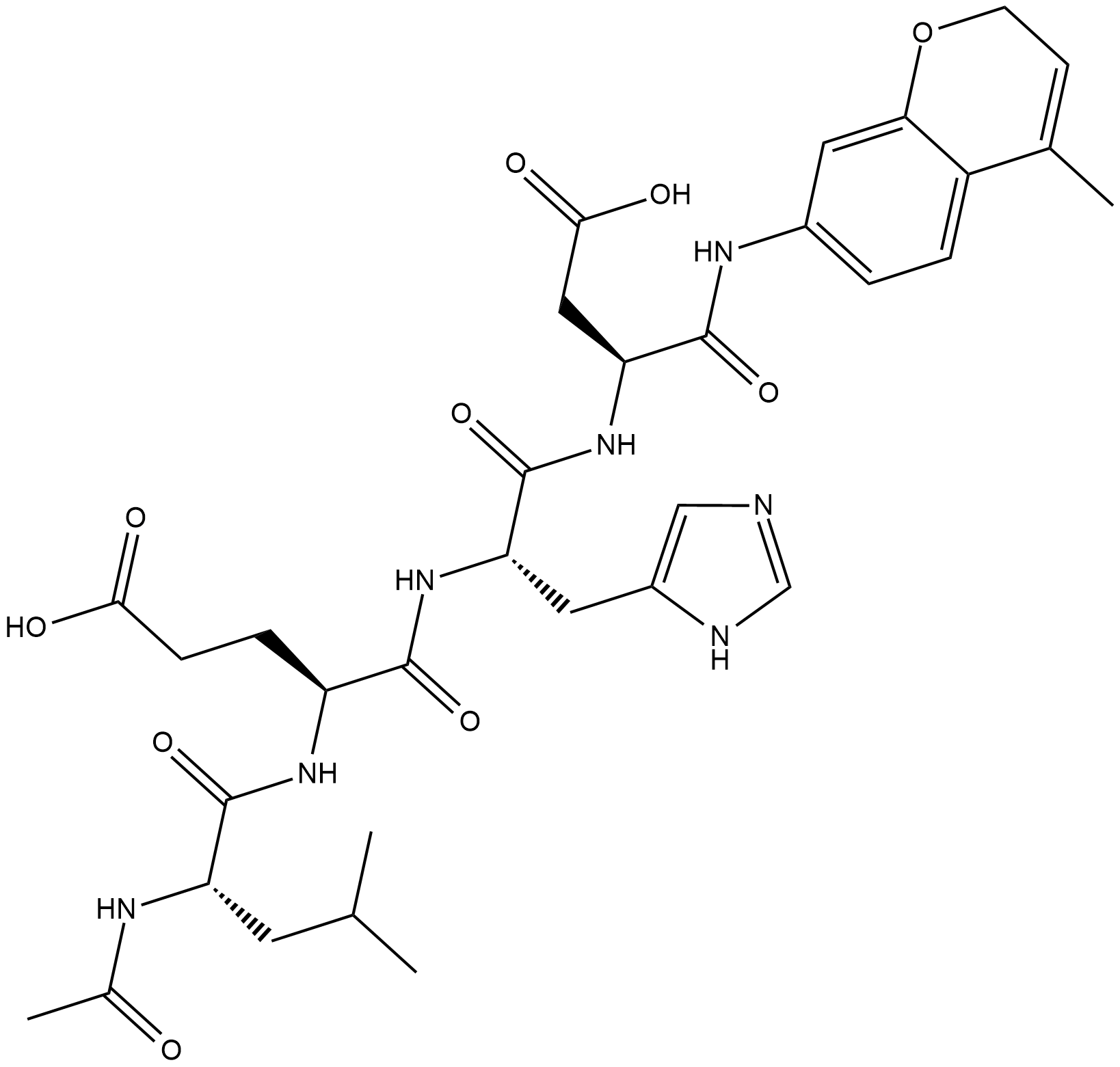
GC18226
Ac-LEHD-AMC (trifluoroacetate salt)
Ac-LEHD-AMC (トリフルオロ酢酸塩) は、カスパーゼ-9 の蛍光発生基質です (励起: 341 nm; 発光: 441 nm)。
-
GC40556
Ac-LETD-AFC
Ac-LETD-AFC は、カスパーゼ 8 蛍光基質です。

-
GC13400
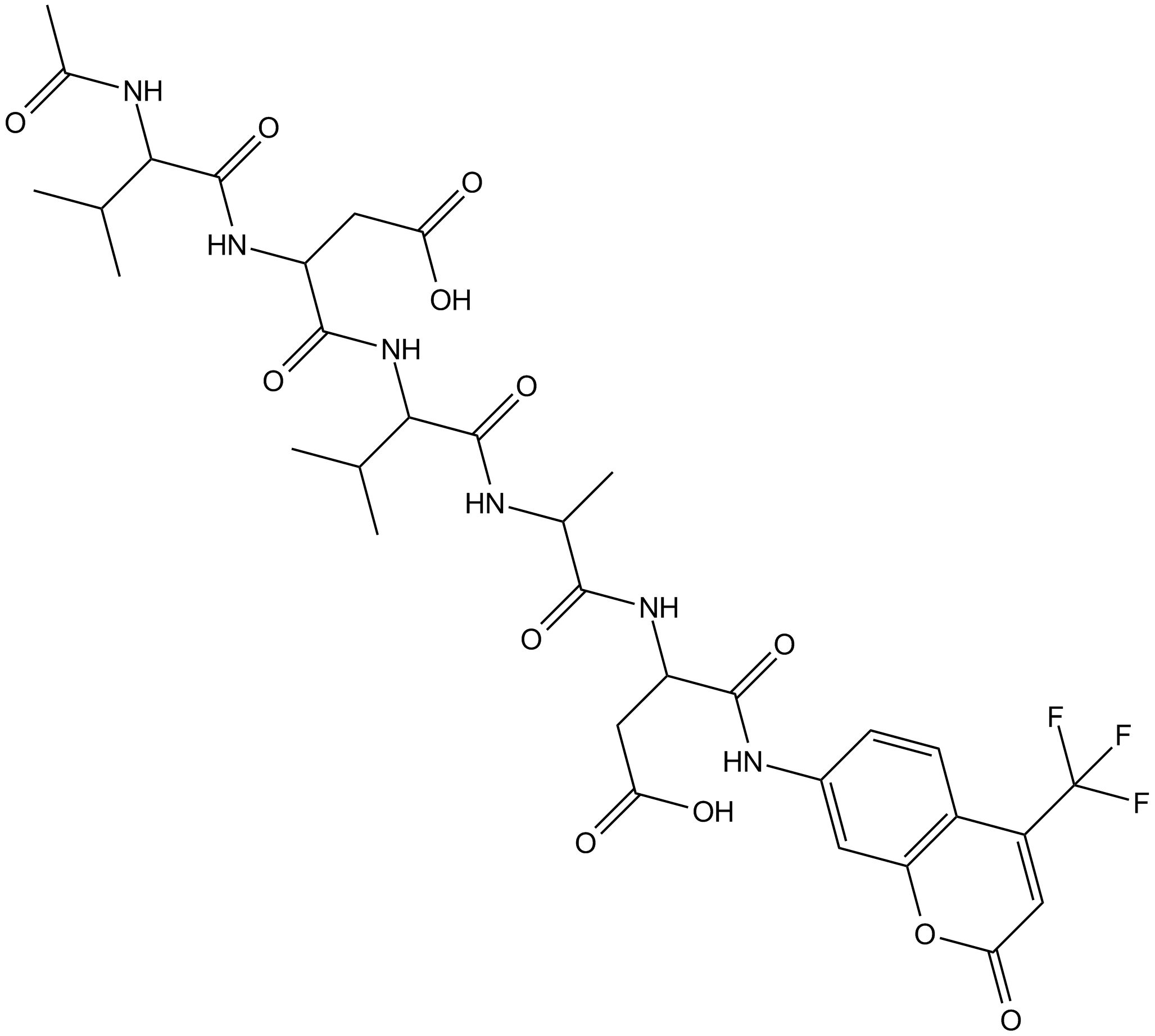
Ac-VDVAD-AFC
Ac-VDVAD-AFC は、カスパーゼ特異的な蛍光基質です。 Ac-VDVAD-AFC は、カスパーゼ 3 様活性とカスパーゼ 2 活性を測定でき、腫瘍やがんの研究に使用できます。
-
GC52372
Ac-VDVAD-AFC (trifluoroacetate salt)
カスパーゼ-2の蛍光基質

-
GC48974
Ac-VEID-AMC (ammonium acetate salt)
カスパーゼ-6蛍光基質

-
GC18021
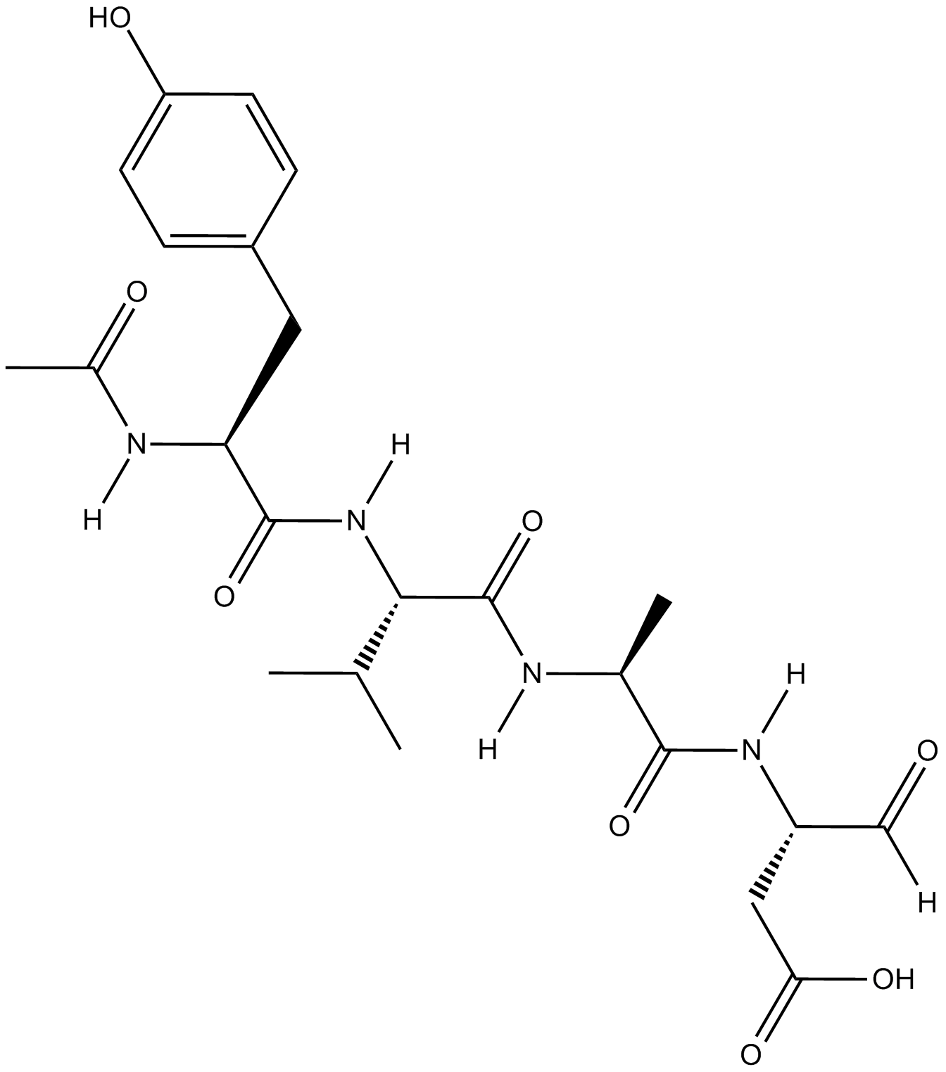
Ac-YVAD-CHO
Ac-YVAD-CHO (L-709049) は、マウスおよびヒトの Ki 値が 3.0 および 0.76 nM である、強力で可逆的な特異的テトラペプチド インターロイキン-1β 変換酵素 (ICE) 阻害剤です。
-
GC42721
Ac-YVAD-CMK
Ac-YVAD-CMKは、カスパーゼ-1の選択的不可逆阻害剤であり(K i=0.8nM)、炎症性サイトカインIL-1βの活性化を防止することができます。 Ac-YVAD-CMKは、炎症反応を減少させ、長期的な神経保護効果を引き起こすことができます。

-
GC35227
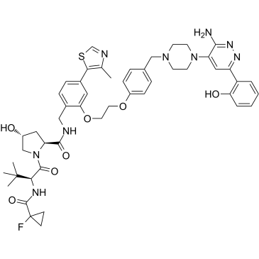
ACBI1
ACBI1 は、強力で協調的な SMARCA2、SMARCA4、および PBRM1 分解剤であり、DC50 はそれぞれ 6、11、および 32 nM です。 ACBI1 は PROTAC デグレーダーです。 ACBI1 は抗増殖活性を示します。 ACBI1 はアポトーシスを誘導します。
-
GN10341
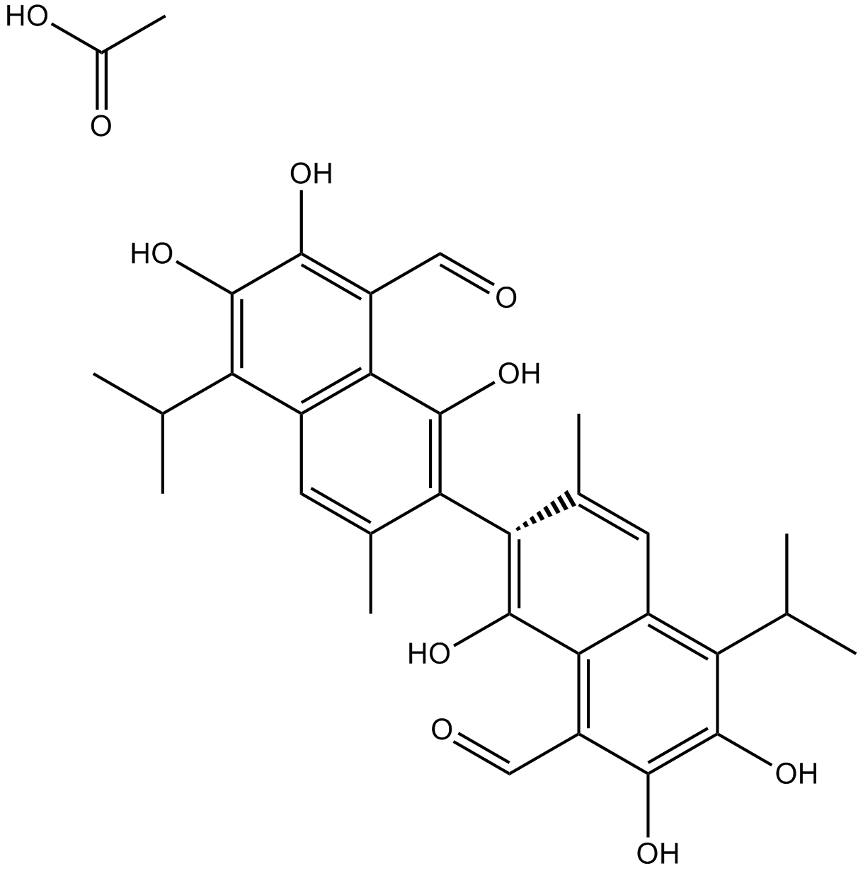
Acetate gossypol
-
GC11786
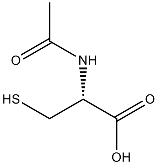
Acetylcysteine
アセチルシステインは、システインのN-アセチル誘導体です。
-
GC17094
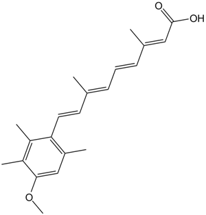
Acitretin
アシトレチン (Ro 10-1670) は、乾癬の治療に使用されてきた第 2 世代の全身性レチノイドです。
-
GC35242
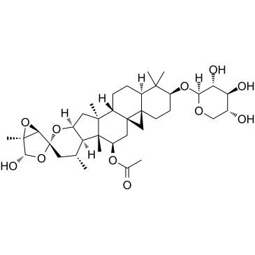
Actein
アクテインは、Cimicifuga foetida の根茎から分離されたトリテルペン配糖体です。アクテインは細胞増殖を抑制し、ROS/JNK 活性化を促進してオートファジーとアポトーシスを誘導し、ヒト膀胱癌の AKT 経路を鈍らせます。アクテインは生体内でほとんど毒性がありません。
-
GC16866
Actinomycin D
抗がん活性を持つDNAと相互作用する転写ブロッカー

-
GC16350
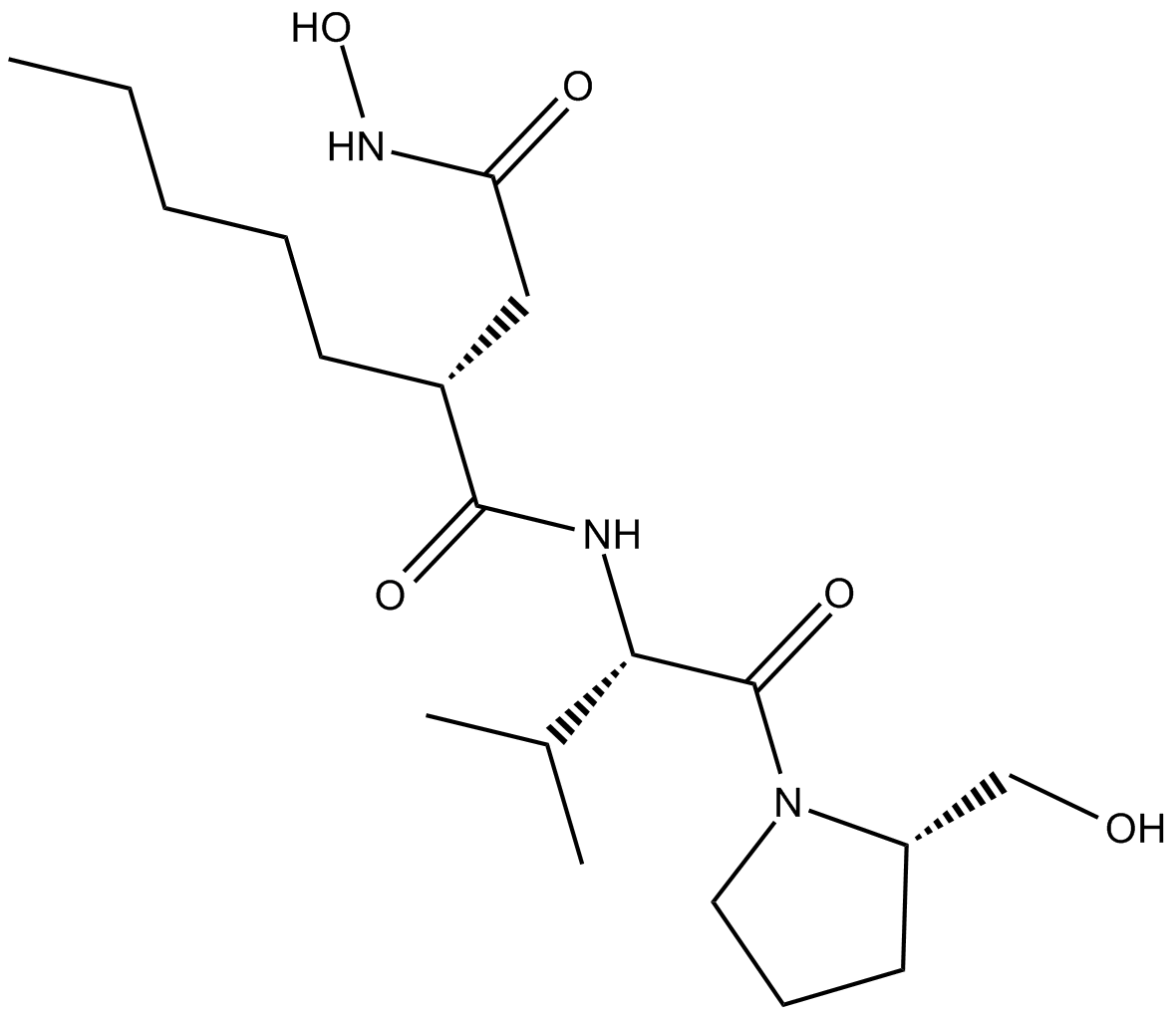
Actinonin
アクチノニン ((-)-アクチノニン) は、放線菌によって産生される天然の抗菌剤です。
-
GC16362
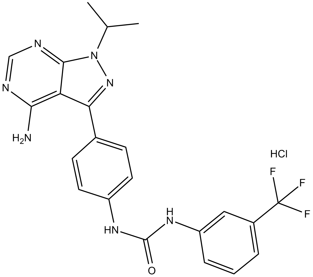
AD57 (hydrochloride)
RETを抑制する多剤作用がん治療薬。
-
GC34214
Adalimumab (Anti-Human TNF-alpha, Human Antibody)
アダリムマブ(抗ヒトTNF-α、ヒト抗体)は、腫瘍壊死因子α(TNF-α)を標的とする人間のモノクローナルIgG1抗体です。


