

MPEG-2000-DSPE (Synonyms: 1,2-DSPE-MPEG(5000), Methyl-PEG2000-DSPE, MPEG-2000-DSPE) |
Katalog-Nr.GC64095
 One-Click Copy Product Info
|

MPEG-2000-DSPE ist ein Phospholipid-PEG-Konjugat, das sowohl hydrophil als auch hydrophob ist. MPEG-2000-DSPE wird bei der Synthese von Liposomen verwendet, erhÖht die Hydrophilie auf der OberflÄche von Liposomen und erhÖht folglich die Langlebigkeit von Liposomen im Blutkreislauf fÜr die Untersuchung von Antikrebs- und Antimalariamitteln.
Products are for research use only. Not for human use. We do not sell to patients.
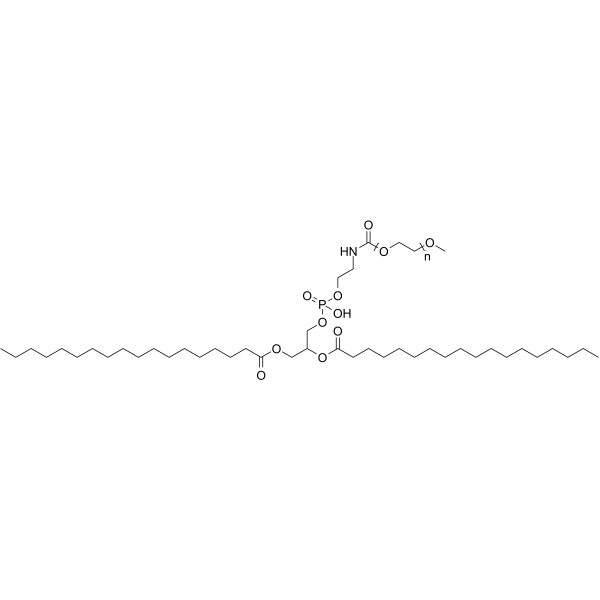
Cas No.: 147867-65-0
Sample solution is provided at 25 µL, 10mM.
_1-3.$$$39978408@51.jpg');)
_1-9.$$$38538795@65.jpg');)
_1-9.$$$39112701@51.jpg');)
_1-7.$$$37316672@70.jpg');)
_366-372.$$$36198801@70.jpg');)
.$$$38565142@65.jpg');)
_1867-1883.$$$33248023@67.jpg');)
_eads6055.$$$39977546@45.jpg');)
_eadm9805.$$$40080571@45.jpg');)
_eadj3020.$$$39666821@45.jpg');)
_1-20.$$$39757301@28.jpg');)
_1-24.$$$38898113@28.jpg');)
_273-287.$$$36806353@46.jpg');)
_1-16.$$$37156877@44.jpg');)
_1-19.$$$37460805@44.jpg');)
_1291-1307.$$$34518654@46.jpg');)
MPEG-2000-DSPE is a phospholipid PEG conjugate, with both hydrophilicity and hydrophobility, MPEG-2000-DSPE is used as a component of liposomes. The polar heads of the phospholipids are located on the outer and inner surfaces of the membranes, in contact with the aqueous environment. And fatty acid chains form the hydrophobic core of the membranes and are protected from water [1]. MPEG-2000-DSPE is often used for drug delivery of hydrophobic or hydrophilic molecules[2-3].
References:
[1] Papachristos A, Pippa N, Ioannidis K, Sivolapenko G, Demetzos C. Liposomal forms of anticancer agents beyond anthracyclines: present and future perspectives. Journal of liposome research. 2015 Apr 3;25(2):166-73.
[2] Rathore SS, Ghosh PC. Effect of surface charge and density of distearylphosphatidylethanolamine-mPEG-2000 (DSPE-mPEG-2000) on the cytotoxicity of liposome-entrapped ricin: Effect of lysosomotropic agents. International journal of pharmaceutics. 2008 Feb 28;350(1-2):79-94.
[3] Cui J, Li C, Wang C, Li Y, Zhang L, Zhang L, Xiu X, Li Y, Wei N. Development of pegylated liposomal vincristine using novel sulfobutyl ether cyclodextrin gradient: is improved drug retention sufficient to surpass DSPE–PEG-induced drug leakage?. Journal of Pharmaceutical Sciences. 2011 Jul 1;100(7):2835-48.
This program only provides a guide, please modify according to your specific needs.
Preparation of tarin (a plant lectin characterized as a hydrophilic globular protein) liposomal nanocapsules[1]
1. Weigh the liposome components using an analytical balance.
|
Liposome Components |
Weight (g) |
Concentration (mM)
|
Final volume
|
|
DOPE |
0.0420 |
5.7 |
10 mL |
|
MPEG-2000-DSPE |
0.1059 |
3.8 |
|
|
CHEMS |
0.0024 |
0.5 |
2. Dissolve the lipid components in chloroform using a 250 mL volumetric flask that fits in a rotary evaporator to avoid loss of material.
3. Stir the mixture at 150 rpm for 15 min.
4. Remove the chloroform using a rotary evaporator under the following conditions:
1. Adjust the volumetric flask mouth to the standard position (25°) for optimal efficiency, while in contact with the water from the heating bath.
NOTE: The equipment arm should be inclined at 25° to maintain contact between the volumetric flask and water bath, while not affecting evaporation efficiency or damaging the sample. The standard position can vary according to the equipment brand.
2. Set the condenser temperature to a minimum of 3 °C.
3. Set the heating bath temperature to 40 ± 1 °C.
4. Set the rotation to 120 rpm.
5. Adjust the vacuum to 207 mbar and boiling point to 20 °C.
6. After ~25 min, remove the flask and discard the evaporated solvent, remaining in the condenser, appropriately.
NOTE: A thin and opaque film, consisting of the liposome components, is formed in this step and can be easily visualized. The evaporated solvent remaining in the condenser must be stored into disposal recipients (chlorinated) to be handled by a specialized company for appropriate discarding.
5. Hydrate the lipid film to reach a 0.01 M lipid concentration in 0.3 M ammonium sulfate solution (pH = 7.4) containing tarin at 1 mg/mL to a final volume of 10 mL.
- Stir the mixture for 40 min and incubate overnight at 4 °C.
NOTE: This step can be considered a stop point. Overnight incubation is not obligatory.
6. After incubation, sonicate the suspension for 1 min at 25 °C (room temperature; RT) to reduce vesicle size and avoid aggregation.
NOTE: Size reduction was performed in an ultrasonic sonicator under the following conditions: 130 W and 40 kHz.
7. Perform a 12-cycle extrusion through a 0.2 µm polycarbonate pore membrane.
NOTE: Before the extrusion process, test the mini-extruder assembly using water to avoid sample leakage. A 0.1 µm pore membrane is also suitable. In this case, pre-heat the mini-extruder holder above the lipid transition temperature to facilitate extrusion, while maintaining the physicochemical characteristics of both lipids and proteins.
- Fit the parts of the mini extruder, as described in the manufacturer's manual.
- Place the polycarbonate membrane between two pre-wet filter supports and place it into the holder.
- Insert a 1 mL empty syringe into the device, fill the other syringe to its total volume with the liposomal suspension, and insert it on the opposite side.
- Perform a 12-cycle extrusion through a 0.2 µm polycarbonate pore membrane. Push the sample from one syringe to another, slowly. Collect the extruded suspension in a pre-cooled tube.
NOTE: The polycarbonate membrane should be replaced only when sample transference from one syringe to another becomes difficult. The liposomal suspension should become clear during the extrusion process as a result of size reduction due to the formation of small unilamellar vesicles (SUVs). About 0.2 mL of the sample can be lost during this step.
8. Separate liposomes by ultracentrifugation.
NOTE: Maintain the samples in an ice bath until the ultracentrifuge is ready to be used. Separate SUVs from the remaining components and ammonium sulfate by ultracentrifugation using an ultracentrifuge with a swing-bucket rotor (see details below).
- Weigh the sample in the titanium tube that fits the rotor and balance the tubes. Check the minimum volume required according to the rotor used to avoid damaging the tubes, and adjust the liposomal suspension volume with ammonium sulphate, if necessary.
NOTE: The swing bucket should always be supported on the stand when outside of the centrifuge to avoid scratching the "zebra stripes" (i.e., the black and white stripes on the bottom), which are used by the centrifuge to determine rotation speed.
2. Turn on the vacuum before using the centrifuge to allow it to refrigerate.
3. Fit the titanium tubes into the swing bucket.
NOTE: Lift the tubes to the position they assume when running to ensure that they are perfectly fitted.
4. Release the vacuum, open the centrifuge door, and place the rotor inside.
NOTE: Pay attention to the circle mark on the bottom of the rotor, which must fit in the opposite direction of the same circle mark into the centrifuge itself.
5. Close the centrifuge door, press Vacuum and wait until the vacuum reaches from 200 to <20 microns or from 26 Pa to <3 Pa.
6. Adjust the parameters in the ultracentrifuge display to 150,000 x g (the equivalent to 29,600 rpm for the aforementioned swing bucket) for 90 min at 4 °C (acceleration: max, deceleration: max).
NOTE: Always convert the speed to x g if the specific centrifuge is set in rpm. Use the centrifuge website to convert the unit according to the rotor used.
7. Press Recall, check the conditions, and press Start to run.
NOTE: Wait until the centrifuge reaches the desired speed.
8. After 90 min, release the vacuum by pressing the Vacuum button and open the centrifuge door when the vacuum reaches 200-700 microns (equivalent to 26-93 Pa).
9. Switch off the centrifuge, remove the rotor from the inside, and leave it on the bench with the buckets to dry.
10. Maintain the ultracentrifuged samples on ice.
9. Carefully, separate supernatant from the pellet by turning the tube upside down into a disposable 15 mL centrifuge tube to separate the supernatant and pellet.
NOTE: Store the supernatant containing the unencapsulated protein at 4 °C. It will be used to determine the encapsulation efficiency. The pellet appears as a translucent jelly.
10. Suspend the pellet containing the encapsulated protein in HEPES buffered saline (3 mL of 1x HBS).
NOTE: The HBS 2x (stock solution) is prepared by diluting the following amounts of reagents in distilled water: 140 mM NaCl, 1.5 mM Na2HPO4, 50 mM HEPES, then adjusting the pH to 7.4 and final volume to 100 mL. Na2HPO4 can be replaced by NaHCO3 or omitted to avoid interference if liposome concentration is to be determined. The HBS 2x stock solution should be diluted in distilled water to obtain HBS 1x before use.
References:
[1] Papachristos A, Pippa N, Ioannidis K, Sivolapenko G, Demetzos C. Liposomal forms of anticancer agents beyond anthracyclines: present and future perspectives. Journal of liposome research. 2015 Apr 3;25(2):166-73.
| Cas No. | 147867-65-0 | SDF | |
| Überlieferungen | 1,2-DSPE-MPEG(5000), Methyl-PEG2000-DSPE, MPEG-2000-DSPE | ||
| Formula | C45H87NNaO11P | M.Wt | 872.14 |
| Löslichkeit | Storage | Store at -20°C, protect from light, stored under nitrogen | |
| General tips | Please select the appropriate solvent to prepare the stock solution according to the
solubility of the product in different solvents; once the solution is prepared, please store it in
separate packages to avoid product failure caused by repeated freezing and thawing.Storage method
and period of the stock solution: When stored at -80°C, please use it within 6 months; when stored
at -20°C, please use it within 1 month. To increase solubility, heat the tube to 37°C and then oscillate in an ultrasonic bath for some time. |
||
| Shipping Condition | Evaluation sample solution: shipped with blue ice. All other sizes available: with RT, or with Blue Ice upon request. | ||
| Prepare stock solution | |||

|
1 mg | 5 mg | 10 mg |
| 1 mM | 1.1466 mL | 5.733 mL | 11.466 mL |
| 5 mM | 229.3 μL | 1.1466 mL | 2.2932 mL |
| 10 mM | 114.7 μL | 573.3 μL | 1.1466 mL |
Step 1: Enter information below (Recommended: An additional animal making an allowance for loss during the experiment)
 g
μL
g
μL
Step 2: Enter the in vivo formulation (This is only the calculator, not formulation. Please contact us first if there is no in vivo formulation at the solubility Section.)
Calculation results:
Working concentration: mg/ml;
Method for preparing DMSO master liquid: mg drug pre-dissolved in μL DMSO ( Master liquid concentration mg/mL, Please contact us first if the concentration exceeds the DMSO solubility of the batch of drug. )
Method for preparing in vivo formulation: Take μL DMSO master liquid, next addμL PEG300, mix and clarify, next addμL Tween 80, mix and clarify, next add μL saline, mix and clarify.
Method for preparing in vivo formulation: Take μL DMSO master liquid, next add μL Corn oil, mix and clarify.
Note: 1. Please make sure the liquid is clear before adding the next solvent.
2. Be sure to add the solvent(s) in order. You must ensure that the solution obtained, in the previous addition, is a clear solution before proceeding to add the next solvent. Physical methods such as vortex, ultrasound or hot water bath can be used to aid dissolving.
3. All of the above co-solvents are available for purchase on the GlpBio website.
Quality Control & SDS
- View current batch:
- Purity: >99.50% Appearance: A solid
- COA (Certificate of Analysis)
- SDS (Safety Data Sheet)
- Datasheet
Average Rating: 5 (Based on Reviews and 33 reference(s) in Google Scholar.)