

MQAE (Synonyms: MQAE) |
Catalog No.GC30035
 One-Click Copy Product Info
|

MQAE는 세포 내 염소 이온 농도([Cl⁻]i)를 측정하는 비비율형 염소 이온(Cl⁻) 끄는 형광 센서로 최대 자극/방출 파장(350/460 nm)을 가진다.
Products are for research use only. Not for human use. We do not sell to patients.
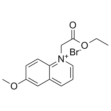
Cas No.: 162558-52-3
Sample solution is provided at 25 µL, 10mM.
MQAE는 세포 내 염소 이온 농도([Cl⁻]i)를 측정하는 비비율형 염소 이온(Cl⁻) 끄는 형광 센서로 최대 자극/방출 파장(350/460 nm)을 가진다.MQAE는 표면 형광 현미경, 공초점 현미경, 2광자 현미경, 유세포 측정기 및 기타 생화학 분석 실험에 적합한다[1-2].[Cl⁻]i의 변화와 상관없이 MQAE 농도가 감소할 때 형광 강도는 감소하고 MQAE 농도가 증가할 때 형광 강도는 증가한다[2].세포 내 MQAE의 분포는 균일하지 않고 삼투압에 의해 유발된 세포 부피 변화에 영향을 받지 않는다[3].MQAE는 알려지지 않은 세포 내 소기관 구조물에 결합할 수 있고 결합된 MQAE는 세포 부피 변화가 있는 경우에도 기도 airway-ciliated세포에서 [Cl⁻]i를 측정할 수 있도록 보여준다[1,4].MQAE는 미토콘드리아 염화 이온을 검출하기 위한 형광 프로브를 생성할 때 사용할 수 있다[5].MQAE 기반 프로브의 형광은 생리학적 pH 범위에서 pH에 영향을 받지 않고 염소 이온에 의해 끄여지고 pH 7.0에서 Stern-Volmer 끄기 상수는 201 M⁻¹이다[5].
References:
[1] Ikeuchi Y, Kogiso H, Hosogi S, et al. Measurement of [Cl−]i unaffected by the cell volume change using MQAE-based two-photon microscopy in airway ciliary cells of mice[J]. The journal of physiological sciences, 2018, 68: 191-199.
[2] Andersson C, Roomans G M. Determination of chloride efflux by X‐ray microanalysis versus MQAE‐fluorescence[J]. Microscopy research and technique, 2002, 59(6): 531-535.
[3] Koncz C, Daugirdas J T. Use of MQAE for measurement of intracellular [Cl−] in cultured aortic smooth muscle cells[J]. American Journal of Physiology-Heart and Circulatory Physiology, 1994, 267(6): H2114-H2123.
[4] Miyazaki H, Shiozaki A, Niisato N, et al. Physiological significance of hypotonicity-induced regulatory volume decrease: reduction in intracellular Cl− concentration acting as an intracellular signaling[J]. American Journal of Physiology-Renal Physiology, 2007, 292(5): F1411-F1417.
[5] Park S H, Shin I, Kim Y H, et al. Mitochondrial Cl–-selective fluorescent probe for biological applications[J]. Analytical Chemistry, 2020, 92(18): 12116-12119.
이 프로토콜은 단지 지침을 제공하고 특정 필요에 따라 수정하시기 바랍니다.
1.MQAE 염색 용액 준비:
Krebs-Hepes 버퍼(20 mM HEPES, 128 mM NaCl, 2.5 mM KCl, 2.7 mM CaCl₂, 1 mM MgCl₂, 16 mM 포도당; pH 7.4)에서 5-10 mM MQAE 작업용액을 준비합니다. 준비 후에는 0.22㎛ 필터 막을 사용해서 여과 및 멸균합니다. MQAE 작업 용액의 농도는 실제 상황에 따라 조정하고 즉시 사용도록 합니다.
2. 소생된 세포 염색 (6웰 판 프로토콜)
(1) 소생된 세포의 경우:
세포 소생된 용액을 1000×g에서 3-5분간 원심분리하고 상청액을 조심스럽게 흡인합니다. Krebs-Hepes 버퍼로 세포를 두 번 세척하고(세척당 5분) 세척 사이에 1000×g에서 원심분리합니다.
(2) 고착세포의 경우:
Krebs-Hepes 버퍼로 고착 세포를 두 번 헹군 후 트립신화를 통해 세포를 분리하고 1000×g에서 3-5분간 원심분리합니다.
(3) 염료 배양:
세포 펠릿을 염료 작업 용액 1 mL로 소생시킵니다. 실온에서 30-60분 동안 어두운 곳에서 배양합니다(세포 유형에 따라 최적의 배양 시간은 달라질 수 있습니다).
(4) 배양 후 처리:
1000×g에서 5분간 원심분리하고 상청액을 조심스럽게 제거합니다. 세척 간격에 원심분리를 하면서 PBS로 세포를 2-3회 세척합니다(세척당 5분).
(5) 소생 및 관찰:
염색된 세포를 무혈청 배양액 또는 PBS로 소생시킨 후 현미경 검사를 진행합니다.
3. 고착 세포 염색:
(1) 세포 배양 준비:
멸균 커버슬립 위에서 고착 세포를 배양해서 원하는 결합성에 도달할 때까지 기릅니다.
(2) 염색 전 처리:
배양액을 조심스럽게 제거하고 커버슬립을 가습된 방에 놓습니다.
(3) 염료 배양:
커버슬립의 한쪽 가장자리에 염료 작업 용액 100㎕를 떨어뜨리고 살짝 기울여 세포 단층을 균일하게 덮도록 합니다. 실온에서 30-60분 동안 어두운 곳에서 배양합니다.
(4) 염색 후 세척:
염료 용액을 흡인하고 PBS로 커버슬립을 2-3회 헹굽니다(세척당 5분).
4. 현미경 분석:
형광 현미경이나 유세포 측정기로 염색된 세포를 검사합니다. MQAE는 최대 자극/방출 파장이 350/460 nm입니다.
References:
[1] Koncz C, Daugirdas J T. Use of MQAE for measurement of intracellular [Cl?] in cultured aortic smooth muscle cells[J]. American Journal of Physiology-Heart and Circulatory Physiology, 1994, 267(6): H2114-H2123.
[2] Miyazaki H, Shiozaki A, Niisato N, et al. Physiological significance of hypotonicity-induced regulatory volume decrease: reduction in intracellular Cl? concentration acting as an intracellular signaling[J]. American Journal of Physiology-Renal Physiology, 2007, 292(5): F1411-F1417.
| Cas No. | 162558-52-3 | SDF | |
| Synonyms | MQAE | ||
| Canonical SMILES | COC1=CC2=CC=C[N+](CC(OCC)=O)=C2C=C1.[Br-] | ||
| Formula | C14H16BrNO3 | M.Wt | 326.19 |
| Solubility | DMSO : ≥ 35 mg/mL (107.30 mM) | Storage | Store at -20°C, protect from light |
| General tips | Please select the appropriate solvent to prepare the stock solution according to the
solubility of the product in different solvents; once the solution is prepared, please store it in
separate packages to avoid product failure caused by repeated freezing and thawing.Storage method
and period of the stock solution: When stored at -80°C, please use it within 6 months; when stored
at -20°C, please use it within 1 month. To increase solubility, heat the tube to 37°C and then oscillate in an ultrasonic bath for some time. |
||
| Shipping Condition | Evaluation sample solution: shipped with blue ice. All other sizes available: with RT, or with Blue Ice upon request. | ||
| Prepare stock solution | |||

|
1 mg | 5 mg | 10 mg |
| 1 mM | 3.0657 mL | 15.3285 mL | 30.657 mL |
| 5 mM | 613.1 μL | 3.0657 mL | 6.1314 mL |
| 10 mM | 306.6 μL | 1.5328 mL | 3.0657 mL |
Step 1: Enter information below (Recommended: An additional animal making an allowance for loss during the experiment)
 g
μL
g
μL
Step 2: Enter the in vivo formulation (This is only the calculator, not formulation. Please contact us first if there is no in vivo formulation at the solubility Section.)
Calculation results:
Working concentration: mg/ml;
Method for preparing DMSO master liquid: mg drug pre-dissolved in μL DMSO ( Master liquid concentration mg/mL, Please contact us first if the concentration exceeds the DMSO solubility of the batch of drug. )
Method for preparing in vivo formulation: Take μL DMSO master liquid, next addμL PEG300, mix and clarify, next addμL Tween 80, mix and clarify, next add μL saline, mix and clarify.
Method for preparing in vivo formulation: Take μL DMSO master liquid, next add μL Corn oil, mix and clarify.
Note: 1. Please make sure the liquid is clear before adding the next solvent.
2. Be sure to add the solvent(s) in order. You must ensure that the solution obtained, in the previous addition, is a clear solution before proceeding to add the next solvent. Physical methods such as vortex, ultrasound or hot water bath can be used to aid dissolving.
3. All of the above co-solvents are available for purchase on the GlpBio website.
Quality Control & SDS
- View current batch:
- Purity: >99.50% Appearance: A solid
- COA (Certificate of Analysis)
- SDS (Safety Data Sheet)
- Datasheet
Average Rating: 5 (Based on Reviews and 18 reference(s) in Google Scholar.)