

MQAE (Synonyms: MQAE) |
Catalog No.GC30035
 One-Click Copy Product Info
|

MQAE functions as a non-ratiometric chloride ion (Cl−) quenching fluorescent sensor that is employed to measure intracellular Cl− concentration ([Cl−]i), with the maximum wavelength excitation/emission(350/460nm).
Products are for research use only. Not for human use. We do not sell to patients.
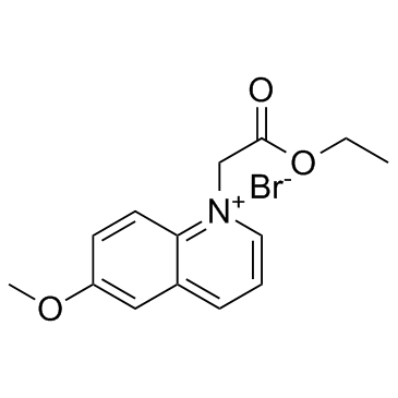
Cas No.: 162558-52-3
Sample solution is provided at 25 µL, 10mM.
MQAE functions as a non-ratiometric chloride ion (Cl−) quenching fluorescent sensor that is employed to measure intracellular Cl− concentration ([Cl−]i), with the maximum wavelength excitation/emission(350/460nm)[1]. MQAE is suitable for surface fluorescence microscopy, confocal microscopy, two-photon microscopy, flow cytometry and other biochemical analysis experiments[1-2].The fluorescence intensity diminishes when MQAE concentration decreases and increases when MQAE concentration increases regardless of any changes in [Cl−]i[2]. The intracellular distribution of MQAE is not uniform and is not affected by changes in cell volume caused by osmotic stress[3]. MQAE can bind to unknown subcellular structures, and these bound MQAE appear to enable the measurement of [Cl−]i in airway-ciliated cells even in the presence of changes in cell volume[1,4]. MQAE can be used to create a fluorescent probe for the detection of mitochondrial chloride ions[5]. The fluorescence of MQAE-based probe is pH-insensitive in the physiological pH range and is quenched by chloride ions, with a Stern-Volmer quenching constant of 201M-1 at pH 7.0[5].
References:
[1] Ikeuchi Y, Kogiso H, Hosogi S, et al. Measurement of [Cl−]i unaffected by the cell volume change using MQAE-based two-photon microscopy in airway ciliary cells of mice[J]. The journal of physiological sciences, 2018, 68: 191-199.
[2] Andersson C, Roomans G M. Determination of chloride efflux by X‐ray microanalysis versus MQAE‐fluorescence[J]. Microscopy research and technique, 2002, 59(6): 531-535.
[3] Koncz C, Daugirdas J T. Use of MQAE for measurement of intracellular [Cl−] in cultured aortic smooth muscle cells[J]. American Journal of Physiology-Heart and Circulatory Physiology, 1994, 267(6): H2114-H2123.
[4] Miyazaki H, Shiozaki A, Niisato N, et al. Physiological significance of hypotonicity-induced regulatory volume decrease: reduction in intracellular Cl− concentration acting as an intracellular signaling[J]. American Journal of Physiology-Renal Physiology, 2007, 292(5): F1411-F1417.
[5] Park S H, Shin I, Kim Y H, et al. Mitochondrial Cl–-selective fluorescent probe for biological applications[J]. Analytical Chemistry, 2020, 92(18): 12116-12119.
This protocol only provides a guideline, and should be modified according to your specific needs.
1. Prepare the MQAE staining solution:
The 5-10mM MQAE working solution is prepared in Krebs-hepes buffer (20mM HEPES, 128mM NaCl, 2.5mM KCl, 2.7mM CaCl2, 1mM MgCl2, 16mM glucose; pH 7.4). After configuration, use a 0.22μm filter membrane to filter and sterilize. Please adjust the concentration of MQAE working solution according to the actual situation, and use it now.
2. Staining of Suspension Cells (6-well plate protocol)
(1) For suspension cells:
Centrifuge the cell suspension at 1000×g for 3-5min and carefully aspirate the supernatant. Wash the cells twice with Krebs-Hepes buffer (5min per wash) with centrifugation at 1000×g between washes.
(2) For adherent cells:
Rinse the adherent cells twice with Krebs-Hepes buffer. Detach the cells using trypsinization, followed by centrifugation at 1000×g for 3-5min.
(3) Dye incubation:
Resuspend the cell pellet in 1mL of dye working solution. Incubate at room temperature for 30-60min in the dark (optimal incubation time may vary depending on cell type).
(4) Post-incubation processing:
Centrifuge at 1000×g for 5min and carefully remove the supernatant. Wash the cells 2-3 times with PBS (5min per wash) with centrifugation between washes.
(5) Resuspension and observation:
Resuspend the stained cells in serum-free culture medium or PBS. Proceed to microscopic examination.
3. Staining of Adherent Cells:
(1) Cell culture preparation:
Grow adherent cells on sterile coverslips until the desired confluency is reached.
(2) Pre-staining treatment:
Carefully remove the culture medium and place the coverslip in a humidified chamber.
(3) Dye incubation:
Apply 100μL of dye working solution to one edge of the coverslip and gently tilt to ensure uniform coverage of the cell monolayer. Incubate at room temperature for 30-60min in the dark.
(4) Post-staining washes:
Aspirate the dye solution and rinse the coverslip 2-3 times with PBS (5min per wash).
4. Microscopic Analysis:
Examine the stained cells using either fluorescence microscopy or flow cytometry. MQAE exhibits maximum excitation/emission at 350/460nm.
References:
[1] Koncz C, Daugirdas J T. Use of MQAE for measurement of intracellular [Cl?] in cultured aortic smooth muscle cells[J]. American Journal of Physiology-Heart and Circulatory Physiology, 1994, 267(6): H2114-H2123.
[2] Miyazaki H, Shiozaki A, Niisato N, et al. Physiological significance of hypotonicity-induced regulatory volume decrease: reduction in intracellular Cl? concentration acting as an intracellular signaling[J]. American Journal of Physiology-Renal Physiology, 2007, 292(5): F1411-F1417.
| Cas No. | 162558-52-3 | SDF | |
| Synonyms | MQAE | ||
| Canonical SMILES | COC1=CC2=CC=C[N+](CC(OCC)=O)=C2C=C1.[Br-] | ||
| Formula | C14H16BrNO3 | M.Wt | 326.19 |
| Solubility | DMSO : ≥ 35 mg/mL (107.30 mM) | Storage | Store at -20°C, protect from light |
| General tips | Please select the appropriate solvent to prepare the stock solution according to the
solubility of the product in different solvents; once the solution is prepared, please store it in
separate packages to avoid product failure caused by repeated freezing and thawing.Storage method
and period of the stock solution: When stored at -80°C, please use it within 6 months; when stored
at -20°C, please use it within 1 month. To increase solubility, heat the tube to 37°C and then oscillate in an ultrasonic bath for some time. |
||
| Shipping Condition | Evaluation sample solution: shipped with blue ice. All other sizes available: with RT, or with Blue Ice upon request. | ||
| Prepare stock solution | |||

|
1 mg | 5 mg | 10 mg |
| 1 mM | 3.0657 mL | 15.3285 mL | 30.657 mL |
| 5 mM | 613.1 μL | 3.0657 mL | 6.1314 mL |
| 10 mM | 306.6 μL | 1.5328 mL | 3.0657 mL |
Step 1: Enter information below (Recommended: An additional animal making an allowance for loss during the experiment)
 g
μL
g
μL
Step 2: Enter the in vivo formulation (This is only the calculator, not formulation. Please contact us first if there is no in vivo formulation at the solubility Section.)
Calculation results:
Working concentration: mg/ml;
Method for preparing DMSO master liquid: mg drug pre-dissolved in μL DMSO ( Master liquid concentration mg/mL, Please contact us first if the concentration exceeds the DMSO solubility of the batch of drug. )
Method for preparing in vivo formulation: Take μL DMSO master liquid, next addμL PEG300, mix and clarify, next addμL Tween 80, mix and clarify, next add μL saline, mix and clarify.
Method for preparing in vivo formulation: Take μL DMSO master liquid, next add μL Corn oil, mix and clarify.
Note: 1. Please make sure the liquid is clear before adding the next solvent.
2. Be sure to add the solvent(s) in order. You must ensure that the solution obtained, in the previous addition, is a clear solution before proceeding to add the next solvent. Physical methods such as vortex, ultrasound or hot water bath can be used to aid dissolving.
3. All of the above co-solvents are available for purchase on the GlpBio website.
Quality Control & SDS
- View current batch:
- Purity: >99.50% Appearance: A solid
- COA (Certificate of Analysis)
- SDS (Safety Data Sheet)
- Datasheet
Average Rating: 5 (Based on Reviews and 18 reference(s) in Google Scholar.)