

Stachyose |
カタログ番号GC18245
 One-Click Copy Product Info
|

Stachyoseはオリゴ糖とプレバイオティクスである。
Products are for research use only. Not for human use. We do not sell to patients.
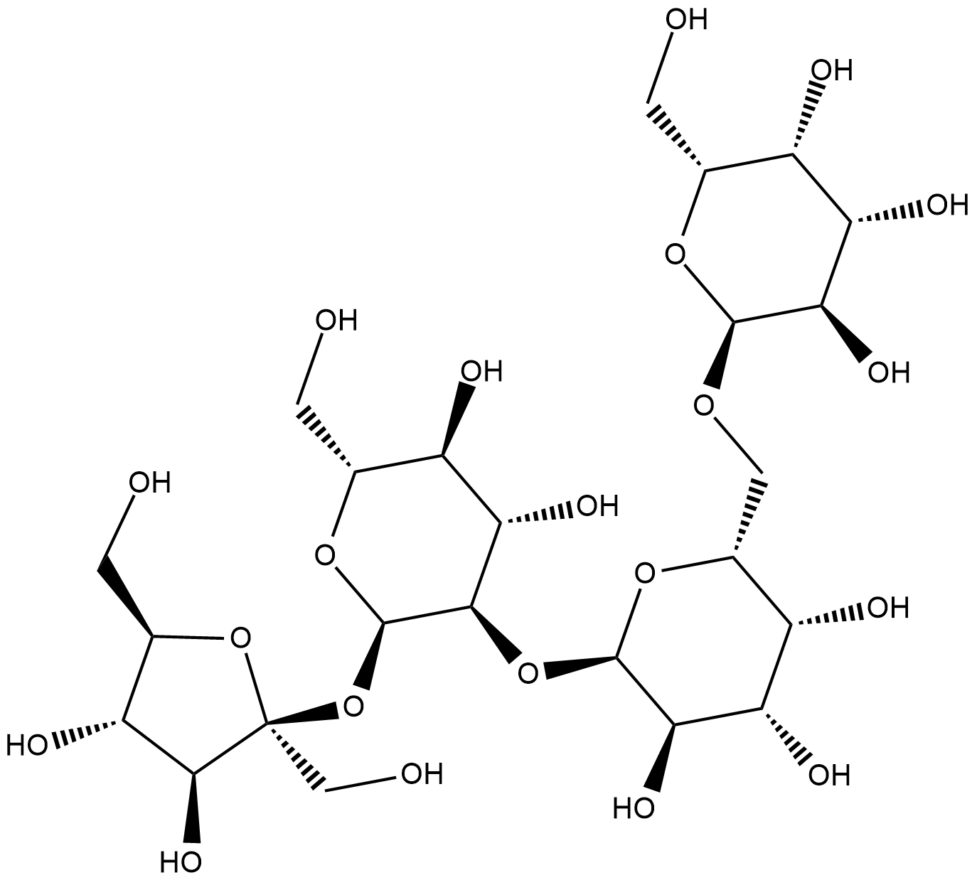
Cas No.: 470-55-3
Sample solution is provided at 25 µL, 10mM.
_1-3.$$$39978408@51.jpg');)
_1-9.$$$38538795@65.jpg');)
_1-9.$$$39112701@51.jpg');)
_1-7.$$$37316672@70.jpg');)
_366-372.$$$36198801@70.jpg');)
.$$$38565142@65.jpg');)
_1867-1883.$$$33248023@67.jpg');)
_eads6055.$$$39977546@45.jpg');)
_eadm9805.$$$40080571@45.jpg');)
_eadj3020.$$$39666821@45.jpg');)
_1-20.$$$39757301@28.jpg');)
_1-24.$$$38898113@28.jpg');)
_273-287.$$$36806353@46.jpg');)
_1-16.$$$37156877@44.jpg');)
_1-19.$$$37460805@44.jpg');)
_1291-1307.$$$34518654@46.jpg');)
Stachyoseはオリゴ糖とプレバイオティクスである[1]。StachyoseはPRDX5 (ペルオキシレドキシン5)の特異的な阻害剤として、PRDX5タンパク質に直接機能し、酵素活性を阻害する[2]。Stachyoseは、去勢耐性前立腺癌、そして2型糖尿病などの代謝性疾患に対する研究に広く使われている[3][4]。
体外で、Stachyose (培地で0.4-3.2mg/mL;24時間)はCaco-2結腸癌細胞の増殖を用量依存的に阻害し、細胞のアポトーシスを誘発し、ミトコンドリア膜電位を減らし、Baxを上方制御し、Bcl-2を下方制御し、シトクロムCの放出を促進し、caspases 3/9を活性化した[5]。
体内で、Stachyose (200-600mg/kg/日;3週間、経口投与)は、DSS誘発の結腸炎マウスモデルで、DSS誘発の結腸炎の症状を用量依存的に軽減し、密着結合タンパク質の発見(Occludin、Claudin-1、ZO-1)を増やし、炎症促進サイトカイン(TNF-α、IL-1β、IL-6)を減らし、抗炎症サイトカインIL-10を増やし、Treg/Th17細胞のバランスを回復させた[6]。Stachyose (1または5g/kg/日;3日間経口投与)は、心筋虚血再潅流損傷のマウスモデルで、心筋梗塞の面積を減らし、心筋機能を改善した[7]。
References:
[1] Li CN, Wang X, Lei L, et al. Berberine combined with stachyose induces better glycometabolism than berberine alone through modulating gut microbiota and fecal metabolomics in diabetic mice. Phytother Res. 2020;34(5):1166-1174.
[2] Wang R, Pan Y, Zhang L, et al. Prebiotic stachyose inhibits PRDX5 activity and castration-resistant prostate cancer development. Int J Biol Macromol. 2024;278(Pt 3):134844.
[3] Wang R, Mi Y, Ni J, et al. Identification of PRDX5 as A Target for The Treatment of Castration-Resistant Prostate Cancer. Adv Sci (Weinh). 2024;11(9):e2304939.
[4] Wu Y, Cao Y, Feng L, et al. The natural compound stachyose targets SGLT2-mediated metabolic reprogramming to ameliorate diabetic kidney disease. Phytomedicine. 2025;147:157182.
[5] Huang G, Mao J, Ji Z, Ailati A. Stachyose-induced apoptosis of Caco-2 cells via the caspase-dependent mitochondrial pathway. Food Funct. 2015;6(3):765-771.
[6] He N, Chen K, Yu S, et al. Stachyose Exerts Anticolitis Efficacy by Re-balancing Treg/Th17 and Activating the Butyrate-Derived PPARγ Signaling Pathway. J Agric Food Chem. 2024;72(21):12171-12183.
[7] Zhang AY, Su JB, Sun HT, et al. Stachyose ameliorates myocardial ischemia-reperfusion injury by inhibiting cardiomyocyte ferroptosis and macrophage pyroptosis. Int Immunopharmacol. 2024;143(Pt 1):113334.
| 細胞実験[1]: | |
細胞株 | Caco-2細胞 |
準備方法 | 30から50の継代のCaco-2細胞をこの実験に使った。サブ合流点に達した後、Caco-2細胞を、PBSで二回洗浄した後、0.25%のトリプシン消化を通じて、培養フラスコから回復させた。培地を2日ごとに交換した。導電率測定に基づく細胞数と細胞生存率を、トリパンブルー試験で評価した。Stachyoseの細胞毒性効果を評価するために、対数増殖期の細胞を96ウェルの細胞培養プレート(104個の細胞/ウェル)に播種し、24時間事前インキュベートした。続いて、異なる濃度(0、0.4、0.8、1.6、3.2mg/mL)のStachyoseが混ざった無血清培地(SFM)で細胞を24時間処理した。市販のプロトコールに従い、MTT法で細胞の生存率を測定した。細胞のアポトーシス用のフローサイトメトリーを、アネキシンV-フルオレセインイソチオシアネート(FITC)/ヨウ化プロピジウム(PI)アポトーシス検出キットで行った。 |
反応条件 | 0.4-3.2mg/mL;24時間 |
アプリケーション | StachyoseはCaco-2結腸癌細胞の増殖を用量依存的に阻害し、細胞のアポトーシスを誘発した。 |
| 動物実験 [2]: | |
動物モデル | 雄C57BL/6Jマウス |
準備方法 | 6週齢の雄C57BL/6Jマウスを、湿度と温度が制御された環境で飼育し、12時間の昼夜循環で、標準飼料と清潔な水を自由に摂取させた。1週間適応させた後、MIRIのマウスモデルに対するStachyoseの効果を評価した。模擬またはMIRI手術の前に、Stachyose (1または5g/kg/日)を三日間連続で経口投与した。MIRIモデルを、清潔の標準条件下で作成した。Vevo 3100LT超音波心電図システムを使い、心エコー検査を行った。心エコー検査を完成した後、マウスを頚髄脱臼で安楽死させ、胸部の皮膚と肋骨を切開し、心尖部を生理食塩水で濯いだ。続いて、心臓を除去し、-20℃で20分間冷凍した。心臓が完全に冷凍されたなら、外科手術ツールで解剖した。そして、37℃、心臓を2,3,5-triphenyltetrazolium chloride (TTC染料)で30分間浸した。壊死の部位は白く見え、生心筋が赤色であった。4%のパラホルムアルデヒドで24時間インキュベートした後、心臓を白いバックグラウンドで撮影した。 |
投与形態 | 1または5g/kg/日;3日間、経口投与 |
アプリケーション | Stachyoseは、心筋虚血-再潅流損傷のマウスモデルで、心筋梗塞の面積を減らし、心筋機能を改善した。 |
References: | |
| Cas No. | 470-55-3 | SDF | |
| Formula | C24H42O21 | M.Wt | 666.58 |
| 溶解度 | H2O : 125 mg/mL (Need ultrasonic) | Storage | Store at RT |
| General tips | Please select the appropriate solvent to prepare the stock solution according to the
solubility of the product in different solvents; once the solution is prepared, please store it in
separate packages to avoid product failure caused by repeated freezing and thawing.Storage method
and period of the stock solution: When stored at -80°C, please use it within 6 months; when stored
at -20°C, please use it within 1 month. To increase solubility, heat the tube to 37°C and then oscillate in an ultrasonic bath for some time. |
||
| Shipping Condition | Evaluation sample solution: shipped with blue ice. All other sizes available: with RT, or with Blue Ice upon request. | ||
| Prepare stock solution | |||

|
1 mg | 5 mg | 10 mg |
| 1 mM | 1.5002 mL | 7.501 mL | 15.002 mL |
| 5 mM | 300 μL | 1.5002 mL | 3.0004 mL |
| 10 mM | 150 μL | 750.1 μL | 1.5002 mL |
Step 1: Enter information below (Recommended: An additional animal making an allowance for loss during the experiment)
 g
μL
g
μL
Step 2: Enter the in vivo formulation (This is only the calculator, not formulation. Please contact us first if there is no in vivo formulation at the solubility Section.)
Calculation results:
Working concentration: mg/ml;
Method for preparing DMSO master liquid: mg drug pre-dissolved in μL DMSO ( Master liquid concentration mg/mL, Please contact us first if the concentration exceeds the DMSO solubility of the batch of drug. )
Method for preparing in vivo formulation: Take μL DMSO master liquid, next addμL PEG300, mix and clarify, next addμL Tween 80, mix and clarify, next add μL saline, mix and clarify.
Method for preparing in vivo formulation: Take μL DMSO master liquid, next add μL Corn oil, mix and clarify.
Note: 1. Please make sure the liquid is clear before adding the next solvent.
2. Be sure to add the solvent(s) in order. You must ensure that the solution obtained, in the previous addition, is a clear solution before proceeding to add the next solvent. Physical methods such as vortex, ultrasound or hot water bath can be used to aid dissolving.
3. All of the above co-solvents are available for purchase on the GlpBio website.
Quality Control & SDS
- View current batch:
- Purity: >80.00% Appearance: A solid
- COA (Certificate of Analysis)
- SDS (Safety Data Sheet)
- Datasheet
Average Rating: 5 (Based on Reviews and 3 reference(s) in Google Scholar.)