

Stachyose |
Catalog No.GC18245
 One-Click Copy Product Info
|

Stachyose는 올리고당이자 프리바이오틱스이다.
Products are for research use only. Not for human use. We do not sell to patients.
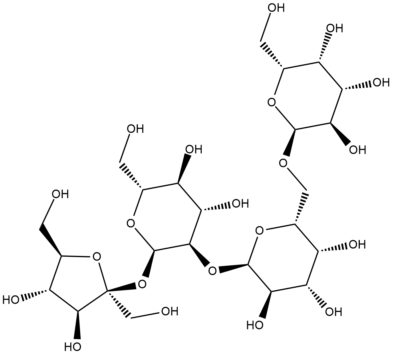
Cas No.: 470-55-3
Sample solution is provided at 25 µL, 10mM.
_1-3.$$$39978408@51.jpg');)
_1-9.$$$38538795@65.jpg');)
_1-9.$$$39112701@51.jpg');)
_1-7.$$$37316672@70.jpg');)
_366-372.$$$36198801@70.jpg');)
.$$$38565142@65.jpg');)
_1867-1883.$$$33248023@67.jpg');)
_eads6055.$$$39977546@45.jpg');)
_eadm9805.$$$40080571@45.jpg');)
_eadj3020.$$$39666821@45.jpg');)
_1-20.$$$39757301@28.jpg');)
_1-24.$$$38898113@28.jpg');)
_273-287.$$$36806353@46.jpg');)
_1-16.$$$37156877@44.jpg');)
_1-19.$$$37460805@44.jpg');)
_1291-1307.$$$34518654@46.jpg');)
Stachyose는 올리고당이자 프리바이오틱스이다. Stachyose는 PRDX5(peroxiredoxin 5)의 특이적 억제제로서 PRDX5 단백질에 직접 작용해서 그 효소 활성을 억제한다 [2]. Stachyose는 거세 저항성 전립선암 및 제2형 당뇨병 등 대사 질환 연구에 일반적으로 사용된다 [3][4].
체외 실험에서 Stachyose(배지 내 0.4–3.2 mg/mL, 24 시간 동안)는 용량 의존적으로 Caco-2 대장암 세포 증식을 억제하고 세포사멸을 유도하고 미토콘드리아 막 전위를 감소시키고 Bax를 상향 조절해서 Bcl-2를 하향 조절하고 사이토크롬 C 방출을 촉진해서 시스테인-3/9를 활성화하였다 [5].
체내 실험에서 Stachyose(200–600 mg/kg/일, 경구 3주)는 DSS 유발 대장염 쥐 모델에서 용량 의존적으로 대장염 증상을 완화시키고 티트 결합 단백(Occludin, Claudin-1, ZO-1) 발현을 증가시켜서 전염증성 사이토카인(TNF-α, IL-1β, IL-6)을 감소시키고 항염증성 사이토카인 IL-10을 증가시키고 Treg/Th17 세포 균형을 회복하였다 [6]. Stachyose(1 또는 5 g/kg/일, 경구 3일)는 심근 허혈-재관류 손상 쥐 모델에서 심근경색 부위를 줄이고 심장 기능을 개선하였다 [7].
References:
[1] Li CN, Wang X, Lei L, et al. Berberine combined with stachyose induces better glycometabolism than berberine alone through modulating gut microbiota and fecal metabolomics in diabetic mice. Phytother Res. 2020;34(5):1166-1174.
[2] Wang R, Pan Y, Zhang L, et al. Prebiotic stachyose inhibits PRDX5 activity and castration-resistant prostate cancer development. Int J Biol Macromol. 2024;278(Pt 3):134844.
[3] Wang R, Mi Y, Ni J, et al. Identification of PRDX5 as A Target for The Treatment of Castration-Resistant Prostate Cancer. Adv Sci (Weinh). 2024;11(9):e2304939.
[4] Wu Y, Cao Y, Feng L, et al. The natural compound stachyose targets SGLT2-mediated metabolic reprogramming to ameliorate diabetic kidney disease. Phytomedicine. 2025;147:157182.
[5] Huang G, Mao J, Ji Z, Ailati A. Stachyose-induced apoptosis of Caco-2 cells via the caspase-dependent mitochondrial pathway. Food Funct. 2015;6(3):765-771.
[6] He N, Chen K, Yu S, et al. Stachyose Exerts Anticolitis Efficacy by Re-balancing Treg/Th17 and Activating the Butyrate-Derived PPARγ Signaling Pathway. J Agric Food Chem. 2024;72(21):12171-12183.
[7] Zhang AY, Su JB, Sun HT, et al. Stachyose ameliorates myocardial ischemia-reperfusion injury by inhibiting cardiomyocyte ferroptosis and macrophage pyroptosis. Int Immunopharmacol. 2024;143(Pt 1):113334.
| 세포 실험 [1]: | |
세포 라인 | Caco-2 세포 |
제조 방법 | 본 실험에는 30–50 패시지의 Caco-2 세포를 사용하였습니다. 세포가 아경합 상태에 도달하면 PBS로 2회 세척한 후 0.25 % 트립신 소화로 배양 플라스크에서 회수하였습니다. 배지는 2일마다 교체하였습니다. 전도도 측정을 통한 세포 수와 트리판 블루 거부 시험으로 세포 생존율을 평가하였습니다. Stachyose의 세포 독성을 평가하기 위해 로그 상기 세포를 96-well 배양 판에 접종(웰당 10⁴ 세포)하고 24 시간 전 배양하였습니다. 이후 무혈청 배지(SFM)에 0, 0.4, 0.8, 1.6, 3.2 mg/mL 농도의 Stachyose를 혼합해서 24 시간 처리하였습니다. 세포 생존율은 상업적 프로토콜에 따라 MTT 검사로 측정하였습니다. 세포사멸은 V-fluorescein isothiocyanate (FITC)/propidium iodide (PI)를 이용한 유세포 분석법으로 분석하였습니다. |
반응 조건 | 0.4-3.2mg/mL; 24 시간 동안 |
응용 분야 | Stachyose는 용량 의존적으로 Caco-2 대장암 세포 증식을 억제하고 세포사멸을 유도하였습니다. |
| 동물 실험 [2]: | |
동물 모형 | 수컷 C57BL/6J 쥐 |
제조 방법 | 6주령 수컷 C57BL/6J 쥐를 온습도가 통제된 환경에 12 h 광암 주기로 사육하였고 표준 사료와 깨끗한 식수를 자유 섭취하게 하였습니다. 1주간 적응 후 MIRI(심근 허혈-재관류 손상) 쥐 모델에서 Stachyose의 효과를 평가하였습니다. 가식 또는 MIRI 수술 전 3일간 경구로 Stachyose(1 또는 5 g/kg/일)을 투여하였습니다. MIRI 모델은 청결하고 표준화된 조건에서 수립하였습니다. 심장초음파는 Vevo 3100LT 초음파 심장촬영 시스템을 이용해서 시행하였습니다.심장초음파 검사가 끝난 후 쥐는 경추 탈구로 안락사시켰고 흉부 피부와 늑골을 절개하고 심첨을 생리식염수로 세척하였습니다. 이후 심장을 적출해서 –20 ℃에서 20 분 동결하였습니다. 완전하게 동결된 심장은 외과 도구로 절개하였습니다. 심장을 37 ℃의 2,3,5-트리페닐테트라졸륨(TTC) 염료 용액에 30 분 침지하였습니다: 괴사 부위는 흰색으로 생존 심근은 빨간색으로 나타났습니다. 4 % 파라포르말데히드에서 24 시간 고정한 후 흰색 배경에 대해 촬영하였습니다. |
제형 | 1 또는 5g/kg/일; 3일 동안 구강 투여 |
응용 분야 | Stachyose는 심근 허혈-재관류 손상 쥐 모델에서 심근경색 면적을 줄이고 심장 기능을 개선하였습니다. |
References: | |
| Cas No. | 470-55-3 | SDF | |
| Formula | C24H42O21 | M.Wt | 666.58 |
| Solubility | H2O : 125 mg/mL (Need ultrasonic) | Storage | Store at RT |
| General tips | Please select the appropriate solvent to prepare the stock solution according to the
solubility of the product in different solvents; once the solution is prepared, please store it in
separate packages to avoid product failure caused by repeated freezing and thawing.Storage method
and period of the stock solution: When stored at -80°C, please use it within 6 months; when stored
at -20°C, please use it within 1 month. To increase solubility, heat the tube to 37°C and then oscillate in an ultrasonic bath for some time. |
||
| Shipping Condition | Evaluation sample solution: shipped with blue ice. All other sizes available: with RT, or with Blue Ice upon request. | ||
| Prepare stock solution | |||

|
1 mg | 5 mg | 10 mg |
| 1 mM | 1.5002 mL | 7.501 mL | 15.002 mL |
| 5 mM | 300 μL | 1.5002 mL | 3.0004 mL |
| 10 mM | 150 μL | 750.1 μL | 1.5002 mL |
Step 1: Enter information below (Recommended: An additional animal making an allowance for loss during the experiment)
 g
μL
g
μL
Step 2: Enter the in vivo formulation (This is only the calculator, not formulation. Please contact us first if there is no in vivo formulation at the solubility Section.)
Calculation results:
Working concentration: mg/ml;
Method for preparing DMSO master liquid: mg drug pre-dissolved in μL DMSO ( Master liquid concentration mg/mL, Please contact us first if the concentration exceeds the DMSO solubility of the batch of drug. )
Method for preparing in vivo formulation: Take μL DMSO master liquid, next addμL PEG300, mix and clarify, next addμL Tween 80, mix and clarify, next add μL saline, mix and clarify.
Method for preparing in vivo formulation: Take μL DMSO master liquid, next add μL Corn oil, mix and clarify.
Note: 1. Please make sure the liquid is clear before adding the next solvent.
2. Be sure to add the solvent(s) in order. You must ensure that the solution obtained, in the previous addition, is a clear solution before proceeding to add the next solvent. Physical methods such as vortex, ultrasound or hot water bath can be used to aid dissolving.
3. All of the above co-solvents are available for purchase on the GlpBio website.
Quality Control & SDS
- View current batch:
- Purity: >80.00% Appearance: A solid
- COA (Certificate of Analysis)
- SDS (Safety Data Sheet)
- Datasheet
Average Rating: 5 (Based on Reviews and 3 reference(s) in Google Scholar.)