

Adenosine (Synonyms: NSC 7652) |
Catalog No.GC14106
 One-Click Copy Product Info
|

Adenosine은 아데닌을 함유하고 있어 푸란당 분자와 β-N9-글리코시드 결합을 통해 연결된 내원성 신경 조절 물질이다.
Products are for research use only. Not for human use. We do not sell to patients.
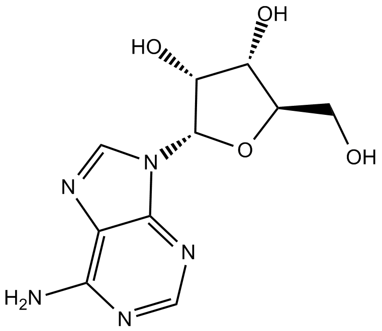
Cas No.: 58-61-7
Sample solution is provided at 25 µL, 10mM.
_1-3.$$$39978408@51.jpg');)
_1-9.$$$38538795@65.jpg');)
_1-9.$$$39112701@51.jpg');)
_1-7.$$$37316672@70.jpg');)
_366-372.$$$36198801@70.jpg');)
.$$$38565142@65.jpg');)
_1867-1883.$$$33248023@67.jpg');)
_eads6055.$$$39977546@45.jpg');)
_eadm9805.$$$40080571@45.jpg');)
_eadj3020.$$$39666821@45.jpg');)
_1-20.$$$39757301@28.jpg');)
_1-24.$$$38898113@28.jpg');)
_273-287.$$$36806353@46.jpg');)
_1-16.$$$37156877@44.jpg');)
_1-19.$$$37460805@44.jpg');)
_1291-1307.$$$34518654@46.jpg');)
Adenosine은 아데닌을 함유하고 있어 푸란당 분자와 β-N9-글리코시드 결합을 통해 연결된 내원성 신경 조절 물질이다. Adenosine은 신경 활동, 혈관 기능, 혈소판 응집, 혈구 생성 조절을 포함한 세포 생리의 거의 모든 측면에 영향을 미친다. Adenosine은 상심실성 빈맥을 치료하기 위해 사용할 수 있다[2].
체외 실험에서 Adenosine (1.0-4.0 mM; 12, 24, 48 시간)은 시간과 농도 의존적으로 HepG2 세포의 생존율을 억제하고 GRP78/BiP, PERK, ATF4, CHOP, 시스테인-3 그리고 사이토크롬 c의 mRNA와 단백질 수준을 현저하게 증가시켰고 미토콘드리아 막 전위를 감소시키고 소포체 스트레스를 활성화시켰다[3]. Adenosine (0.5-4.0 mM; 36 시간)은 EC109 세포에서 시스테인-3의 발현을 농도 의존적으로 현저하게 증가시켰고 세포 사멸과 NF-κB 활성을 유도했다[4].
체내 실험에서 Adenosine (1-10mg/kg/일; 복강주사)을 강제 수영 시험 (FST)과 꼬리 현수 시험 (TST)에 사용된 쥐에게 단일 용량으로 투여해서 FST에서의 부동 시간을 현저하게 단축시키고 TST에서 유사한 항우울제와 같은 효과를 나타냈다[5]. 쥐에서 Adenosine (5mg/kg/일; 복강주사; 6회, 1시간 간격) 처리는 염증 반응성 별세포 마커 (Lcn 2)의 발현 수준을 증가시키고 동시에 Ccl 2, Cxcl 1, Ilia와 Tnf와 같은 여러 가지 다른 염증 유발 사이토카인/사이토카인 화학 인자 유전자들의 발현을 강화시켰다[6].
References:
[1] Fredholm B B, Chen J F, Cunha R A, et al. Adenosine and brain function[J]. Int Rev Neurobiol, 2005, 63(1): 191-270.
[2] Borea PA, Gessi S, Merighi S, et al. Pharmacology of Adenosine Receptors: The State of the Art. Physiol Rev. 2018 Jul 1;98(3):1591-1625.
[3] Zhou XT, Pu ZJ, Liu LX,,et al.. Inhibition of autophagy enhances adenosine‑induced apoptosis in human hepatoblastoma HepG2 cells. Oncol Rep. 2019 Feb;41(2):829-838.
[4] Wu L F, Wei B L, Guo Y T, et al. Apoptosis induced by adenosine involves endoplasmic reticulum stress in EC109 cells[J]. International journal of molecular medicine, 2012, 30(4): 797-804.
[5] Kaster MP, Rosa AO, Rosso MM, et al. Adenosine administration produces an antidepressant-like effect in mice: evidence for the involvement of A1 and A2A receptors. Neurosci Lett. 2004;355(1-2):21-24.
[6] Guo Q, Gobbo D, Zhao N, et al. Adenosine triggers early astrocyte reactivity that provokes microglial activation and drives the pathogenesis of sepsis-associated encephalopathy[J]. bioRxiv, 2023: 2023.10. 30.563169.
| 세포 실험 [1]: | |
세포 라인 | 인간 간모세포종 HepG2세포 라인 |
제조 방법 | HepG2 세포는 37°C에서 5% CO₂를 포함하는 습윤된 대기 중에서 96웰 판 (웰당 5×10³ 세포)에 시드되고 단독으로 Adenosine (0, 1.0, 2.0, 3.0, 4.0 mM)으로 12, 24, 48 시간 동안 처리되었지만 단독으로 10 µM LY 294002 또는 2.0 mM Adenosine과 10 µM LY 294002를 조합해서 12, 24, 36, 48 시간 동안 처리되었습니다. 그리고 각 웰에 10 µl MTT (5 mg/ml)를 추가하고 세포를 추가로 4 시간 동안 배양했습니다. 상청액을 제거한 후 DMSO (웰당 100 µl)를 추가하여 HepG2 세포에서 MTT로 변환된 파란색 formazan 결정을 녹였습니다. 560 nm의 광학 밀도에서 효소 마커 (Wellscan K3; KHB Labsystems, 헬싱키, 핀란드)를 사용해서 세포 생존력이 평가되었습니다. 이 실험은 세 번 반복되었습니다. |
반응 조건 | 0, 1, 2, 3및 4mM; 12, 24 및 48 시간 동안 |
응용 분야 | Adenosine은 시간과 농도 의존적으로 세포 생존력을 억제하였습니다. |
| 동물 실험 [2]: | |
동물 모형 | 스위스 쥐 |
제조 방법 | 30-40g의 체중을 가진 다른 6마리의 스위스 쥐는 상수 온도 (22-27°C)에서 자유롭게 물과 음식에 접근할 수 있도록 유지되었고 12:12 시간의 빛:어두운 주기 (07:00h에 불이 켜짐)하에서 관리되었습니다. 모든 조작은 09:00에서 17:00 사이에 이루어졌고 각 동물은 한 번만 사용되었습니다. Adenosine과 커피인은 생리식염수에 녹였습니다. 약물은 복강주사 (i.p.) 경로를 통해 10mg/kg 체중의 부피로 투여되었습니다. Adenosine 또는 대조용매는 FST, TST 또는 오픈 필드 테스트 30분 전에 i.p.로 투여되었습니다. 대안적으로 그 중추 효과를 평가하기 위해 Adenosine은 FST 또는 오픈 필드 테스트 15분 전에 뇌실 주사(i.c.v.) 경로를 통해 투여되었습니다. I.c.v. 주사는 경증 에테르 마취하에 이루어졌습니다. 생리식염수나 Adenosine은 5μl의 부피로 30초 동안 주사되었고 캐뉼러는 추가 30초 동안 그대로 남겨졌습니다. |
제형 | 1-10mg/kg; i.p. |
응용 분야 | Adenosine은 복강주사 (i.p.)와 뇌실주사 (i.c.v.)를 통해FST에서의 부동 시간을 현저하게 줄였습니다. |
References: | |
| Cas No. | 58-61-7 | SDF | |
| Synonyms | NSC 7652 | ||
| Chemical Name | (2R,3R,4S,5R)-2-(6-aminopurin-9-yl)-5-(hydroxymethyl)oxolane-3,4-diol | ||
| Canonical SMILES | C1=NC2=C(C(=N1)N)N=CN2C3C(C(C(O3)CO)O)O | ||
| Formula | C10H13N5O4 | M.Wt | 267.24 |
| Solubility | ≥ 50mg/mL in DMSO with gentle warming | Storage | Store at 2-8°C |
| General tips | Please select the appropriate solvent to prepare the stock solution according to the
solubility of the product in different solvents; once the solution is prepared, please store it in
separate packages to avoid product failure caused by repeated freezing and thawing.Storage method
and period of the stock solution: When stored at -80°C, please use it within 6 months; when stored
at -20°C, please use it within 1 month. To increase solubility, heat the tube to 37°C and then oscillate in an ultrasonic bath for some time. |
||
| Shipping Condition | Evaluation sample solution: shipped with blue ice. All other sizes available: with RT, or with Blue Ice upon request. | ||
| Prepare stock solution | |||

|
1 mg | 5 mg | 10 mg |
| 1 mM | 3.742 mL | 18.7098 mL | 37.4195 mL |
| 5 mM | 748.4 μL | 3.742 mL | 7.4839 mL |
| 10 mM | 374.2 μL | 1.871 mL | 3.742 mL |
Step 1: Enter information below (Recommended: An additional animal making an allowance for loss during the experiment)
 g
μL
g
μL
Step 2: Enter the in vivo formulation (This is only the calculator, not formulation. Please contact us first if there is no in vivo formulation at the solubility Section.)
Calculation results:
Working concentration: mg/ml;
Method for preparing DMSO master liquid: mg drug pre-dissolved in μL DMSO ( Master liquid concentration mg/mL, Please contact us first if the concentration exceeds the DMSO solubility of the batch of drug. )
Method for preparing in vivo formulation: Take μL DMSO master liquid, next addμL PEG300, mix and clarify, next addμL Tween 80, mix and clarify, next add μL saline, mix and clarify.
Method for preparing in vivo formulation: Take μL DMSO master liquid, next add μL Corn oil, mix and clarify.
Note: 1. Please make sure the liquid is clear before adding the next solvent.
2. Be sure to add the solvent(s) in order. You must ensure that the solution obtained, in the previous addition, is a clear solution before proceeding to add the next solvent. Physical methods such as vortex, ultrasound or hot water bath can be used to aid dissolving.
3. All of the above co-solvents are available for purchase on the GlpBio website.
Quality Control & SDS
- View current batch:
- Purity: >99.50% Appearance: A solid
- COA (Certificate of Analysis)
- SDS (Safety Data Sheet)
- Datasheet
Average Rating: 5 (Based on Reviews and 32 reference(s) in Google Scholar.)