


Enzalutamide
Enzalutamide that is also known as MDV3100, is a small molecule. The complete two-dimensional view of the molecular structure of the Enzalutamide compound is diagrammatically presented in Figure 1. The trifluoromethyl substituted anilide is the most important structure feature of the overall Enzalutamide molecule as it is crucial to its biological activity.
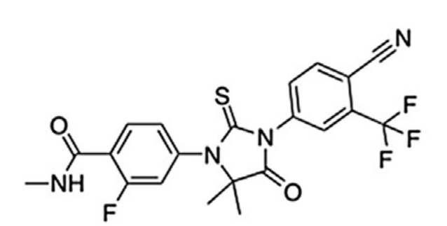
Figure 1 The two-dimensional view of the molecular structure of the Enzalutamide compound which is also known as MDV3100
Enzalutamide specifically inhibits the signalling of the Androgen Receptor; thus, it is regarded as Androgen Receptor antagonist in the pharmaceutical context and it is FDA approved for the treatment of prostate cancer.
The primary structure of the Androgen Receptor (AR) protein molecule is composed of a linear sequence of covalently bounded, i.e., specifically via the peptide linkages, 919 amino acids. These amino acids are divided into three major functional domains in such a manner, starting from the N-terminal domain (NTD) having 1 to 554 amino acids, through the DNA binding domain (DBD) having 555 to 628 amino acids and all the way to ligand binding domain (LBD) having 671 to 919 amino acids along with a small hinge domain. This is illustrated with the help of a schematic diagram in Figure 2.
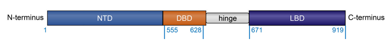
Figure 2 The Major functional domains of the Androgen Receptor protein molecule: NTD stands for N-terminal domain, DBD stands for DNA binding domain, LBD stands for ligand binding domain. Schematic diagram of a molecule of the Androgen Receptor protein highlighting all the significant structural motifs and the functional domains
Although the primary structure of the N-terminal domain (NTD) is known but the crystal structure, i.e., the secondary structure as well as the tertiary structures, for the N-terminal domain (NTD) has not been estimated as the N-terminal domain (NTD) itself exhibits a structural plasticity thus allowing it to interact with a wide spectrum of other molecules. Nevertheless, there is an Activation Function region (AF-1) in the N-terminal domain (NTD), which is further composed of two separate transcription activation regions namely TAU-1 and TAU-5. TAU-1 contains two distinct motifs namely LKDIL motif and LX7LL motif; the former motif is somewhat alike to the nuclear receptor box sequence of the nuclear receptor co-regulator proteins and critical to the activity of the Androgen Receptor (AR) protein molecule, while the latter motif is evolutionarily conserved motif and critical for the de-repression of genes as a response to the inflammatory cytokine signaling. TAU-5 also contains WHTLF motif which performs transactivation, i.e., the process by which a gene or a group of genes are activated meaning transcribed and ultimately translated due to the presence of another gene, in the absence of the natural ligands of the Androgen Receptor (AR) protein molecule. On top of that this WHTLF motif also regulates an intramolecular interaction between the N-terminal and C-terminal of the same through binding the Activation Function region (AF-2) of the ligand binding domain (LBD). This indicates that accessibility of this WHTLF motif is regulated. A closer look at the N-terminal domain (NTD) in Androgen Receptor (AR) protein molecule is given in Figure 3.
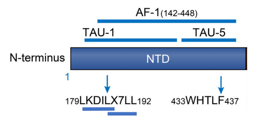
Figure 3 The Structural analysis of the N-terminal domain (NTD) in Androgen Receptor (AR) protein molecule in terms of regions and motifs
Taking about the DNA binding domain (DBD), it has two separate spheres of residual Cystine amino acids keeping a coordinate covalently bounded zinc ion in the center in the form of two fingers. In first finger, proximal box or P box is present that recognizes the Androgen Response Elements (ARE) on the DNA molecule and binds to them which is the core function of this particular domain. In second finger, distal box or D box is present which is involved in the homodimerization of the Androgen Receptor (AR) protein molecule. A closer look at the DNA binding domain (DBD) in Androgen Receptor (AR) protein molecule is given in Figure 4. The homodimerized DNA binding domain (DBD) bounded to the Androgen Response Elements (ARE) of the DNA molecule, suggesting the P Box as well as the D Box are active, is presented in Figure 5.
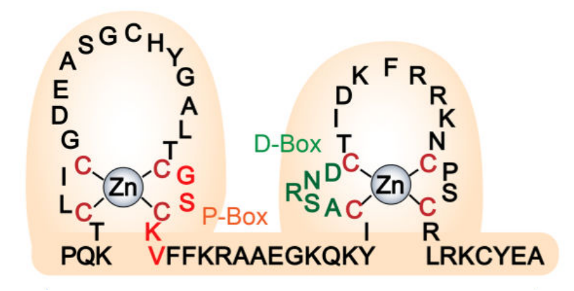
Figure 4 The Structural analysis of the DNA binding domain (DBD) of the Androgen Receptor (AR) protein molecule. Overall, the DBD is composed two bulging regions which are called as fingers, as both have a zinc ion they are termed as zinc fingers. Moreover, the four residual amino acids within the P Box are indicated in red; the five residual amino acids within the D Box are indicated in green
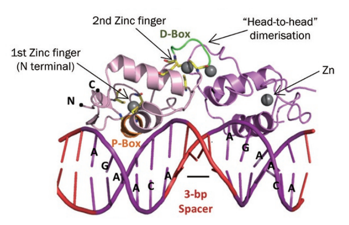
Figure 5 The dimerized DNA binding domain (DBD)and also bound to the Androgen Response Elements (ARE) of the double-stranded DNA molecule
The crystal structure of the ligand binding domain (LBD) is composed of three layers, as indicated in Figure 6, such that there are two antiparallel α-helices which sandwich the central pocket where the natural ligand, primarily the dihydrotestosterone (DHT), binds and is hydrophobic in nature.
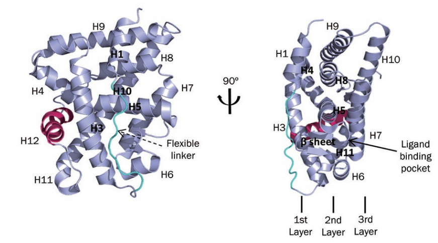
Figure 6 The crystal structure of the ligand binding domain (LBD) from two different orientations
In the human male body, testosterone is brought to the prostrate tissue by the activity of serum sex hormone binding globulin (SHBG). Upon entry in the cells, testosterone gets converted into dihydrotestosterone (DHT) by the enzymatic activity of 5-α-reductase in the cytosol; this dihydrotestosterone (DHT) then binds to the Androgen Receptor (AR) protein molecule in the cytosol and while separating it from the heat-shock proteins (HSPs) and also interacting with importin-α so that can Androgen Receptor (AR) protein be localized into nucleus. Followed by this, dihydrotestosterone (DHT) bound Androgen Receptor (AR) protein get localized into the cell nucleus. There, it gets dimerized and get bound to the Androgen Response Elements (ARE) of the DNA molecule, mainly on the promotor sites of the target gene, i.e., prostate-specific antigen (PSA) and transmembrane protease serine 2 (TMPRSS2). On the same site, the Androgen Receptor (AR) protein molecule is also able to bring some of the protein members of the basal transcription machinery for example, TATA-box-binding protein (TBP) and transcription factor IIF (TFIIF) along with some of the coregulator protein, for example, members of the p160 family of coactivators and cAMP-response element-binding protein (CREB)-binding protein (CBP). All this sequence of events leads to overall a normal survival, growth and differentiation of the cells within the tissues of the prostate gland; a quick overview of all these events is presented with the help of a cartoon diagram in Figure 7. In short, the Androgen Receptor (AR) protein regulates the transcription step, i.e., the process of formation of mRNA sequence according to the gene sequence, of the gene expression for a number of other genes; thus, any change in the rate of the activity of the Androgen Receptor (AR) protein would directly cause the dysregulation in all the other genes for which Androgen Receptor (AR) protein acts as transcriptional factor. On a bigger picture, the most vital cellular processes, i.e., growth and differentiation along with survival, would be negatively affected.
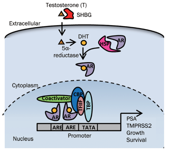
Figure 7 The mechanism of Androgen Receptor (AR) protein activity within the prostrate tissues
In the pathological condition, i.e., prostate cancer, the Androgen Receptor (AR) protein exhibits hyperactivity and it could be inhibited effectively via Enzalutamide.


 Chemical Structure")


Kommentare