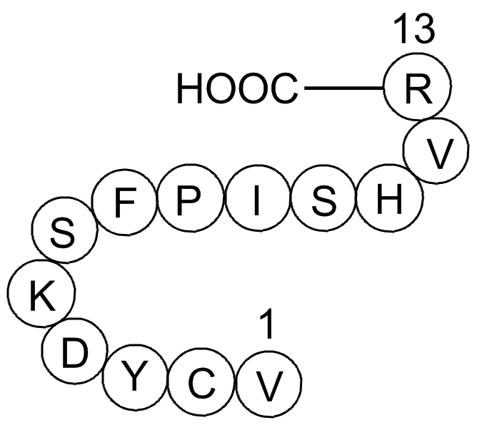
Gap 26 (Synonyms: Val-Cys-Tyr-Asp-Lys-Ser-Phe-Pro-Ile-Ser-His-Val-Arg ) |
| Katalog-Nr.GP10107 |
Products are for research use only. Not for human use. We do not sell to patients.

Cas No.: 197250-15-0
Sample solution is provided at 25 µL, 10mM.
GlpBio Produkte in seriösen Veröffentlichungen zitiert
Product Documents
Quality Control & SDS
- View current batch:
- Purity: >99.50%
- COA (Certificate Of Analysis)
- SDS (Safety Data Sheet)
- Datasheet
Protocol
| Cell experiment: [1] | |
|
Cell lines |
ECV304 cells |
|
Preparation method |
The solubility of this peptide in sterile water is >10 mM. Stock solution should be splited and stored at -80°C for several months. |
|
Reaction Conditions |
0.25mg/ml, 30min |
|
Applications |
Preventing the InsP3-triggered calcium increase by ester loading the cells with the calcium chelator BAPTA reduced the InsP3-triggered ATP release back to the control level. Incubation of the cells with gap 26 completely abolished the InsP3-triggered ATP response and reduced the ATP release to below the control level, indicating that the basal ATP release is also affected. |
| Animal experiment: [2] | |
|
Animal models |
Female Sprague-Dawley rats |
|
Dosage form |
300 μM, 45 min |
|
Applications |
The rats were prepared with closed cranial windows 24 h before the study. A 10-mm-diameter craniotomy was performed over the skull midline. The dura was removed carefully to keep the sagittal sinus intact. An 11-mm-diameter glass window outfitted with three ports was glued to the skull using cyanoacrylate. The skin overlying the window was sutured, and the animals were permitted to recover. On the day of study, three stainless steel screws were inserted into the skull, along the periphery of the cranial window, for electroencephalogram (EEG) recording. Cannulae were then connected to the three ports. The rats were subjected to one of two neuronal activation paradigms: SNS or bicuculline-induced seizure. Following the initial measurement of pial arteriolar diameter changes during SNS or during bicuculline exposure, baseline conditions were reestablished. After 20 min, a suffusion of gap-26 was initiated. Forty-five minutes later, the neural activation was repeated. Exposure to the Cx40/Cx37 inhibitory peptide, gap-26 (300 μM), was without effect on bicuculline- or SNS-induced pial arteriolar dilations. |
|
Other notes |
Please test the solubility of all compounds indoor, and the actual solubility may slightly differ with the theoretical value. This is caused by an experimental system error and it is normal. |
|
References: [1] Braet K, Vandamme W, Martin P E M, et al. Photoliberating inositol-1, 4, 5-trisphosphate triggers ATP release that is blocked by the connexin mimetic peptide gap 26. Cell calcium, 2003, 33(1): 37-48. [2] Xu H L, Mao L, Ye S, et al. Astrocytes are a key conduit for upstream signaling of vasodilation during cerebral cortical neuronal activation in vivo. American Journal of Physiology-Heart and Circulatory Physiology, 2008, 294(2): H622-H632. | |
| Cas No. | 197250-15-0 | SDF | |
| Überlieferungen | Val-Cys-Tyr-Asp-Lys-Ser-Phe-Pro-Ile-Ser-His-Val-Arg | ||
| Formula | C70H107N19O19S | M.Wt | 1550.79 |
| Löslichkeit | ≥77.55 mg/mL in DMSO with ultrasonic and warming, <7.75 mg/mL in EtOH, ≥155.1 mg/mL in Water with ultrasonic | Storage | -20°C, protect from light |
| General tips | Please select the appropriate solvent to prepare the stock solution according to the
solubility of the product in different solvents; once the solution is prepared, please store it in
separate packages to avoid product failure caused by repeated freezing and thawing.Storage method
and period of the stock solution: When stored at -80°C, please use it within 6 months; when stored
at -20°C, please use it within 1 month. To increase solubility, heat the tube to 37°C and then oscillate in an ultrasonic bath for some time. |
||
| Shipping Condition | Evaluation sample solution: shipped with blue ice. All other sizes available: with RT, or with Blue Ice upon request. | ||
Complete Stock Solution Preparation Table
| Prepare stock solution | |||

|
1 mg | 5 mg | 10 mg |
| 1 mM | 0.6448 mL | 3.2242 mL | 6.4483 mL |
| 5 mM | 0.129 mL | 0.6448 mL | 1.2897 mL |
| 10 mM | 0.0645 mL | 0.3224 mL | 0.6448 mL |
In vivo Formulation Calculator (Clear solution)
Step 1: Enter information below (Recommended: An additional animal making an allowance for loss during the experiment)
 g
g
 μL
μL

Step 2: Enter the in vivo formulation (This is only the calculator, not formulation. Please contact us first if there is no in vivo formulation at the solubility Section.)
Calculation results:
Working concentration: mg/ml;
Method for preparing DMSO master liquid: mg drug pre-dissolved in μL DMSO ( Master liquid concentration mg/mL, Please contact us first if the concentration exceeds the DMSO solubility of the batch of drug. )
Method for preparing in vivo formulation: Take μL DMSO master liquid, next addμL PEG300, mix and clarify, next addμL Tween 80, mix and clarify, next add μL ddH2O, mix and clarify.
Method for preparing in vivo formulation: Take μL DMSO master liquid, next add μL Corn oil, mix and clarify.
Note: 1. Please make sure the liquid is clear before adding the next solvent.
2. Be sure to add the solvent(s) in order. You must ensure that the solution obtained, in the previous addition, is a clear solution before proceeding to add the next solvent. Physical methods such as vortex, ultrasound or hot water bath can be used to aid dissolving.
3. All of the above co-solvents are available for purchase on the GlpBio website.
Related Products
- GP10028 tumor protein p53 binding protein fragment [Homo sapiens]/[Mus musculus]
- GP10037 Large T antigen - rhesus polyomavirus 560-568
- GP10010 COG 133
- GP10088 Luteinizing hormone releasing hormone human acetate salt (LHRH)
- GP10016 Lamin fragment
- GP10111 β-Pompilidotoxin
- GP10103 Laminin (925-933)
- GP10049 Amyloid Beta-Peptide (12-28) (human)
- GP10012 Angiotensin 1/2 (1-8) amide
- GP10076 Agouti-related Protein (AGRP) (25-82), human
- GP10046 Amyloid Precursor C-Terminal Peptide
- GP10017 [Ser25] Protein Kinase C (19-31)
- GP10060 a-MSH, amide
- GP10087 Angiotensin I (human, mouse, rat)
- GP10039 eukaryotic translation initiation factor 3
- GP10160 Oritavancin(LY-333328)
- GP10143 Cytochrome c - pigeon (88-104)
Bewertungen
Average Rating: 5 ★★★★★ (Based on Reviews and 30 reference(s) in Google Scholar.)
GLPBIO products are for RESEARCH USE ONLY. Please make sure your review or question is research based.
Required fields are marked with *


