


CFDA-SE
Chemicals known as CFDA-SE (5-(and-6)-Carboxyfluorescein diacetate succinimidyl ester) are frequently employed in biological and biochemical studies. The fluorophore carboxyfluorescein, which releases green fluorescence when stimulated by light, is the source of CFDA-SE. CFDA-SE is reactive to primary amines due to the succinimidyl ester group, which enables it to label amino group-containing compounds such as proteins. Because of this characteristic, CFDA-SE is a useful tool in research on cell and molecular biology. It is a member of the fluorescent dye class and is frequently used in cellular and molecular experiments to investigate biological processes. The molecular structure of CFDA-SE is shown in Figure 1.

Figure 1: The molecular structure of CFDA-SE.
Due to the presence of two acetoxy groups, carboxyfluorescein dicetate succinimidyl ester (CFDA-SE) is a non-polar molecule that is not fluorescent. Because of its lipophilicity, it can freely flow across the membrane of the cell. Two acetoxy groups are cut off by intracellular esterase cleavage, resulting in the formation of a fluorescent carboxyfluorescein succinimidyl ester (CFSE). This process also greatly lowers the rate of passage across the cell membrane and reduces lipophilicity . As a result, this gives its covalent bonding to intracellular molecules more time. The intracellular molecule's amino group is conjugated to carboxyfluorescein (CF) by a stable amide bond to generate CFR1 or (and) CFR2, as the succinimide side chain of CFSE is extremely reactive with the amino group. While CFR2 is durable and unable to penetrate the cell membrane, CFR1 rapidly degrades or diffuses out of the cell (Figure 2), resulting in stable CFSE staining .
Initially, the CFDA-SE was investigated as a long-term lymphocyte migratory tracer dye. It has been immediately apparent that CFSE is advantageous in detecting cell division since its fluorescence intensity is half with the cell division series. Furthermore, CFSE has been shown to have uses for monitoring cell fusion and has been widely utilized in studies of cell proliferation both in vitro and in vivo.
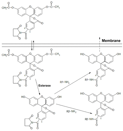
Figure 2: The staining mechanism of CFDA-SE.
Antibody synergy between immobilized CD3 and CD28 can mimic, to some extent, the normal process of antigen-presenting cells (APCs) activating lymphocytes. Immobilized CD3 and CD28 antibodies provide more cross-linking for receptors to activate intracellular signaling pathways and promote cell proliferation when compared to the use of free CD3 and CD28 antibodies. Free CD3 and CD28 antibodies can be immobilized by attaching them to solid surfaces, microspheres, or immunocompetent cells . The use of sulphate latex beads, CD3/CD28 antibodies with Dynabeads beads, and human t-activator CD3/CD28 Dynabeads has been documented in the literature for causing immobilization in Treg cell suppression experiments. Nevertheless, for this experiment, no MACSiBeadsTM Particles from the Treg expansion kit were utilized. This work examined the use of MACSiBeadsTM Particles in conjunction with CFDA-SE for in vitro Treg cell suppression experiments . Using magnetically stimulated cell sorting, CD8+ T cells and CD4+CD25+ Treg cells were segregated. To find the ideal staining concentration, CFDA-SE labeling was applied to CD8+ T cells. In suppression experiments, MACSi-BeadsTM Particles, a part of the Treg expansion kit, were employed as stimulators. Five experimental groups—stimulated CD8+ cells without CFDA-SE staining, unstimulated CD8+ cells with CFDA-SE staining, stimulated CD8+ cells with CFDA-SE staining, and co-cultured with Treg cells at a ratio of 1:0.25, 1:0.125, and 1:0—were established based on the conditions of MACSiBeadsTM Particles, CFDA-SE staining, and the cell composition of the co-culture system. BD FACS CantoTM was used for flow cytometry, and FCS Express 4 Plus software was used to analyze the flow data.
For the purpose of measuring the mean fluorescence intensity (MFI) of CFSE, various PBMC cell groups were chosen. The CFSE MFI of the cells was substantially correlated with the cell type at the same CFDA-SE staining dose (Figure 3). When the outcomes of six separate trials were examined, it was found that the three cell populations, R1, R2, and R3, had significantly different MFIs (Figure 4).
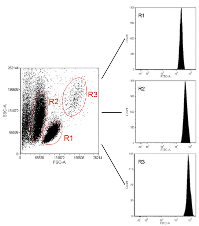
Figure 3: Fluorescence intensity of various cell groups.
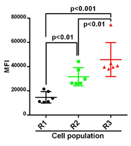
Figure 4: Mean fluorescence intensity comparison of various cell groups.
Following cell penetration by CFDA-SE, two fluorescent compounds were produced: CFR1 or (and) CFR2. CFR1 diffuses or breaks down quickly inside the cell. As a result, following CFDA-SE labeling, cells' fluorescence intensity will gradually decline. We carried out the experiment with various CFDA-SE staining concentrations and cultures in vitro without stimulation in order to ascertain the ideal staining concentration. After 1, 2, and 7 days, we saw variations in the fluorescence intensity of the cells (Figure 5). The day after staining, the CFSE MFI saw the biggest decrease. The alterations then progressively slowed down (Figure 6), which is in line with behavior described in the literature .
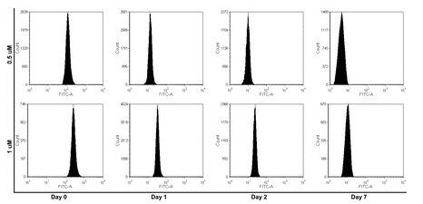
Figure 5: Fluorescence intensity decay of undivided cells with a final concentration 0.5 μM and 1.0 μM.
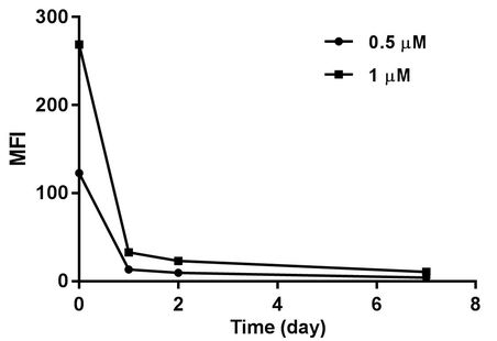
Figure 6: Mean fluorescence intensity of undivided cells with a final concentration 0.5 μM and 1.0 μM.
Co-cultured the two cell types at various ratios to ascertain the inhibitory effect of Treg cells on CD8+ T cells in vitro. A 4:1 ratio was used to add the CD8+ T cells and MACSibeadsTM Particles. The cells were taken out for identification after being co-cultured for 72 hours. Proliferation was not seen when the ratio of CD8+ T cells to Treg cells was 1:0.5. Consequently, in order to assess the inhibitory function of Treg cells, we selected the latter two ratios for the ensuing tests. Figure 7 displays the analysis of proliferation. We computed the CD8+ T cell division and proliferation indices, respectively. According to Figure 8, the differences were statistically significant. Consequently, it is verified that Treg cells have an inhibiting influence on the growth of CD8 cells .
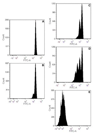
Figure 7: CD8+ T cell proliferation. (A) Fluorescence intensity of unstimulated CD8+ T cells with CFDA-SE staining; (B-D) Fluorescence intensity of stimulated CD8+ T cells with CFDA-SE staining co-cultured with Treg cells.
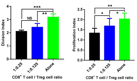
Figure 8: Split index and proliferation index for statistical analysis of Treg suppression assays.
CFDA-SE has numerous benefits over the traditional [3H] thymidine blending experiment and has been utilized extensively in the investigation of Treg cell in vitro suppression experiments. While Treg cell proliferation was relatively low and did not significantly affect the 3H-TdR blending value, the results of the classical [3H] thymidine blending experiment may have underestimated the Treg inhibitory activity. Furthermore, it was not possible to examine the reactions and phenotypes of different cells in the co-culture system independently. Cell membranes or intracellular specific markers, along with CFSE fluorescence, can be used to identify CFDA-SE stained cells from other types of cells . Depending on the experimental design, judgments concerning cell viability, proliferation, protein expression, or other characteristics can be made from the fluorescence data collected from CFDA-SE-labeled samples.
In research, CFDA-SE is a fluorescent dye that is widely used and flexible. It can be used for tagging proteins, measuring cellular dynamics, and evaluating the viability of cells. Because CFDA-SE can offer quantitative data in real time, it is a useful tool for researchers looking into many areas of molecular processes and cell biology. CFDA-SE and related fluorophores will probably become more important as technology and research techniques develop in order to further our comprehension of intricate biological systems. For the most recent details on the usage and applications of CFDA-SE, researchers should always consult the most recent publications and procedures.


Kommentare