

HBC620 |
Katalog-Nr.GC60186
 One-Click Copy Product Info
|

HBC620 ist ein HBC-ähnlicher Fluorophor, der dem GFP-Fluorophor ähnelt und eine maximale Anregungswellenlänge von 577 nm und eine maximale Emissionswellenlänge von 620 nm hat.
Products are for research use only. Not for human use. We do not sell to patients.
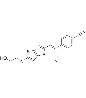
Cas No.: 2530162-07-1
Sample solution is provided at 25 µL, 10mM.
HBC620 ist ein HBC-ähnlicher Fluorophor, der dem GFP-Fluorophor ähnelt und eine maximale Anregungswellenlänge von 577 nm und eine maximale Emissionswellenlänge von 620 nm hat[1]. HBC620 kann als RNA-Tag verwendet werden, das hell, stabil und farbenfroh ist. Es kann auch verschiedene RNAs in vitalen Zellen abbilden[2]. Der einzigartige Fluoreszenz-Bildgebungs-Kanal von HBC620 wird meistens bei der Entwicklung luminometrischer Sensor-Plattformen für die Visualisierung einiger miRNA-Moleküle verwendet[3]. HBC620 kann zur In-situ-Lokalisierung tumorspezifischer Proteasen und für optische In-vivo-Bildanalyseexperimente an Tieren eingesetzt werden[4].
References:
[1] Chen X, Zhang D, Su N, et al. Visualizing RNA dynamics in live cells with bright and stable fluorescent RNAs[J]. Nature Biotechnology, 2019, 37(11): 1287-1293.
[2] Tang H, Peng J, Peng S, et al. Live-cell RNA imaging using the CRISPR-dCas13 system with modified sgRNAs appended with fluorescent RNA aptamers[J]. Chemical Science, 2022, 13(47): 14032-14040.
[3] Yin P, Ge M, Xie S, et al. A universal orthogonal imaging platform for living-cell RNA detection using fluorogenic RNA aptamers[J]. Chemical Science, 2023, 14(48): 14131-14139.
[4] Xie X, Sun Y, Peng J, et al. Collagen Anchoring Protein-Nucleic Acid Chimeric Probe for In Situ In Vivo Mapping of a Tumor-Specific Protease[J]. Analytical Chemistry, 2023, 95(50): 18487-18496.
HBC620-basierte In-vivo-Kartierung einer tumor-spezifischen Protease
Das Protokoll basiert auf Literaturangaben. Passen Sie bitte es an Ihre spezifischen Bedürfnisse an.
1. Etablierung des Xenograft-Tumor-Mausmodells
(1) Männliche BALB/c-Nacktmäuse(6 Wochen alt; 18-20 g Körpergewicht); 1×10⁶ B16-Zellen wurden subkutan in die rechte Achselhöhle jeder Maus injiziert. Das Tumorvolumen erreichte 80-100mm3 (nach etwa 7-8 Tagen).
(2) Chimäre Sonden wurden in die Schwanzvene von Mäusen verabreicht: CBD-miRFP-DCV-Chimärensonden wurden in 200 μl PBS gelöst und 30 s lang langsam bei 29 G der Schwanzvene hineingedrückt (1 nM/20 g).
2. Herstellung und Anwendung von HBC620
(1) 5μM HBC620-Arbeitslösung aus 0.5mM HBC620-Lösung(in PBS gelöst, 0.22 μm filtriert). Sofort verwenden.
(2) Nach der Anästhesie wurden die Mäuse enthaart und 200 μl HBC620-Arbeitslösung (Enddosis 1 nM/20 g, Molverhältnis 1:1 zur Sonde) in die Schwanzvene injiziert.
3. In-vivo-Bildgebung
(1) Echtzeit-Bildgebung: 0 Minute, 5 Minuten, 15 Minuten, 30 Minuten, 1 Stunde, 2 Stunden, 4 Stunden, 8 Stunden, 24 Stunden; Parameter: Anregungswellenlänge 570 nm, Emissionswellenlänge 650-680 nm, Belichtungszeit 500 ms, Blende 2, Sichtfeld 12cm×12cm.
(2) Quantifizierung: Tumor-ROI und ipsilateraler Haut-ROI zur Berechnung des T/N-Verhältnisses(Tumor/Normalgewebe-Verhältnis).
4. Hinweise
(1) Dunkelbetrieb: Die HBC620-Arbeitslösung hat eine Halbwertszeit von 4 Stunden bei 4 °C und wird bei Bedarf zubereitet und verwendet.
(2) Die Laserleistung war ≤0,5 W/cm2, um thermische Gewebeschäden zu vermeiden.
(3) Wenn die Bildgebung longitudinal wiederholt werden muss, beträgt das Intervall ≥48 Stunden, um sicherzustellen, dass der erste Farbstoff entfernt wurde (24-Stunden-Urinausscheidung > 85 %).
References:
[1] Xie X, Sun Y, Peng J, et al. Collagen Anchoring Protein-Nucleic Acid Chimeric Probe for In Situ In Vivo Mapping of a Tumor-Specific Protease[J]. Analytical Chemistry, 2023, 95(50): 18487-18496.
| Cas No. | 2530162-07-1 | SDF | |
| Canonical SMILES | OCCN(C)C1=CC(S2)=C(S1)C=C2/C=C(C#N)/C3=CC=C(C#N)C=C3 | ||
| Formula | C19H15N3OS2 | M.Wt | 365.47 |
| Löslichkeit | DMSO:16.67 mg/mL (45.61 mM) | Storage | Store at -20°C,unstable in solution, ready to use. |
| General tips | Please select the appropriate solvent to prepare the stock solution according to the
solubility of the product in different solvents; once the solution is prepared, please store it in
separate packages to avoid product failure caused by repeated freezing and thawing.Storage method
and period of the stock solution: When stored at -80°C, please use it within 6 months; when stored
at -20°C, please use it within 1 month. To increase solubility, heat the tube to 37°C and then oscillate in an ultrasonic bath for some time. |
||
| Shipping Condition | Evaluation sample solution: shipped with blue ice. All other sizes available: with RT, or with Blue Ice upon request. | ||
| Prepare stock solution | |||

|
1 mg | 5 mg | 10 mg |
| 1 mM | 2.7362 mL | 13.681 mL | 27.362 mL |
| 5 mM | 547.2 μL | 2.7362 mL | 5.4724 mL |
| 10 mM | 273.6 μL | 1.3681 mL | 2.7362 mL |
Step 1: Enter information below (Recommended: An additional animal making an allowance for loss during the experiment)
 g
μL
g
μL
Step 2: Enter the in vivo formulation (This is only the calculator, not formulation. Please contact us first if there is no in vivo formulation at the solubility Section.)
Calculation results:
Working concentration: mg/ml;
Method for preparing DMSO master liquid: mg drug pre-dissolved in μL DMSO ( Master liquid concentration mg/mL, Please contact us first if the concentration exceeds the DMSO solubility of the batch of drug. )
Method for preparing in vivo formulation: Take μL DMSO master liquid, next addμL PEG300, mix and clarify, next addμL Tween 80, mix and clarify, next add μL saline, mix and clarify.
Method for preparing in vivo formulation: Take μL DMSO master liquid, next add μL Corn oil, mix and clarify.
Note: 1. Please make sure the liquid is clear before adding the next solvent.
2. Be sure to add the solvent(s) in order. You must ensure that the solution obtained, in the previous addition, is a clear solution before proceeding to add the next solvent. Physical methods such as vortex, ultrasound or hot water bath can be used to aid dissolving.
3. All of the above co-solvents are available for purchase on the GlpBio website.
Quality Control & SDS
- View current batch:
- Purity: >98.00% Appearance: A solid
- COA (Certificate of Analysis)
- SDS (Safety Data Sheet)
- Datasheet
Average Rating: 5 (Based on Reviews and 9 reference(s) in Google Scholar.)