>>Peptides>> Tag Peptides>>3X FLAG Peptide

3X FLAG Peptide Sale

(Synonyms: H-Met-Asp-Tyr-Lys-Asp-His-Asp-Gly-Asp-Tyr-Lys-Asp-His-Asp-Ile-Asp-Tyr-Lys-Asp-Asp-Asp-Asp-Lys-OH) 카탈로그 번호: GP10149

합성 펩타이드 태그

Products are for research use only. Not for human use. We do not sell to patients.
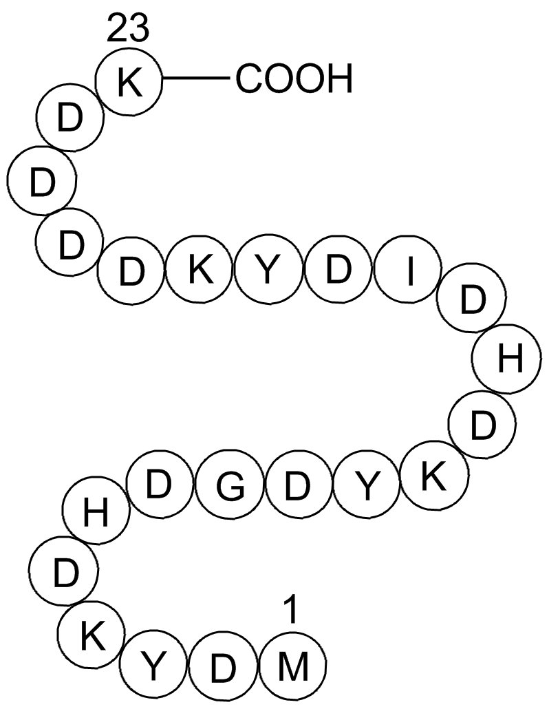
3X FLAG Peptide 화학 구조

Cas No.:402750-12-3

Size 가격 재고 수량
5mg
US$65.00
재고 있음
25mg
US$264.00
재고 있음
100mg
US$740.00
재고 있음
500mg
US$2,218.00
재고 있음
1g
US$3,548.00
재고 있음

고객 리뷰

고객 리뷰를 기반으로 합니다.

전화: (626) 353-8530 이메일 주소: sales@glpbio.com

Sample solution is provided at 25 µL, 10mM.

Product has been cited by 10 publications

Product Documents

품질 관리 및 안전 데이터 시트

현재 배치 보기

Protocol

Protocol for Protein expression and purification by 3×FLAG peptide [1]:

  1. The plasmids of FLAG-tagged genes were transfected into 293T cells for 2 days.
  2. The Cells were harvested by centrifugation at 14,000 rpm for 10 min at 4℃ and lysed with HEPES buffer (150 mM sodium chloride, 50 mM HEPES, pH 7.4, 5 mM EDTA, 1% (w/v) sodium deoxycholate, 1% (v/v) Triton X-100, 0.2% sodium fluoride, 0.1% sodium orthovanadate, and protease Inhibitor cocktail).
  3. Anti-FLAG M2 gel beads were then added and incubated on a rotary shaker at 4 C for 2 h. M2 gel beads were harvested by centrifugation at 3,000 rpm for 1 min and washed four times in HEPES buffer.
  4. The FLAG-tagged proteins were purified by the competition of 3×FLAG peptide. Briefly, M2 beads were resuspended with 1.5 mg/mL 3×FLAG peptide buffer and incubated at 4℃ for 2 h.
  5. After centrifugation at 5,000 rpm for 1 min, the supernatant further purified by gel filtration using a SuperdexTM 200 increase column equilibrated in store buffer (50 mM Tris-HCl, 37 mM NaCI, 1 mM EDTA, 5 mM DTT).
  6. All the purified proteins were concentrated by centrifugal filtration and stored in aliquots at 80℃.
  7. The purified protein was quantified using a ND-2000 NanoDrop spectrophotometer with OD 280 and verified by Coomassie staining.

This protocol only provides a guideline, and should be modified according to your specific needs.

Protocol for Co-IP and co-IP–MS analyses using 3×FLAG peptide [2]:

  1. Cell lysates were centrifuged at 13,300 r.p.m. for 15 min at 4℃ to remove intact cells.
  2. The supernatant was either incubated with control immunoglobulin G (IgG) or primary antibody overnight in IP buffer (150 mM NaCl, 20 mM HEPES at pH 7.4, 1% Triton X-100, 12.5 mM β-glycerophosphate, 1.5 mM MgCl2, 2 mM EGTA with phosphatase and protease inhibitors), followed by incubation with 20 μl of resuspended volume of Protein A/G beads for 2 h at 4℃ to pull down bound proteins.
  3. For sequential IP, bound protein was eluted from the beads by incubation with 5 packed gel volumes of 3×FLAG peptide elution solution (150 ng/ml final concentration) at 4℃ for 2 h and then immunoprecipitated with the second antibody overnight at 4℃.
  4. Beads were centrifuged at 1,000g for 5 min at 4℃ to remove the supernatant, washed 4 times with the IP buffer and boiled for 20 min at 95℃. Samples were run on SDS PAGE gel, followed by Coomassie Brilliant Blue R-250 staining.
  5. Afterwards, gel bands were excised, destained, trypsinized and subjected to MS analysis to identify individual proteins using liquid chromatography MS.

This protocol only provides a guideline, and should be modified according to your specific needs.

References:

[1].  Gao XK, Rao XS, et al. Phase separation of insulin receptor substrate 1 drives the formation of insulin/IGF-1 signalosomes. Cell Discov. 2022 Jun 28;8(1):60. doi: 10.1038/s41421-022-00426-x. PMID: 35764611; PMCID: PMC9240053.

[2].  Cong M, Wang Y, et al. MTSS1 suppresses mammary tumor-initiating cells by enhancing RBCK1-mediated p65 ubiquitination. Nat Cancer. 2020 Feb;1(2):222-234. doi: 10.1038/s43018-019-0021-y. Epub 2020 Jan 20. PMID: 35122005.

배경

FLAG-tag 시스템은 관심 단백질에 융합된 짧고 친수성인 8개의 아미노산 펩타이드를 이용합니다. FLAG 펩타이드는 항체 M1에 결합합니다. 결합이 칼슘 의존적인 방식2 또는 독립적인 방식3 인지 여부는 논란이 있습니다. 이 시스템의 단점은 모노클로날 항체 정제 매트릭스가 다른 것들보다 안정성이 낮다는 것입니다. 일반적으로 작은 태그는 특정한 모노클로날 항체로 감지됩니다.
FLAG 태그 검출을 개선하기 위해 3x FLAG 시스템이 개발되었습니다. 이 세 가닥의 FLAG 에피토프는 친수성으로, 22개의 아미노산으로 구성되어 있으며, 발현 융합 단백질을 최대 10 fmol까지 감지할 수 있습니다. Pyrococcus furiosus의 maltodextrin-binding protein에 FLAG 태그가 지원된 경우, 결정의 질은 untagged protein결정과 매우 유사합니다4.
마지막으로, enterokinase 처리를 통해 C-종단 다섯 개 아미노산 서열에 특이적인 FLAG 태그를 제거할 수 있습니다5.

참고문헌:
1. Hopp TP, Prickett KS, Price VL, Libby RT, March CJ, Ceretti DP, Urdal DL, Conlon PJ (1988) 재조합 단백질 식별과 정제에 유용한 짧은 폴리펩타이드 마커 시퀀스. Bio/Technology 6:1204-1210.
2. Hopp TP, Gallis B, Prikett KS (1996) 칼슘 의존적인 모노클로날 항체의 금속 결합 특성. Mol Immunol 33:601-608.
3. Einhauer A, Jungbauer A (2000) FLAG 펩타이드를 대상으로 한 모노클로날 항체 M1의 운동학 및 열역학적 특성. 단백질 분리 국제 심포지엄(ISPPP), 슬로베니아 루블랴나에서 개최된 제20회(2000년 11월 5일-8일).
4. Bucher MH, Evdokimov AG,Waugh DS (2002) Pyrococcus furiosus maltodextrin-binding protein 결정화에 대한 짧은 친화력 태그의 차이점 영향.Biol Cryst58:392-397.
5.Maroux S., Baratti J., Desnuelle P. (1971) 돼지 엔테로키나제의 정제 및 특이성. J Biol Chem 246:5031-5039.

화학적 특성

Cas No. 402750-12-3 SDF
동의어 H-Met-Asp-Tyr-Lys-Asp-His-Asp-Gly-Asp-Tyr-Lys-Asp-His-Asp-Ile-Asp-Tyr-Lys-Asp-Asp-Asp-Asp-Lys-OH
Chemical Name 3X FLAG Peptide
Canonical SMILES CCC(C)C(C(=O)NC(CC(=O)O)C(=O)NC(CC1=CC=C(C=C1)O)C(=O)NC(CCCCN)C(=O)NC(CC(=O)O)C(=O)NC(CC(=O)O)C(=O)NC(CC(=O)O)C(=O)NC(CC(=O)O)C(=O)NC(CCCCN)C(=O)O)NC(=O)C(CC(=O)O)NC(=O)C(CC2=CNC=N2)NC(=O)C(CC(=O)O)NC(=O)C(CCCCN)NC(=O)C(CC3=CC=C(C=C3)O)NC(=O)C(CC(=O)O)NC(
Formula C120H169N31O49S M.Wt 2861.87
용해도 ≥143.1mg/mL in DMSO, <14.35mg/mL in EtOH, ≥143.4mg/mL in Water with gentle warming Storage Desiccate at -20°C
일반적인 팁 높은 용해도를 얻기 위해 튜브를 37℃에서 데운 후 초음파 욕조에서 잠시 흔들어 주십시오. 원액은 -20℃ 이하에서 수개월간 보관할 수 있습니다.
배송 조건 평가 샘플 솔루션: 블루아이스와 함께 배송합니다. 다른 모든 가능한 크기: 요청 시 RT 또는 블루아이스와 함께 배송합니다.

In vivo Formulation Calculator (Clear solution)

Step 1: Enter information below (Recommended: An additional animal making an allowance for loss during the experiment)

mg/kg g μL

Step 2: Enter the in vivo formulation (This is only the calculator, not formulation. Please contact us first if there is no in vivo formulation at the solubility Section.)

% DMSO % % Tween 80 % saline
%DMSO %

Calculation results:

Working concentration: mg/ml;

Method for preparing DMSO master liquid: mg drug pre-dissolved in μL DMSO ( Master liquid concentration mg/mL, Please contact us first if the concentration exceeds the DMSO solubility of the batch of drug. )

Method for preparing in vivo formulation: Take μL DMSO master liquid, next addμL PEG300, mix and clarify, next addμL Tween 80, mix and clarify, next add μL saline, mix and clarify.

Method for preparing in vivo formulation: Take μL DMSO master liquid, next add μL Corn oil, mix and clarify.

Note: 1. Please make sure the liquid is clear before adding the next solvent.
2. Be sure to add the solvent(s) in order. You must ensure that the solution obtained, in the previous addition, is a clear solution before proceeding to add the next solvent. Physical methods such as vortex, ultrasound or hot water bath can be used to aid dissolving.
3. All of the above co-solvents are available for purchase on the GlpBio website.

  • 몰 농도 계산기

  • 희석 계산기

  • Molecular Weight Calculator

질량
=
농도
x
용량
x
메가와트


**원액을 준비할 때는 항상 바이알 라벨과 MSDS/CoA(온라인에서 사용 가능)에 있는 제품의 배치별 분자량을 사용하십시오.

계산하다

Related Posts

리뷰

Review for 3X FLAG Peptide

Average Rating: 5 ★★★★★ (Based on Reviews and 29 reference(s) in Google Scholar.)

5 Star
100%
4 Star
0%
3 Star
0%
2 Star
0%
1 Star
0%
Review for 3X FLAG Peptide

GLPBIO products are for RESEARCH USE ONLY. Please make sure your review or question is research based.

Required fields are marked with *

You may receive emails regarding this submission. Any emails will include the ability to opt-out of future communications.