

Golgi-Tracker Red |
Catalog No.GC19881
 One-Click Copy Product Info
|

Golgi red fluorescent probe, one of the sphingolipid fluorescent probes, which can be used for Golgi-specific fluorescent staining of living cells.
Products are for research use only. Not for human use. We do not sell to patients.
Sample solution is provided at 25 µL, 10mM.
Golgi-Tracker Red is a Golgi red fluorescent probe, one of the sphingolipid fluorescent probes, which can be used for Golgi-specific fluorescent staining of living cells.
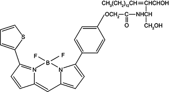
Golgi-Tracker Red is C5-ceramide fluorescently labeled with BODIPY TR from Molecular Probes. Ceramide or its analogs can selectively bind to Golgi, so fluorescently labeled ceramide can be used as a Golgi-specific fluorescent probe. Golgi-Tracker Red can be used for Golgi fluorescent labeling of living cells, but it is not suitable for labeling fixed cells. Refer to Figure 1 for the chemical structure of Golgi-Tracker Red.

Figure 1. The chemical structure of Golgi-Tracker Red.
Golgi-Tracker Red has red fluorescence with a maximum excitation wavelength of 589nm and a maximum emission wavelength of 617nm. Refer to Figure 2 for the excitation and emission spectra of Golgi-Tracker Red.

Figure 2. Excitation and emission spectra of Golgi-Tracker Red.
Golgi-Tracker Red is often used in the study of lipid transportation and metabolism in living cells. Compared with the traditional probe of the same type, NBD C6-ceramide, Golgi-Tracker Red exhibits a higher molar absorption coefficient and light quantum yield, and has stronger light stability. In addition to the above applications, this probe can also be used to determine the rate of lipid synthesis in Schwann cells, and to mark cell contours for observation of morphological movement under a confocal microscope.
The Golgi-Tracker Red probe has formed a complex with BSA. After the sphingolipid fluorescent probe belongs to this product and BSA form a complex, the labeling of the Golgi of living cells will be more efficient.
This kit provides Golgi-Tracker Red diluent, which makes the use of Golgi-Tracker Red more convenient.
Dilute according to the ratio of 1:100 to prepare 3ml of Golgi-Tracker Red working solution; dilute according to the ratio of 1:200 to prepare 6ml of Golgi-Tracker Red working solution.
This plan only provides a guide, please modify it to meet your specific needs.
1. Prepare dyeing solution
(1) This product is provided in the form of 33.3mg/ml (~47mM) DMSO solution. It is recommended that when using it for the first time, the product should be concentrated at the bottom through low-speed centrifugation, and then packaged into small specifications according to the single dosage, and stored at -20℃ or -80℃ away from light and avoid repeated freezing and thawing.
(2) Prepare the working solution: Use a suitable buffer (such as PBS) to dilute the stock solution at a ratio of 1:100, and mix it to become the working solution.
Notice:
① Please adjust the concentration of the working fluid according to the actual situation and make it ready for use;
② The working concentration of Golgi-Tracker Red can be adjusted appropriately according to the actual situation. The recommended dilution ratio range is 1:50~1:200.
2. Cell suspension staining
(1) Suspension cells: Centrifuge at 1000g for 3-5 minutes, discard the supernatant, and wash twice with an appropriate amount of solution such as Hanks balanced salt solution (containing calcium and magnesium) or other buffers, for 5 minutes each time .
(2) Adherent cells: Wash twice with a suitable buffer, add trypsin to digest the cells, and centrifuge at 1000g for 3-5 minutes after digestion is completed.
(3) Use freshly prepared Golgi-Tracker Red staining working solution to resuspend the cells and incubate at 4°C in the dark for 30 minutes.
(4) Aspirate away the staining solution, wash the cells about 3 times with pre-cooled cell culture medium, replace with fresh culture medium, and incubate in the dark at 37°C for 30 minutes. ,
(5) Wash again with fresh culture medium, and then observe with a fluorescence microscope or confocal microscope.
3. Cell adhesion staining
(1) Culture adherent cells on sterile coverslips.
(2) Remove the coverslip from the culture medium, aspirate the excess culture medium, and place the coverslip in a humid environment.
(3) Remove the cell culture medium and use an appropriate amount of solution such as Hanks balanced salt solution (containing calcium and magnesium) or other buffers to wash the cells growing on the coverslip.
(4) Aspirate the rinse solution, add freshly prepared Golgi-Tracker Red dyeing working solution, and incubate at 4°C in the dark for 30 minutes.
(5) Aspirate away the staining solution, wash the cells about 3 times with pre-cooled cell culture medium, replace with fresh culture medium, and incubate in the dark at 37°C for 30 minutes.
(6) Wash again with fresh culture medium, and then observe with a fluorescence microscope or confocal microscope.
4. Microscopic examination: After the staining is completed, the Golgi apparatus shows bright and strong fluorescence staining, while other membrane systems in the cell show relatively weak fluorescence staining. The maximum excitation/emission light of Golgi-Tracker Red is 589/617 nm respectively.
Precautions:
1) Fluorescent dyes all have quenching problems. Please avoid light as much as possible to slow down fluorescence quenching.
2) For your safety and health, please wear a lab coat and disposable gloves.
| Cas No. | SDF | ||
| Formula | C39H50BF2N3O4S | M.Wt | 705.71 |
| Solubility | Storage | Store at -20°C, protect from light | |
| General tips | Please select the appropriate solvent to prepare the stock solution according to the
solubility of the product in different solvents; once the solution is prepared, please store it in
separate packages to avoid product failure caused by repeated freezing and thawing.Storage method
and period of the stock solution: When stored at -80°C, please use it within 6 months; when stored
at -20°C, please use it within 1 month. To increase solubility, heat the tube to 37°C and then oscillate in an ultrasonic bath for some time. |
||
| Shipping Condition | Evaluation sample solution: shipped with blue ice. All other sizes available: with RT, or with Blue Ice upon request. | ||
| Prepare stock solution | |||

|
1 mg | 5 mg | 10 mg |
| 1 mM | 1.417 mL | 7.0851 mL | 14.1701 mL |
| 5 mM | 283.4 μL | 1.417 mL | 2.834 mL |
| 10 mM | 141.7 μL | 708.5 μL | 1.417 mL |
Step 1: Enter information below (Recommended: An additional animal making an allowance for loss during the experiment)
 g
μL
g
μL
Step 2: Enter the in vivo formulation (This is only the calculator, not formulation. Please contact us first if there is no in vivo formulation at the solubility Section.)
Calculation results:
Working concentration: mg/ml;
Method for preparing DMSO master liquid: mg drug pre-dissolved in μL DMSO ( Master liquid concentration mg/mL, Please contact us first if the concentration exceeds the DMSO solubility of the batch of drug. )
Method for preparing in vivo formulation: Take μL DMSO master liquid, next addμL PEG300, mix and clarify, next addμL Tween 80, mix and clarify, next add μL saline, mix and clarify.
Method for preparing in vivo formulation: Take μL DMSO master liquid, next add μL Corn oil, mix and clarify.
Note: 1. Please make sure the liquid is clear before adding the next solvent.
2. Be sure to add the solvent(s) in order. You must ensure that the solution obtained, in the previous addition, is a clear solution before proceeding to add the next solvent. Physical methods such as vortex, ultrasound or hot water bath can be used to aid dissolving.
3. All of the above co-solvents are available for purchase on the GlpBio website.
Quality Control & SDS
- View current batch:
- Appearance: A liquid
- COA (Certificate of Analysis)
- SDS (Safety Data Sheet)
- Datasheet
Average Rating: 5 (Based on Reviews and 37 reference(s) in Google Scholar.)