

Streptavidin MagBeads |
Catalog No.GC26178
 One-Click Copy Product Info
|

Streptavidin MagBeads are superparamagnetic beads covalently coated with streptavidin, suitable for the purification of biotinylated proteins, peptides, antibodies and oligonucleotides, protein-protein interaction studies, etc.
Products are for research use only. Not for human use. We do not sell to patients.
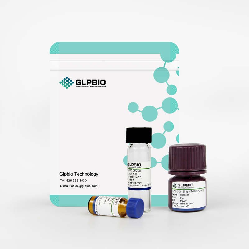
Sample solution is provided at 25 µL, 10mM.
Streptavidin MagBeads are superparamagnetic beads with a diameter of about 45-100μm covalently coated with high-purity streptavidin. The magnetic bead part consists of agarose and a magnetite core encapsulated therein, and streptavidin is covalently bound to the surface of the magnetic bead.
Streptavidin has a molecular weight of 53kDa and each molecule has four biotinylation binding sites. When mixed with a sample containing a biotinylated molecule, Streptavidin MagBeads can bind to the biotin on the molecule in a short incubation time, and then the magnetic beads bound to the molecule are separated by a magnetic separation rack, thereby indirectly capturing the target molecule. Streptavidin and biotin have very high affinity (Kd=10-15) and are widely used in the biological field.
Streptavidin MagBeads are provided in the form of a 25% phosphate buffered saline (PBS) slurry, pH7.4, containing 20% ethanol.
The recommended storage temperature of Streptavidin MagBead is 2–8℃. Do not freeze the product. During storage and use, Streptavidin MagBeads must be kept in liquid suspension. Drying will seriously interfere with the performance and binding capacity of the magnetic beads.
Instructions for use (taking 100μl of pure magnetic beads as an example):
1. Preparation of Streptavidin MagBeads:
(1) Shake or vortex the vial to completely resuspend the magnetic beads.
(2) Transfer 100μl of magnetic beads to a new tube (it is recommended to cut the tip of the pipette at an angle to enlarge the entrance).
(3) Place the test tube on a magnetic rack to collect the magnetic beads and discard the supernatant.
(4) Add 0.5 ml of wash buffer to the test tube and invert the test tube several times to mix. Use a magnetic rack to collect the magnetic beads and discard the supernatant. Repeat this step twice.
Note: When applied to nucleic acids, TES Buffer is recommended for washing buffer; when applied to antibodies/proteins, PBS (pH 7.4) is recommended for washing buffer.
2. Fixation of biotinylated molecules
(1) Related reagents
① Usage ratio: 2-3 mg of biotinylated sample: 1 ml of pure magnetic beads.
② Binding/washing buffer: nucleic acid: TES buffer; protein/antibody: PBS (pH 7.4).
③ Elution buffer: 8 M guanidine•HCl (pH 1.5).
(2) Operation steps
① Resuspend the magnetic beads in 100μl binding/washing buffer.
② Add the biotinylated sample to the magnetic beads prepared in step 1 and gently invert to mix.
③ Incubate the test tube at room temperature for one hour with shaking (oscillator or vortexer).
④ Transfer the test tube to a magnetic rack, collect the magnetic beads, and discard the supernatant. (If necessary, retain the supernatant for analysis)
⑤ Add 1 ml binding/washing buffer to the test tube and mix thoroughly, collect the magnetic beads using a magnetic rack and discard the supernatant, and repeat the wash 3 times.
⑥ Resuspend in an appropriate buffer to the desired concentration for downstream use.
3. Antigen purification
(1) Relevant reagents
① Ratio: 2-3 mg biotinylated antibody: 1 ml pure magnetic beads
② Binding/washing buffer: 0.1 M phosphate buffer (containing 0.15 M NaCl, pH 7.2)
③ Elution buffer: 0.1 M glycine•HCl (pH 2.5-2.8)
④ Neutralization buffer: 1 M Tris•HCl (pH 8.5)
(2) Operation steps
① Add 100 μl binding/washing buffer to the magnetic beads prepared in step 1 and resuspend.
② Add the biotinylated antibody solution to the magnetic beads and gently invert to mix.
③ Incubate the test tube at room temperature for one hour (on an oscillator or vortexer).
④ Transfer the test tube to the magnetic rack, collect the magnetic beads, and discard the supernatant. (If necessary, keep the supernatant for analysis)
⑤ Add 1 ml of binding/washing buffer to the test tube and mix thoroughly, collect the magnetic beads using a magnetic stand and discard the supernatant, and repeat the wash 3 times.
⑥ Add 100μl of binding/washing buffer to resuspend the magnetic beads.
⑦ Add the antigen sample to the test tube, gently invert to mix, and incubate at room temperature for 30 minutes or at 4°C overnight.
⑧ Add 1 ml of binding/washing buffer to the test tube to wash the magnetic beads, collect the magnetic beads using a magnetic stand and discard the supernatant, and repeat the wash 3 times.
⑨ Add 100μl of elution buffer to the test tube, mix thoroughly, and incubate at room temperature for 5 minutes.
⑩ Collect the magnetic beads using a magnetic stand and save the supernatant containing the target antigen.
⑪ Add neutralization buffer to adjust the pH, add 5 μl of neutralization buffer to every 50μl of elution.
Note:
1. The recommended storage temperature of Streptavidin MagBead is 2–8°C. Do not freeze this product!
2. During storage and use, Streptavidin MagBead must be kept in liquid suspension. Drying will seriously interfere with the performance and binding ability of the magnetic beads.
3. Please resuspend the magnetic beads before use.
4. Be careful during the use process to avoid bacterial/fungal contamination.
| Cas No. | SDF | ||
| Formula | M.Wt | ||
| Solubility | Storage | 2-8°C | |
| General tips | Please select the appropriate solvent to prepare the stock solution according to the
solubility of the product in different solvents; once the solution is prepared, please store it in
separate packages to avoid product failure caused by repeated freezing and thawing.Storage method
and period of the stock solution: When stored at -80°C, please use it within 6 months; when stored
at -20°C, please use it within 1 month. To increase solubility, heat the tube to 37°C and then oscillate in an ultrasonic bath for some time. |
||
| Shipping Condition | Evaluation sample solution: shipped with blue ice. All other sizes available: with RT, or with Blue Ice upon request. | ||
Step 1: Enter information below (Recommended: An additional animal making an allowance for loss during the experiment)
 g
μL
g
μL
Step 2: Enter the in vivo formulation (This is only the calculator, not formulation. Please contact us first if there is no in vivo formulation at the solubility Section.)
Calculation results:
Working concentration: mg/ml;
Method for preparing DMSO master liquid: mg drug pre-dissolved in μL DMSO ( Master liquid concentration mg/mL, Please contact us first if the concentration exceeds the DMSO solubility of the batch of drug. )
Method for preparing in vivo formulation: Take μL DMSO master liquid, next addμL PEG300, mix and clarify, next addμL Tween 80, mix and clarify, next add μL saline, mix and clarify.
Method for preparing in vivo formulation: Take μL DMSO master liquid, next add μL Corn oil, mix and clarify.
Note: 1. Please make sure the liquid is clear before adding the next solvent.
2. Be sure to add the solvent(s) in order. You must ensure that the solution obtained, in the previous addition, is a clear solution before proceeding to add the next solvent. Physical methods such as vortex, ultrasound or hot water bath can be used to aid dissolving.
3. All of the above co-solvents are available for purchase on the GlpBio website.
Quality Control & SDS
- View current batch:
- Appearance: A solution
- COA (Certificate of Analysis)
- SDS (Safety Data Sheet)
- Datasheet
Average Rating: 5 (Based on Reviews and 30 reference(s) in Google Scholar.)