
IF 23
PMID: 42489575 ↗2-NBDG
Catalog No.: GC10289Purity:>99.50%Synonyms: NBDGlucose
2-NBDG is a fluorescence-labeled 2-deoxy-glucose analog useful as a tracer for evaluation of cellular glucose metabolism (Ex/Em: 475/550 nm).
Products are for research use only. Not for human use. We do not sell to patients.
Cas No.: 186689-07-6
10mM (in 1mL Water)In Stock
$165.00
1
1mgIn Stock
$52.00
1
5mgIn Stock
$101.00
1
10mgIn Stock
$166.00
1
500mgIn Stock
$2,756.00
1
1gIn Stock
$4,568.00
1
| Pack Size | Price | Availability | Quantity | Action |
|---|---|---|---|---|
| 10mM (in 1mL Water) | $165.00 | In Stock | 1 | |
| 1mg | $52.00 | In Stock | 1 | |
| 5mg | $101.00 | In Stock | 1 | |
| 10mg | $166.00 | In Stock | 1 | |
| 500mg | $2,756.00 | In Stock | 1 | |
| 1g | $4,568.00 | In Stock | 1 |
Tel: (909) 407-4943 / (909) 689-7124Email: sales@glpbio.com
Citations
38 publicationsPublications from researchers using this productIF 20
PMID: 42174143 ↗IF 17
PMID: 38228638 ↗IF 16
PMID: 41644549 ↗IF 16
PMID: 39846120 ↗IF 11
PMID: 39167196 ↗Description
2-NBDG is a fluorescence-labeled 2-deoxy-glucose analog useful as a tracer for evaluation of cellular glucose metabolism (Ex/Em: 475/550 nm).
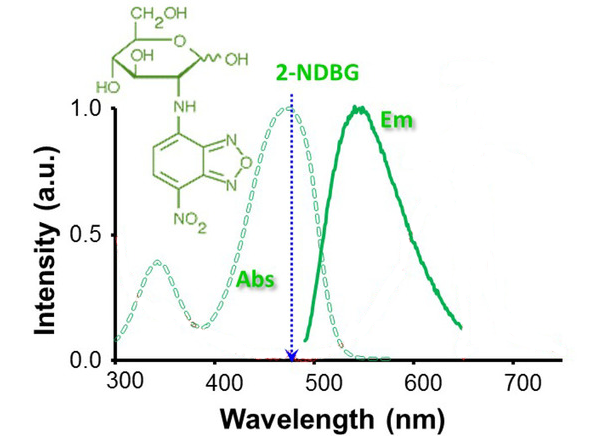
Glucose is a necessary source of energy for sustaining cell activities and homeostasis in tissues. Glucose metabolism is an important target in many diseases and changed with the pathological condition, therefore, evaluation of glucose metabolism can be a significant indication in disease progressions.
2 NBDG can be used in many kinds of cells in vitro, such as HepG2 human hepatocarcinoma cells, L6 rat skeletal muscle cells, MCF-7 breast cancer epithelial cells and astrocytes, it is also used in disease models, epilepsy rat, hyperglycemia, diabetes or mouse xenograft model of cancer.
2-NBDG enters cells through glucose transporters and is subsequently phosphorylated by hexokinase and trapped inside cells. Flow cytometric detection of fluorescence produced by cells can be performed to examine 2 NBDG uptake into living cells, and the intracellular concentration of transported 2-NBDG can be measured with a fluorescence microplate assay. 2 NBDG can be detected with a fluorescence imaging microscopy or CCD camera simply as well.
2-NBDG is a fluorescently labeled glucose tracer that is transported into cells by the same glucose transporter (GLUT) as glucose. Once 2-NBDG is taken up by cells, it is phosphorylated at the C-6 position to give 2-NBDG-6-phosphate, which is well retained in the cell. Compared to other glucose tracers such as 2-DG or FDG, 2-NBDG enables in situ measurement of 2-NBDG with high temporal and spatial resolution at the single-cell level. (suitable for fluorescence microscopy and flow cytometry detection)
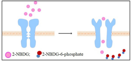
Rationale for 2-NBDG glucose uptake assay in cells: Once 2 NBDG is taken up by cells, it is phosphorylated at the C-6 position to generate 2-NBDG-6-phosphate in 2 NBDG metabolism, which is well retained in the cell, the fluorescence intensity is proportional to the cellular glucose uptake activity.
References:
[1]. Zou C, Wang Y, Shen Z. 2-NBDG as a fluorescent indicator for direct glucose uptake measurement[J]. Journal of biochemical and biophysical methods, 2005, 64(3): 207-215.
[2]. O’Neil R G, Wu L, Mullani N. Uptake of a fluorescent deoxyglucose analog (2-NBDG) in tumor cells[J]. Molecular Imaging and Biology, 2005, 7(6): 388-392.
[3]. Tsytsarev V, Maslov K I, Yao J, et al. In vivo imaging of epileptic activity using 2-NBDG, a fluorescent deoxyglucose analog[J]. Journal of neuroscience methods, 2012, 203(1): 136-140.
[4]. Yan Chen, Junjian Zhang, Xiang-yang Zhang, 2-NBDG as a Marker for Detecting Glucose Uptake in Reactive Astrocytes Exposed to Oxygen-Glucose Deprivation In Vitro. J Mol Neurosci (2015) 55:126–130.
[5]. Tsytsarev V, Maslov K I, Yao J, et al. In vivo imaging of epileptic activity using 2-NBDG, a fluorescent deoxyglucose analog[J]. Journal of neuroscience methods, 2012, 203(1): 136-140.
Protocol
Procedure for 2-NBDG uptake assay for MEFs [1]
1. Mouse embryonic fibroblasts (MEFs) are isolated from the embryos of C57BL/6 WT mouse (13.5 days).
2. Culture the MEF cells until reaching 80-90% confluence in 10 cm Petri dishes with DMEM growth medium in a humidified cell culture incubator (37 °C, 5% CO2).
Note: Don’t use MEFs beyond passage 3. MEFs usually become senescent at about passage 4 to 5.
3. Remove culture medium and wash cells one time with 10 ml 1x PBS.
4. Trypsinize cells using 4 ml of 0.05% trypsin-EDTA for 3 min at 37 °C.
Note: Use room temperature or pre-warmed 1x PBS from Step A3 to Step A9. Using chilled 1x PBS after Step A9.
5. Transfer cells to 15 ml polystyrene centrifuge tubes.
6. Harvest cells at 200 x g for 5 min by centrifugation.
7. Wash pelleted cells one time with 5 ml 1x PBS.
8. Count cells using a hemocytometer chamber.
9. Incubate 1 x 10⁶ MEF cells in a 37 °C water bath for 2 h with 1 ml of PBS containing 100 μM 2-NBDG. Incubate the same number of MEFs in the water bath with 1 ml PBS without 2-NBDG as a negative control.
10. Pellet the cells at 200 x g for 5 min by centrifugation. After washing the cells with chilled 1x PBS, the cells are pelleted at 200 x g for 5 min by centrifugation.
11. Resuspend cells in 0.5 ml of ice-cold 1x PBS with 2% FBS.
Note: Always keep cells on ice after this step.
12. Filter cells through a 35 µm nylon mesh (the cell-strainer cap of the 5 ml round-bottom polystyrene tubes) to obtain a uniform single-cell suspension in a 5 ml tube.
13. Keep the samples on ice until analysis on a flow cytometer.
14. Perform flow cytometric analysis. Acquire 10,000 single-cell events per reaction.
15. Analyze fluorescence intensity
Procedure for 2-NBDG uptake assay for breast cancer cells [1]
Using the same procedure as MEFs’ uptake assay except incubating 1 x 10⁶ MCF7 cells in a 37 °C water bath only for 30 min (instead of two hours for MEFs) with 1 ml of PBS containing 100 μM 2-NBDG.
10 mM stock of 2-NBDG: Dissolve 5 mg 2-NBDG in 1.46 ml PBS. Store at -20 °C in the dark
This protocol only provides a guideline, and should be modified according to your specific needs
References:
[1]. Dong, S., Baranwal, S., Garcia, A., Serrano-Gomez, S. J., Eastlack, S., Iwakuma, T., Mercante, D., Mauvais-Jarvis, F. and Alahari, S. K. (2017). Nischarin inhibition alters energy metabolism by activating AMP-activated protein kinase. J Biol Chem 292(41): 16833-16846.
Product Documents
Purity:>99.50%Appearance:A solid
Biological Data
1 / 8
Chemical Properties
CAS No.
186689-07-6
Synonyms
NBDGlucose
Chemical Name
(3R,4R,5S,6R)-6-(hydroxymethyl)-3-((7-nitrobenzo[c][1,2,5]oxadiazol-4-yl)amino)tetrahydro-2H-pyran-2,4,5-triol
SMILES
OC[C@](O1)([H])[C@](O)([H])[C@@](O)([H])[C@](NC2=CC=C(N(=O)=O)C3=NON=C23)([H])C1([H])O
Molecular Formula
C12H14N4O8
Molecular Weight
342.26
Solubility
≥ 17.1mg/mL in Water with ultrasonic
Storage
-20°C, protect from light
General tips
Select a suitable solvent according to the product solubility data. Once prepared, aliquot the stock solution to avoid repeated freeze-thaw cycles.
Use stock solutions within 6 months when stored at -80°C or within 1 month when stored at -20°C.
To improve solubility, warm the tube to 37°C and sonicate it for an appropriate period.
Use stock solutions within 6 months when stored at -80°C or within 1 month when stored at -20°C.
To improve solubility, warm the tube to 37°C and sonicate it for an appropriate period.
Shipping Condition
Evaluation samples are shipped with blue ice. Other available sizes are shipped at room temperature or with blue ice upon request.
CalculatorsMolarity / Dilution / Molecular Weight / Unit Conversion / In Vivo Formulation / Solubility
g/mol
Reviews
Review for 2-NBDG
Rating: 5
- 5 Star100%
- 4 Star0%
- 3 Star0%
- 2 Star0%
- 1 Star0%