

FerroOrange (Fe2+ indicator)
|
Catalog No.GC19943
 One-Click Copy Product Info
|


FerroOrange (Fe2+ indicator) is a fluorescent probe for detecting ferrous ions Fe2+ in living cells. It is not suitable for dead cells. The maximum excitation/emission wavelengths are 543nm/580nm.
Products are for research use only. Not for human use. We do not sell to patients.
Sample solution is provided at 25 µL, 10mM.
FerroOrange (Fe2+ indicator) is a fluorescent probe for detecting ferrous ions Fe2+ in living cells. Ferroorange is not suitable for dead cells. The maximum excitation/emission wavelengths are 543nm/580nm. The increase in fluorescence intensity after FerroOrange reacts with Fe2+ is irreversible and can be detected by fluorescence microscopy, fluorescence microplate reader, flow cytometer, etc. Iron is one of the most abundant transition metal elements in organisms and is involved in a variety of physiological processes. In recent years, researchers have paid extensive attention to the existence of free iron in living cells. Free iron exists in a stable redox state, mainly as ferrous ions (Fe2+) and ferric ions (Fe3+). In studying the intracellular reducing environment, metal transporters, and the water solubility of Fe2+, it is more important to understand the mechanism of Fe2+ than Fe3+.
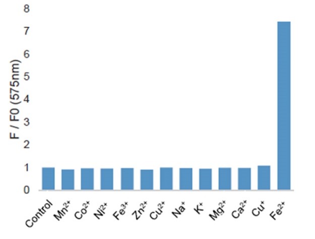
Figure 3. Fluorescence intensity measurement of 2 μl of 1 mM FerroOrange and 2 μl of 10 mM various metal ions added to 1 ml of 50 mM HEPES buffer (pH 7.4) and reacted for 1 hour at room temperature.
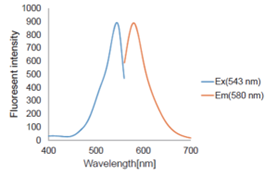
Fig. 4 Fluorescence properties of FerroOrange(Ex/Em:543/580 nm)
1. Probe preparation:
1.1 Remove FerroOrange from the refrigerator and allow it to thaw at room temperature. Place FerroOrange in a low-speed microcentrifuge and centrifuge it until the powder settles at the bottom of the vial, then open the lid.
1.2 Add 35μL of DMSO to the tube containing 24μg of FerroOrange, and mix thoroughly by pipetting up and down until it is completely dissolved to prepare a 1mM FerroOrange storage solution. If the storage solution cannot be used up in one experiment, aliquot and store FerroOrange in the dark at -20℃ for up to one month.
1.3 Before the actual experiment, dilute the 1mM FerroOrange storage solution to the desired working concentration (e.g. 1uM) with neutral buffer or serum-free culture medium. The working solution should be freshly prepared and used as soon as possible.
Note: acidic solutions will oxidize FerroOrange, severely affecting the efficiency of the probe.
2. Fluorescence microscopy operating procedure:
2.1. Seed cells in a fluorescence culture dish and culture overnight in a 37℃, 5% CO2 incubator.
2.2. Discard the supernatant and wash cells three times with HBSS or serum-free culture medium.
2.3. Replace with drug-containing culture medium and culture in a 37℃, 5% CO2 incubator.
Note: Optimize the incubation time based on the characteristics of the drug.
2.4. Add 1 μM working solution of FerroOrange and culture in a 37℃, 5% CO2 incubator for 30min.
Note: Observe after adding FerroOrange dye, without washing.
2.5. Observe cells under a fluorescence microscope.
3. Operating procedure for flow cytometry:
3.1. Seed 1 x 10⁵ HeLa cells (in MEM culture medium containing 10% fetal bovine serum and 1% penicillin-streptomycin) into a 6-well plate and culture overnight in a 37℃, 5% CO2 incubator.
3.2. Wash cells three times with serum-free culture medium (2 mL).
3.3. Add serum-free culture medium (1 mL) to the control group cells, and add 10 mM ammonium iron sulfate (10μL) (final concentration: 100μM) to the ammonium iron sulfate(II)-treated group cells in serum-free culture medium.
3.4. Incubate cells in a 37℃ incubator for 20 minutes, and wash cells three times with HBSS (1 mL).
3.5. Digest cells with trypsin (250 µL) and stop the reaction with serum-containing culture medium (1 mL), then transfer 1.25 mL of cell suspension to a microcentrifuge tube.
3.6. Centrifuge the cell suspension at 1,500 rpm for 3 minutes.
3.7. Discard the supernatant and add HBSS (1 mL) to the microcentrifuge tube, mix by shaking.
3.8. Centrifuge the cell suspension at 1,500 rpm for 3 minutes, and discard the supernatant.
3.9. Add 1μM FerroOrange and MEM containing serum-free culture medium (300μL) to the cells.
3.10. Incubate the cells at 37℃ for 15-30 minutes.
3.11. Filter cells through a cell strainer, and analyze the sample using a flow cytometer.
4. Fluorescence detection:
4.1. For fluorescence microscopy: use universal G excitation filters such as those used for Cv3 detection.
4.2. For laser microscopy and flow cytometry: use 532nm, 514nm or 561nm lasers for excitation. If not available, a 488nm laser can also be used. The emission wavelength is 580nm.
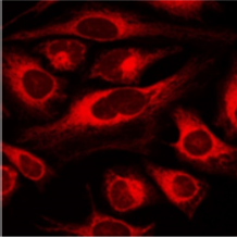
Fig. 1 FerroOrange staining of HeLa cells treated with ammonium iron sulfate.
5. Operating procedure for fluorescence microplate reader:
5.1. Set up groups:
Sample A: HeLa cells with no additives.
Sample B: HeLa cells with addition of iron chelator 2,2'-bipyridyl (Bpy)(GC61754).
Sample C: HeLa cells with addition of iron (ammonium iron sulfate)(GC20135).
5.2. Seed 100 µl of HeLa cell suspension into each well of a 96-well black plate (with a clear bottom) to achieve a density of 10,000 cells/well, and culture overnight in a 37℃, 5% CO2 incubator.
5.3. Wash the cells in Sample C three times with 100 µl of MEM (without FBS).
5.4. Add 100 µl of ammonium iron sulfate/MEM (without FBS) (final concentration: 100 µM) to Sample C and let it sit for 30 minutes in a 37℃, 5% CO2 incubator.
5.5. Wash all wells with 100 µl of HBSS three times.
5.6. Add 100 µl of 1 µM FerroOrange working solution to Samples A and C, and add 100 µl of HBSS solution containing FerroOrange (final concentration: 1 µM) and Bpy (final concentration: 100 µM) to Sample B. Incubate all samples for 30 minutes in a 37℃, 5% CO2 incubator.
5.7. Measure the fluorescence intensity of each sample using a multifunctional microplate reader (Ex: 543 nm, Em: 580 nm).
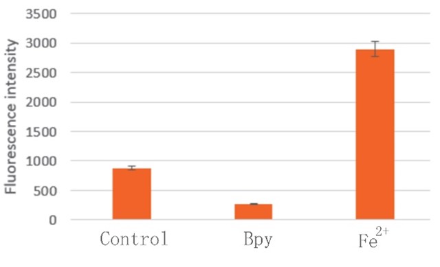
Fig. 2 Results of fluorescence microplate reader detection.
This protocol only provides a guideline, and should be modified according to your specific needs.
| Cas No. | SDF | ||
| Formula | M.Wt | ||
| Solubility | Storage | Store at 2-8°C, protect from light | |
| General tips | Please select the appropriate solvent to prepare the stock solution according to the
solubility of the product in different solvents; once the solution is prepared, please store it in
separate packages to avoid product failure caused by repeated freezing and thawing.Storage method
and period of the stock solution: When stored at -80°C, please use it within 6 months; when stored
at -20°C, please use it within 1 month. To increase solubility, heat the tube to 37°C and then oscillate in an ultrasonic bath for some time. |
||
| Shipping Condition | Evaluation sample solution: shipped with blue ice. All other sizes available: with RT, or with Blue Ice upon request. | ||
Step 1: Enter information below (Recommended: An additional animal making an allowance for loss during the experiment)
 g
μL
g
μL
Step 2: Enter the in vivo formulation (This is only the calculator, not formulation. Please contact us first if there is no in vivo formulation at the solubility Section.)
Calculation results:
Working concentration: mg/ml;
Method for preparing DMSO master liquid: mg drug pre-dissolved in μL DMSO ( Master liquid concentration mg/mL, Please contact us first if the concentration exceeds the DMSO solubility of the batch of drug. )
Method for preparing in vivo formulation: Take μL DMSO master liquid, next addμL PEG300, mix and clarify, next addμL Tween 80, mix and clarify, next add μL saline, mix and clarify.
Method for preparing in vivo formulation: Take μL DMSO master liquid, next add μL Corn oil, mix and clarify.
Note: 1. Please make sure the liquid is clear before adding the next solvent.
2. Be sure to add the solvent(s) in order. You must ensure that the solution obtained, in the previous addition, is a clear solution before proceeding to add the next solvent. Physical methods such as vortex, ultrasound or hot water bath can be used to aid dissolving.
3. All of the above co-solvents are available for purchase on the GlpBio website.
Quality Control & SDS
- View current batch:
- Purity: >99.50% Appearance: A solid
- COA (Certificate of Analysis)
- SDS (Safety Data Sheet)
- Datasheet
Average Rating: 5 (Based on Reviews and 13 reference(s) in Google Scholar.)