Fluorescent Dye
Products for Fluorescent Dye
- Cat.No. Product Name Information
-
GC19943
FerroOrange (Fe2+ indicator)
FerroOrange (Fe2+ indicator) is a fluorescent probe for detecting ferrous ions Fe2+ in living cells. It is not suitable for dead cells. The maximum excitation/emission wavelengths are 543nm/580nm.
-
GC74204
β-Ala-Lys(AMCA)
β-Ala-Lys(AMCA) is a multifunctional dye.

-
GC45306
1,6-Diphenyl-1,3,5-hexatriene
Diphenylhexatriene, DPH, NSC 90479
1,6-Diphenyl-1,3,5-hexatriene is a fluorescent probe for lipid monolayers and bilayer membranes (with maximum excitation and emission wavelengths of 360/430nm).
-
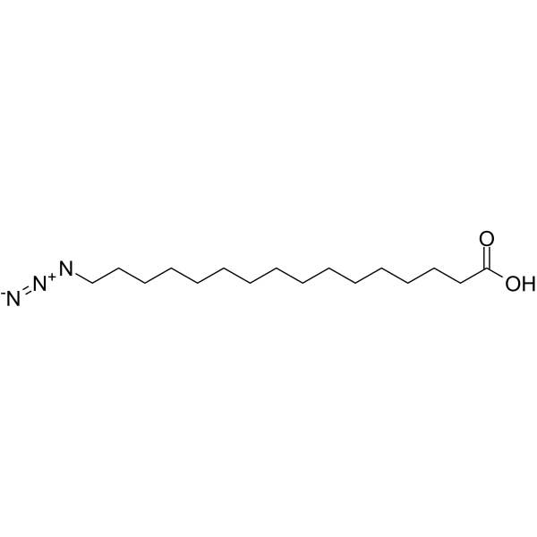
GC65988
16-Azidohexadecanoic acid
16-azidohexadecanoic acid can be a substrate for observing the degradation of fatty acids (FAs).
-
GC74213
3,5-DiBr-PAESA sodium
3,5-DiBr-PAESA (sodium) is an ultrasensitive chelating agent used for the determination of silver (I) in water in the absence of interfering ions.

-
GC78732
6-Carboxyfluorescein dipivalate
6-Carboxyfluorescein dipivalate is a non-fluorescent derivative of 6-Carboxyfluorescein.

-
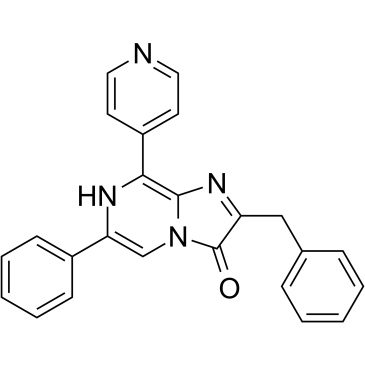
GC39334
8pyDTZ
8pyDTZ is a pyridyl diphenylterazine (DTZ) analog designed for bioluminescent imaging, specifically optimized for use with the LumiLuc luciferase system.
-
GC18326
9-Amino-6-chloro-2-methoxyacridine
ACMA
9-Amino-6-chloro-2-methoxyacridine (ACMA) is a multifunctional, cell-permeable fluorescent probe and bioactive molecule that can be utilized as a DNA intercalator and a pH/membrane potential-sensitive probe.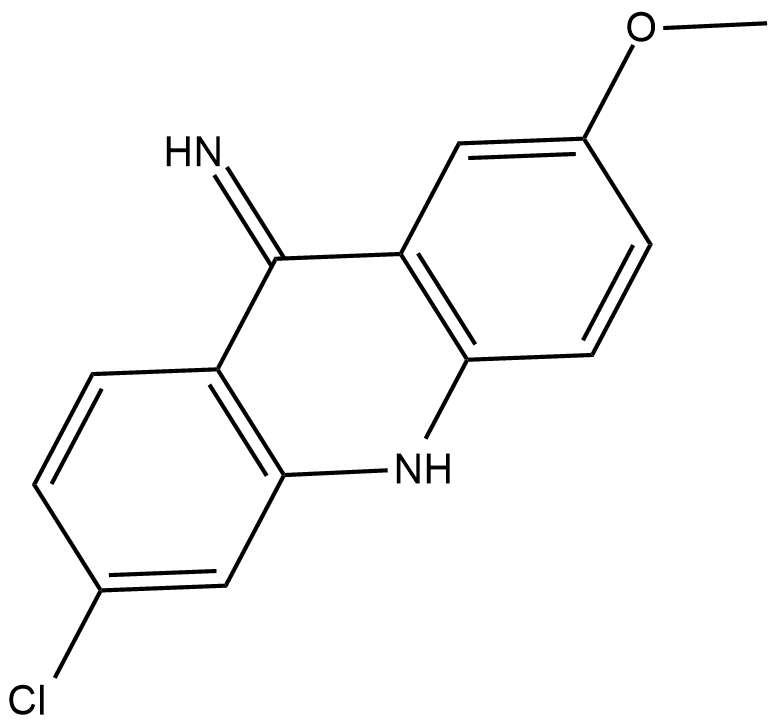
-
GC42711
Acridinium NHS ester
Acridinium NHS ester (AE NHS) is a protein and nucleic acid label at primary amines in biomolecules and produce chemiluminescence in the presence of hydrogen peroxide (H2O2)

-
GC20271
Actin-Tracker Green-488
Actin-Tracker Green-488 is an Actin-specific green fluorescent probe used for staining Actin in cultured cells or tissue slices.
-
GB30275
ADHP(Amplex Red)
A 6550, ADHP, Amplex Red
Amplex Red is utilized to measure hydrogen peroxide concentrations or peroxidase activities.

-
GC74228
AF 488 maleimide
AF 488 maleimide is a thiol-reactive dye for labeling of protein SH groups, and be used to attach AF 488 fluorophore to proteins and peptides containing cysteine residues, as well as to other thiolated molecules.

-
GC74217
AF 568 DBCO
AF 568 DBCO is a fluorescent dye that reacts with azide-labeled molecules or biomolecules via copper-free click chemistry.

-
GC74216
AF488 DBCO ditriethylamine
AF488 DBCO ditrietlamine is the ditrietlamine salt form of AF488 DBCO.

-
GC59158
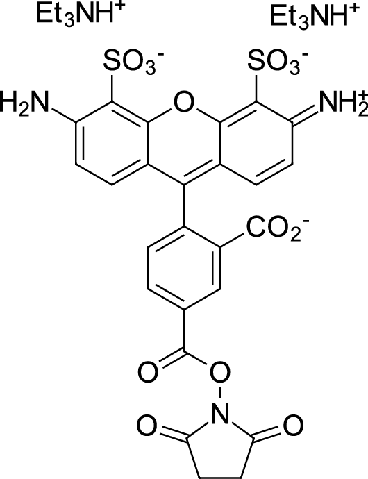
AF488 NHS ester
AF488 NHS ester is an amine-specific fluorescent probe with optimal excitation wavelength of 488nm and the emission wavelength of 525nm.
-
GC74218
Ag2S-PEG-NH2 (1200 nm)
Ag2S-PEG-NH2 (1200 nm) is a fluorescent dyes.

-
GC74219
AgAuSe-PEG-NH2 (1000 nm)
AgAuSe-PEG-NH2 (1000 nm) is a fluorescent quantum dot that emits fluorescence in the near-infrared II region, with an emission peak reaching 1000 nm that can be used in bioimaging.

-
GC74220
AgAuSe-PEG-NH2 (1120 nm)
AgAuSe-PEG-NH2 (1120 nm) is a fluorescent quantum dot that emits fluorescence in the near-infrared II region, with an emission peak reaching 1120 nm that can be used in bioimaging.

-
GC39336
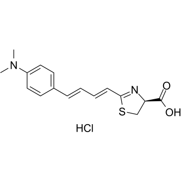
AkaLumine hydrochloride
AkaLumine hydrochloride is a kind of luciferin analog, emits light in the near-infrared (NIR) wavelength range (λmax=677nm) in reactions with native firefly luciferase.
-
GC74295
Aleuria Aurantia Lectin
AAL
Aleuria Aurantia Lectin (AAL) is a plant lectin that can be used as a probe to specifically bind biomolecules (such as polysaccharides, peptides, etc.
-
GC74214
Alexa fluor 647 NHS ester
Alexa fluor 647 NHS ester can be used to label Alexa fluor 647 to the primary amines (R-NH2) of proteins, amine-modified oligonucleotides, and other amine-containing molecules.

-
GC74287
Avidin-Cy3
Avidin-Cy3 is Cy3-labeled Avidin.

-
GC74690
BAPTA-TMFM
BAPTA-TMFM is a fluorescent chelating indicator used to study the role of cytosolic free calcium.

-
GC74205
Basic Red 46
Cationic Red X-GRL
Basic Red 46 is an alkaline dye that can be removed from aqueous solutions in intermittent adsorption systems using tree ferns as biosorbents.
-
GC59057
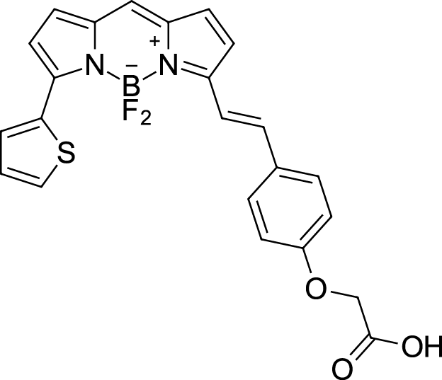
BDP 630/650 carboxylic acid
BDP 630/650 carboxylic acid is a bright far-red fluorophore (Ex=630nm; Em=650nm) based on a boron dipyrrolemethylene backbone.
-
GC59059
BDP R6G maleimide
BDP R6G maleimide is a high-performance, thiol-reactive fluorescent dye with an excitation maximum of 530nm and an emission maximum of 548nm, offering exceptional brightness and photostability.

-
GC50503
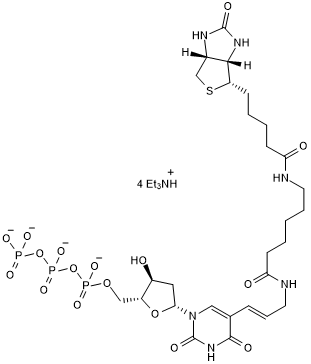
Biotin-11-dUTP
Biotin-11-dUTP is a fluorescent alternative to dTTP that enzymatically non-radioactively labels DNA during PCR, nick translation, cDNA synthesis, random primer labeling, or primer extension.
-
GC66466
Biotin-4-aminophenol
Biotin-4-aminophenol is a compound that fuses biotin with 4-aminophenol. Biotin-4-aminophenol is widely used as a bioconjugate for labeling proteins and nucleic acids, and Biotin-4-aminophenol is a key player in a variety of biochemical assays and molecular biology techniques.

-
GC13376
Biotin-tyramide
Biotin-Phenol,Biotin Phenol
Biotin-tyramide is a biotin-labeled tyramine derivative and is the core reagent of the Tyramide Signal Amplification (TSA) technology.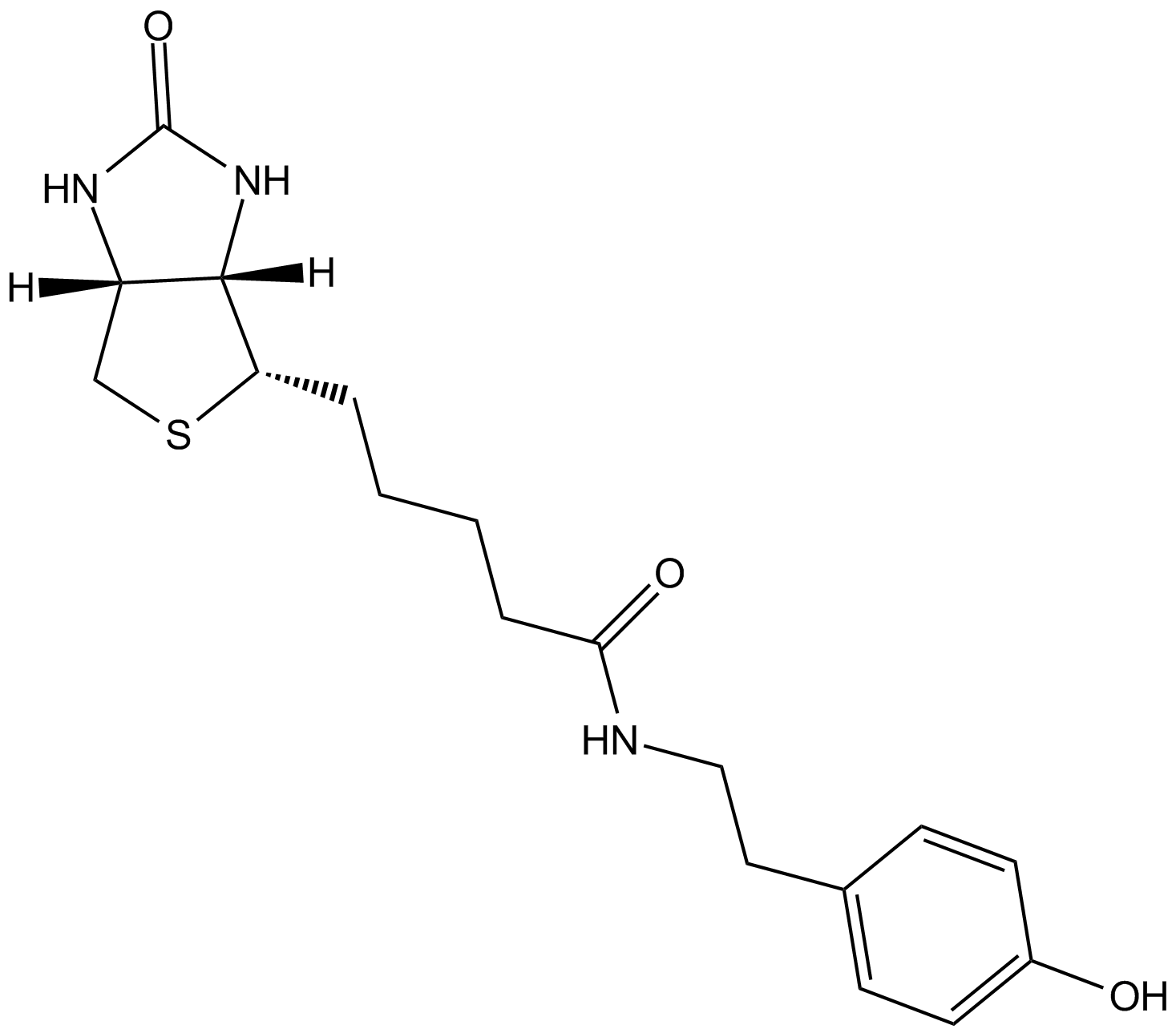
-
GC12033
Biotin-XX Tyramide Reagent
biotin-LC-LC-tyramide
Biotin-XX Tyramide Reagent is a biotin derivative for enzyme-mediated tyramide signal amplification (TSA).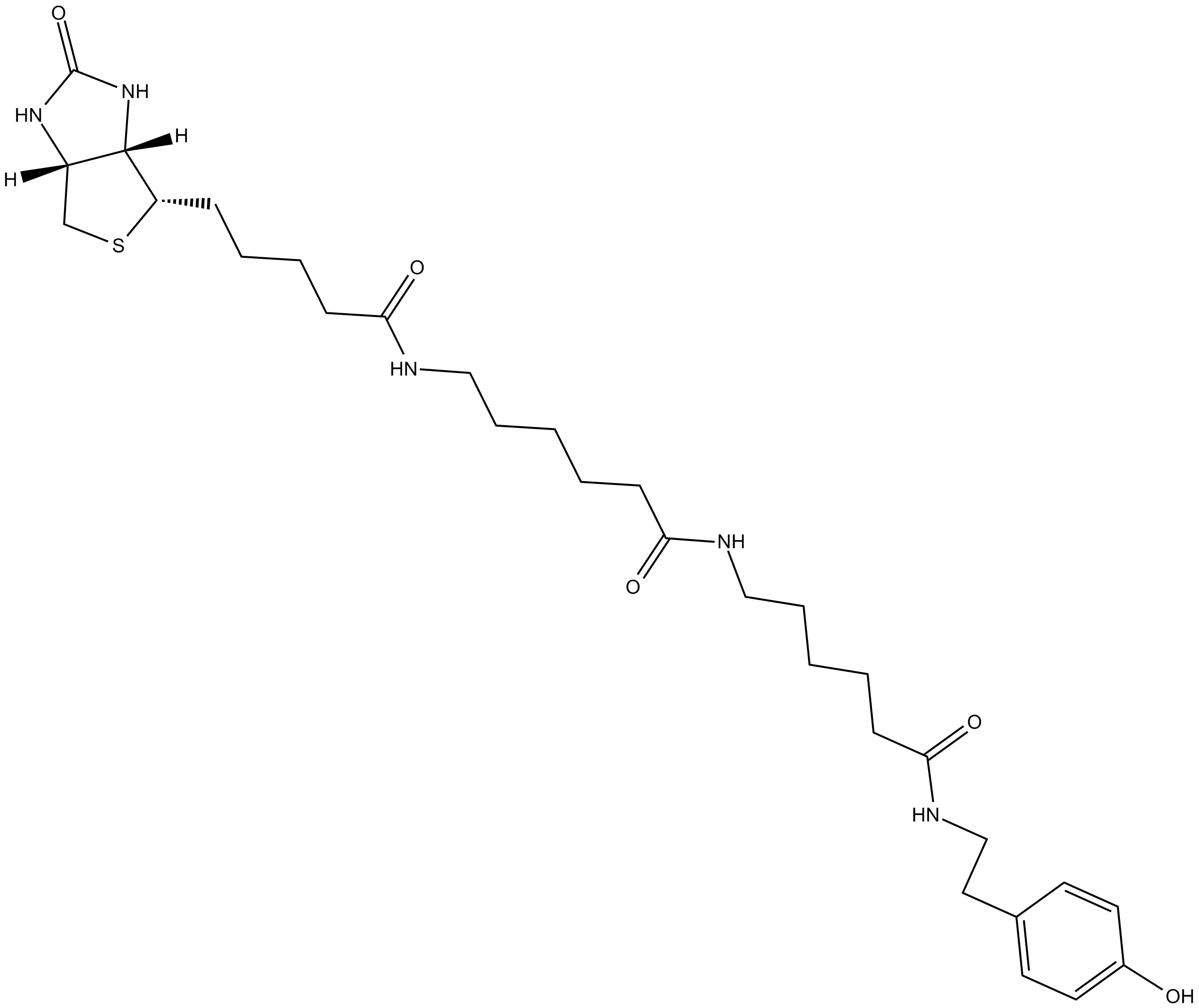
-
GC74378
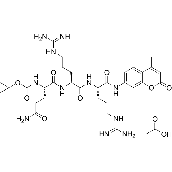
Boc-Gln-Arg-Arg-AMC acetate
Boc-Gln-Arg-Arg-AMC acetate is a fluorogenic substrate for the determination of protease activity.
-
GC68792
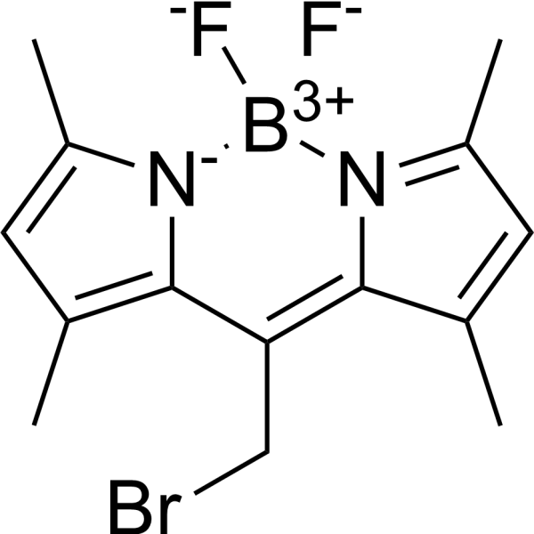
BODIPY 493/503 methyl bromide
BODIPY 493/503 methyl bromide is a fluorescent fatty acid analog with maximum excitation/emission wavelengths at 493/503nm.
-
GC26173
BODIPY 500/510 C1, C12
BODIPY 500/510 C1, C12 is a fluorescent fatty acid analog with maximum excitation/emission wavelengths at 500/510nm.

-
GC27791
BODIPY 505/515-8-C3-COOH
BODIPY 505/515-8-C3-COOH is a green fluorescing derivative, as a fluorescent dye for imaging lipid droplets in nannochloropsis. BODIPY 505/515-8-C3-COOH can be used for the research of flow cytometric high-throughput screening and cell sorting.
-
GC19993
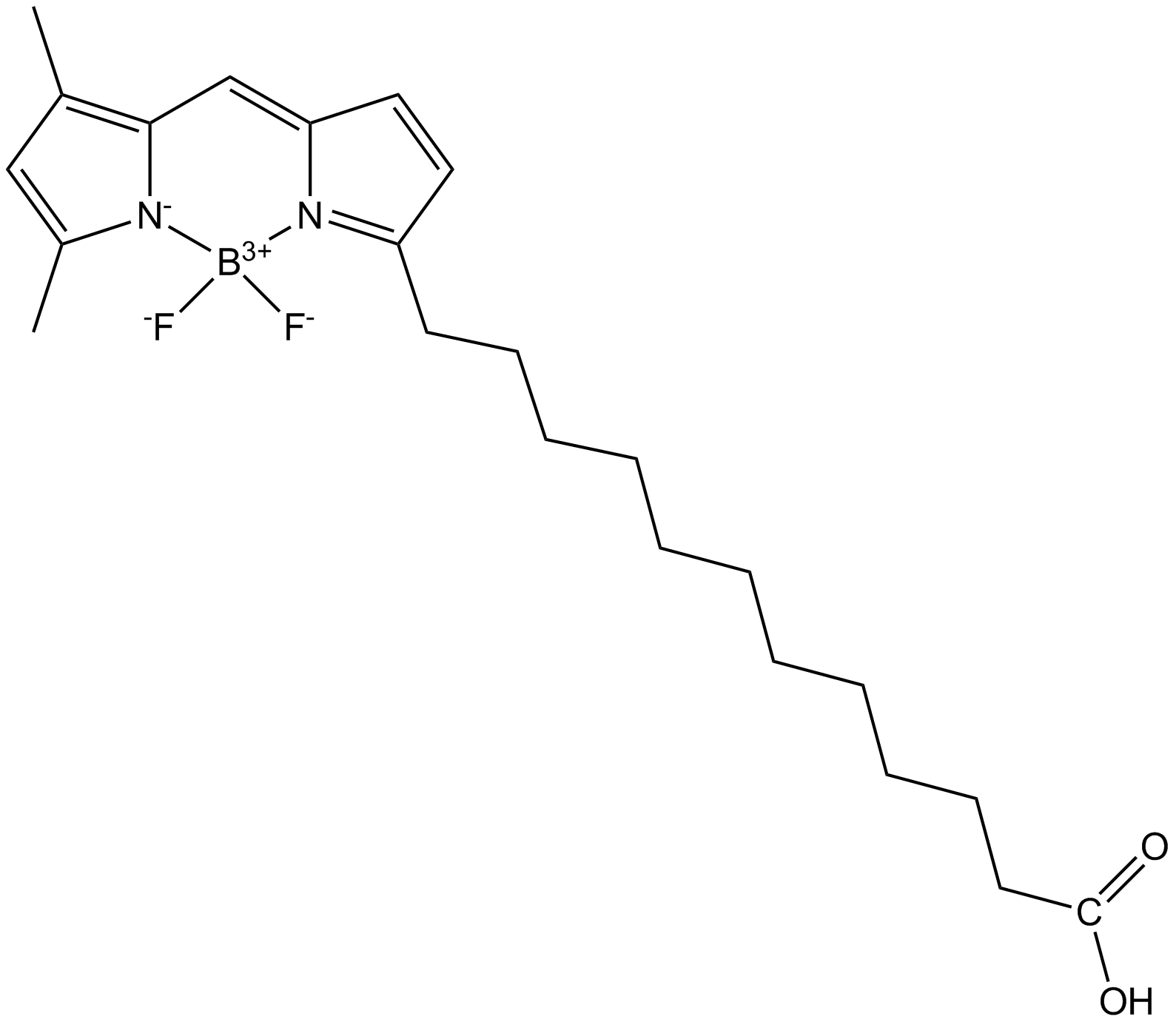
BODIPY FL C12
BODIPY FL C12 is a long-chain fatty acid analogue with a strong fluorescent group, and can be used as an effective tracer for lipid transport (Ex=485nm; Em=525nm).
-
GC68793
BODIPY FL C5-Ceramide
BODIPY FL C5-Ceramide is a Golgi-specific green fluorescent dye, with Ex/Em=505nm/512nm.

-
GC74294
Bovine Sermu Albumin-Cy5.5
BSA-Cy5.5
Bovine Sermu Albumin-Cy5.5 is Cy5.5-labeled Bovine Sermu Albumin (BSA).

-
GC26406
BSA-FITC
BSA-FITC is a fluorescently labeled protein conjugate. BSA-FITC retains the protein-binding properties of BSA while providing a strong green fluorescent signal, with an excitation wavelength of 490-495nm and an emission wavelength of 515-520nm.
-
GC74201
C.I. Disperse blue 79
C.I. Disperse blue 79 is a blue dye commonly used for fiber dyeing.

-
GC26327
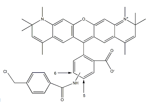
Cell-Tracker Red CMTPX
CellTracker Red CMTPX Dye
Cell Tracker fluorescent probes are excellent tools for monitoring cell movement, localization, proliferation, migration, chemotaxis, and invasion.
-
GC20191
CellTracker Blue CMAC
CellTracker Blue CMAC is a live cell tracking probe (Ex/Em: 353/466nm) that emits bright blue fluorescence.

-
GC74209
CH1055 triethylamine
CH1055 trietlamine is a NIR-II fluorophore based on a synthetic 970-Da organic molecule.

-
GC73290
Copper Fluor-4
CF4
Copper Fluor-4 (CF4) is a Cu+-specific fluorescent probe based on a rhodol dye scaffold.
-
GC74215
CQ-Lyso
CQ-Lyso is a lysosome-targeting chromenoquinoline.

-
GC26723
Cy3-cholesterol
Cyanine3-Cholesterol; Cy3-CLS
Cy3-cholesterol (λex = 554nm, λem = 568nm) is an orange-fluorescent cholesterol labeled with Cyanine3, which has cholesterol-like binding properties.
-
GC26496
CY3.5-Dextran
-
GC26899
CY5-Dextran (MW60-80k)
CY5-Dextran is a fluorescent dye CY5- labeled Dextran. CY5 exhibits a maximal excitation wavelength of 633 nm, and a maximal emission wavelength of 670 nm.

-
GC74203
Cyanine 3.18 TEA
Cyanine 3.18 TEA belongs to the cyanine dye series and is a common fluorescent marker for biomolecules that can interact with biomolecules.

-
GC74207
Cyanine3 amine TFA
Cyanine3 amine (TFA), an analog of Cyanine3 amine, is a potent green fluorescent dye.

-
GC61840
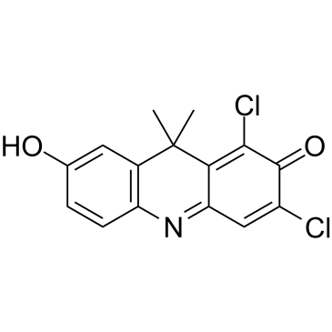
DDAO
DDAO is a near-infrared (NIR) red fluorescent probe with tunable excitation wavelength (600-650nm) and long emission wavelength (λem=656nm) and a relatively low pKa (~5).
-
GD11069
DEAE-dextran (MW 500000)

-
GC74660
DFAME
DFAME is a red fluorophore (Ex=508 nm, Em=641 nm).

-
GC71754
Dibenzocyclooctyne-Cy5.5
Dibenzocyclooctyne-Cy5.5 is a click chemiluminescent probe formed by coupling Dibenzocyclooctyne (DBCO) with Cy5.5 (Ex=673nm, Em=707nm) near-infrared fluorescent dye.

-
GC26813
DQ-BSA-RED
DQ-BSA-RED is a red fluorescent lysosomal activity molecular probe based on bovine serum albumin (BSA). After being degraded by lysosomes, DQ-BSA-RED emits bright red fluorescence (excitation/emission wavelengths approximately 590/620nm).

-
GC74299
FAM-SAMS TFA
FAM-SAMS TFA is a 5-FAM labeled SAMS.

-
GC66319
Fast Red Violet LB
Fast Red Violet LB is an azo dye that can be used to detect tartrate-resistant acid phosphatase (TRAP) and alkaline phosphatase (ALP).

-
GC20134
FeRhoNox-1(Fe2+ indicaton)
FeRhoNox-1(Fe2+ indicaton) is a highly sensitive red fluorescent probe for detecting ferrous ion levels in living cells.
-
GC73909
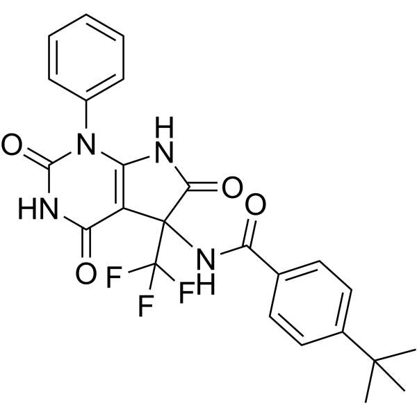
Firefly luciferase-IN-3
Firefly luciferase-IN-3 (CID: 17087944) is an inhibitor of ATP-dependent luciferase (Firefly luciferase) and has nM-level inhibitory activity against NanoLuc (pIC50=7.5).
-
GC73804
Firefly luciferase-IN-4
Firefly luciferase-IN-4 (CID: 3987260) is an inhibitor of ATP-dependent luciferase (Firefly luciferase).

-
GC73911
Firefly luciferase-IN-5
Firefly luciferase-IN-5 (CID: 16031203) is an inhibitor of ATP-dependent luciferase (Firefly luciferase).

-
GC12317
FITC, Fluorescein isothiocyanate
5-FITC, FITC isomer I, 5-Isothiocyanatofluorescein
FITC (Fluorescein isothiocyanate) is the most commonly used green fluorescent dye derivative in biological research and is widely used in flow cytometry experiments to stain proteins, cell nuclei and subnuclear particles. The maximum excitation/emission wavelengths of FITC are 495/519 nm respectively.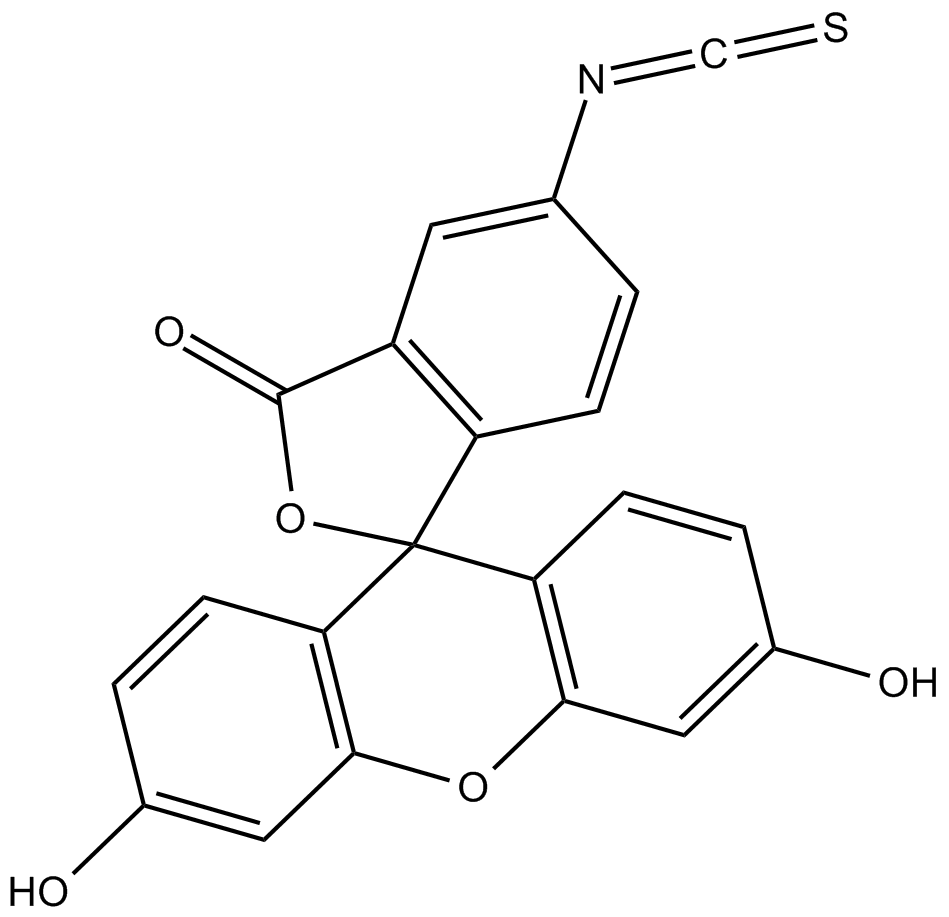
-
GC26553
FITC-Alginate
FITC-Alginate is a fluorescently labeled polysaccharide material derived from sodium alginate (Ex/Em=488/525nm).

-
GC36048
FITC-Dextran (MW 10000)
FITC-Dextran (MW 10000) is a fluorescent probe (Ex=495nm; Em=525nm) that serves as a measure of blood-brain barrier permeability.

-
GC26141
FITC-Dextran (MW 20000)
FITC-Dextran (MW 20000) is a compound belonging to the class of fluorescent dyes.
-
GC26248
FITC-Dextran (MW 2000000)
FITC-Dextran (MW 2000000) is a high-molecular-weight fluorescent tracer composed of fluorescein isothiocyanate (FITC) and dextran, with an Ex/Em of approximately 490/520nm.
-
GC26412
FITC-Dextran (MW 3000-5000)
FITC-Dextran (MW 3000-5000) is a fluorescent probe for fluorescein isothiocyanate (FITC) dextran (Ex=495 nm; Em=525 nm).

-
GC18871
FITC-Dextran (MW 4000)
FITC-Dextran (MW 4000) (FD4) is a large molecule of dextran (MW ~4000) attached to a fluorescein isothiocyanate tag, which makes it fluorescent (Ex=495 nm; Em=525 nm).
-
GC19938
FITC-Dextran (MW 40000)
FITC-Dextran
-
GC20084
FITC-dextran (MW 500,000)
Fluorescein isothiocyanate-dextran 500;FITC-dextran 500
-
GC19939
FITC-Dextran (MW 70000)
FITC-Dextran
FITC-Dextran (MW 70000) is a fluorescein isothiocyanate (FITC) dextran fluorescent probe with Ex/Em=495nm/525nm.
-
GC74223
Flipper-TR probe
Flipper-TR probe (Compound FliptR probe) is a fluorescent probe specifically designed to measure cell membrane tension.

-
GC30141
Fluorescein
Uranine
Fluorescein is a low-molecular-weight synthetic dye that absorbs blue light at 470–490nm and fluoresces green-yellow at 520–530nm.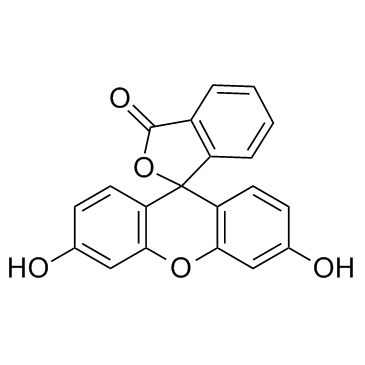
-
GC50456
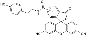
Fluorescein Tyramide
Fluorescein Tyramide is a green fluorescent labeling reagent (λabs: 494nm; λem: 517nm).
-
GC67896
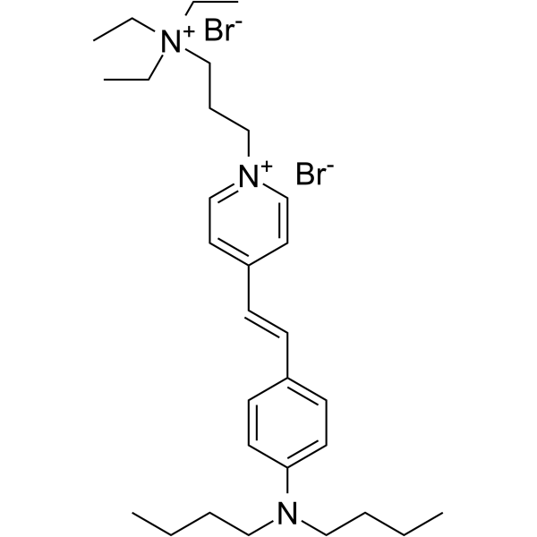
FM1-43
FM1-43 is a water-soluble styryl dye with strong lipophilicity, and FM1-43 is widely used as a fluorescent membrane probe in biological research..
-
GC74283
Griffonia Simplicifolia Lectin I Isolectin B4 (Fluorescein)
GSL I Isolectin B4 (Fluorescein
Griffonia Simplicifolia Lectin I Isolectin B4 (GSL I Isolectin B4) Fluorescein is a plant lectin that can be used as a probe to specifically bind biomolecules (such as polysaccharides, peptides, etc.

-
GA22320
H-D-Val-Leu-Lys-pNA . 2 HCl
D-Val-Leu-Lys-p-nitroanilide, D-VLK-pNA, S 2251
H-D-Val-Leu-Lys-pNA . 2 HCl is a colorimetric substrate for the detection of protease activity, particularly plasmin.
-
GA22334
H-Gln-AMC
H-Gln-AMC is a fluorogenic substrate commenly used to measure the activity of glutaminyl cyclase (QC) enzymes with Km value of 51 ± 3µM at pH 8.0 and kcat value of 5.4 ± 0.1s-1.
-
GC74225
HBmito Crimson
HBmito Crimson is a deep red fluorescent probe (λex: 658 nm, λem: 678 nm) for the inner mitochondrial membrane.

-
GC66439
HMBR
HMBR is a non-fluorescent, cell-permeable fluorogenic dye that binds to the Yellow Fluorescence-Activating and Absorption-Shifting Tag (Y-FAST) and emits yellow fluorescence upon blue light excitation, with Ex/Em of 491/525-539nm.

-
GC74726
ICG-azide
ICG-azide, a near infrared (NIR) fluorescent dye, is a copper (I)-catalyzed azide-alkyne cycloaddition (CuAAC) reagent.

-
GC63438
ICG-carboxylic acid
ICG-carboxylic acid is a near-infrared (NIR) fluorescent probe based on indocyanine green (ICG), characterized by good water solubility, low toxicity, and stability.

-
GC43908
IraZolve-Mito
IraZolve-Mito is a fluorescent probe that can be used to label mitochondria in live cells and tissue sections.

-
GC71797
IRDye 700DX
IRDye 700DX is a near-infrared (NIR) photosensitive dye with a maximum absorption wavelength of 689nm and an emission maximum of 702nm.

-
GC72967
KMG-301AM TFA
KMG-301AM TFA is the acetoxy metl esterified form of KMG-301.

-
GC74284
Lens Culinaris Agglutinin (Biotinylated)
LCA (Biotinylated
Lens Culinaris Agglutinin (LCA) Biotinylated is a plant lectin that can be used as a probe to specifically bind biomolecules (such as polysaccharides, peptides, etc.
-
GC73803
Luciferase-IN-3
Luciferase-IN-3 (compound 5h) is an inhibitor of ATP-dependent luciferase (Firefly luciferase), the major light-emitting enzyme in fireflies and kowtow bugs.

-
GC19882
Lyso-Tracker Red (solution)
LysoTracker Red DND-99
Lyso-Tracker Red is an eosinophilic red fluorescent probe that can penetrate the cell membrane at neutral pH and can be used for lysosome-specific fluorescent staining of living cells and tracing of acidic organelles. The maximum excitation and emission wavelengths of Lyso-Tracker Red are 577/590 nm.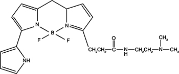
-
GC26409
LysoTracker Blue DND-22
LysoTracker Blue DND-22; LysoTracker Red DND-99; Lysotracker Green
LysoTracker Blue DND-22 is a fluorescent dye (Ex/Em=380/420nm), which targets acidic lysosomes.
-
GC27019
LysoTracker Deep Red
Lysotracker probes are acidotropic fluorescent probes used to label and track acidic organelles within living cells. These probes possess several key features, including highly selective targeting of acidic organelles and efficient labeling of live cells at nanomolar concentrations.
-
GC20116
LysoTracker Green DND-26
LysoTracker Green DND-26 is an acidotropic green fluorescent probe that can be used to label and track lysosomes in living cells, with maximum excitation/emission wavelengths of 504 nm/511 nm. LysoTracker Green is commonly used for live-cell lysosomal staining due to its selective accumulation in acidic compartments.
-
GC74285
Maackia Amurensis Lectin II (Biotinylated)
MAL II (Biotinylated
Maackia Amurensis Lectin II (MAL II) Biotinylated is a plant lectin that can be used as a probe to specifically bind biomolecules (such as polysaccharides, peptides, etc.
-
GC26256
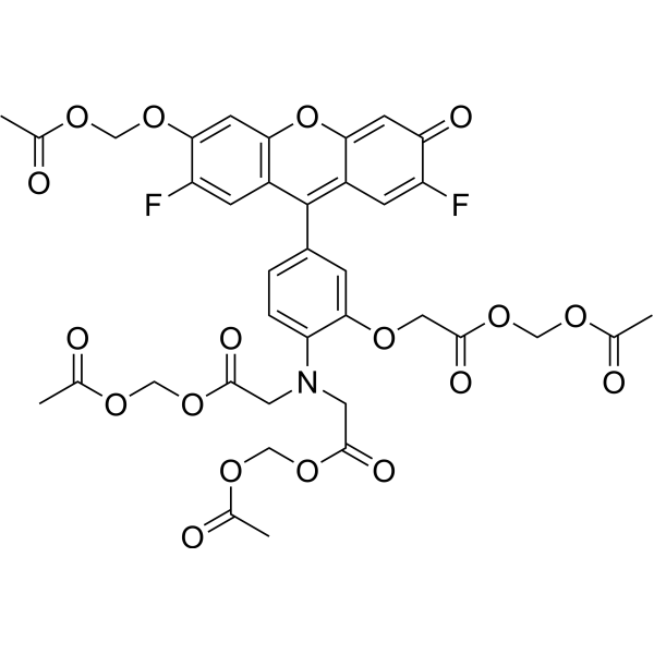
Mag-Fluo-4 AM
Mag-Fluo-4 AM is a cell membrane-permeable fluorescent probe with optimal excitation/emission wavelengths of 494/516nm.
-
GB30290
Mag-Fura-2 AM
Mag-Fura-2 AM is a fluorescent dye which is useful for measuring changes in cytosolic Mg2+ with Ex/Em=336/503nm.

-
GB59294
Merocyanin 540
Merocyanin 540 is a fluorescent membrane probe that selectively stains the membranes of various electrically excitable cells but not non-excitable cells. Its maximum excitation wavelength Ex is 540nm and its maximum emission wavelength Em is 580nm.

-
GC68203
MHI-148
MHI-148 is a near-infrared heptamethine cyanine dye with tumor-targeting properties (excitation/emission wavelengths: 740/950nm).

-
GC20139
Mito-Tracker Deep Red FM
Mito-Tracker Deep Red FM is a red fluorescent dye with an excitation/emission wavelength of 644/665 nm.
-
GC26229
Mito-Tracker Green
Mito-Tracker Green is a green fluorescent probe for mitochondria in living cells with a maximum excitation/emission wavelength of 490/516 nm.

-
GC71774
MitoTracker Orange CMTMRos
MitoTracker Orange CMTMRos is a mitochondrial-specific fluorescent dye probe (excitation/emission: 561nm / 580–620nm) that can penetrate the cell membrane and is commonly used for labeling and imaging mitochondria in living cells.

-
GC17832
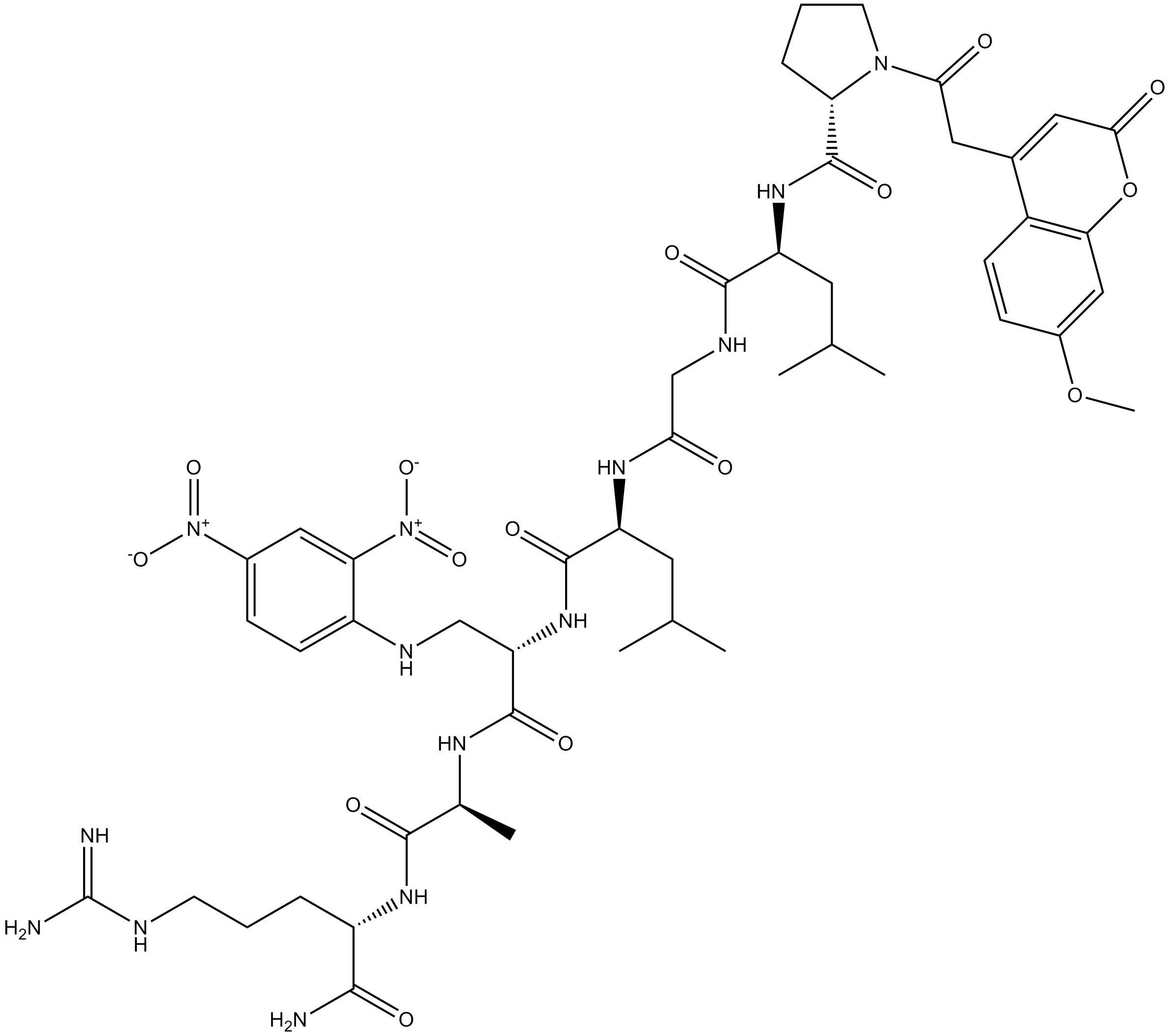
MOCAc-PLGL(Dpa)AR
MOCAc-PLGL(Dpa)AR is a fluorescent substrate specifically designed for the detection of matrix metalloproteinases (MMPs), which is suitable for the activity of MMP-2, MMP-7 and MMP-9.
-
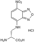
GC50534
NADA-green
NADA hydrochloride
NADA-green is a fluorescent D-amino acid probe suitable for labeling peptidoglycan in living bacteria, with maximum excitation/emission wavelengths of ~450/555nm.