- Cat.No. Nom du produit Informations
-
GC12913
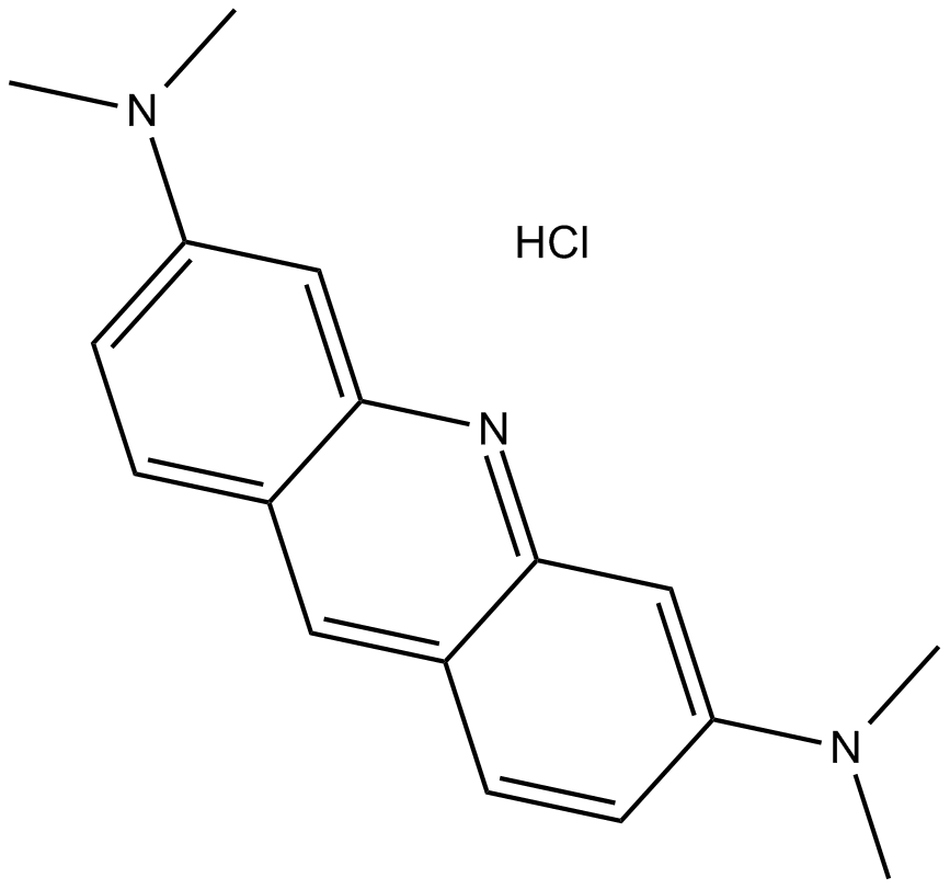
Acridine Orange hydrochloride
Cell and organelle membrane permeable nucleic acid binding dye
-
GC63515
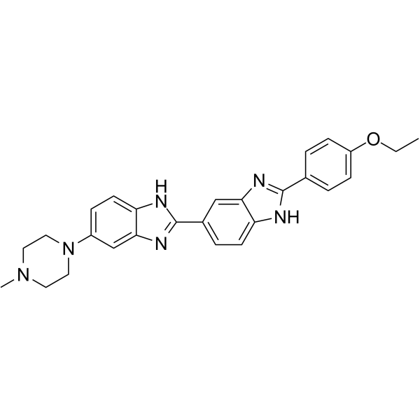
Hoechst 33342
The nucleic acid stain Hoechst 33342 (Ex/Em: 350/461 nm) is frequently utilized as a cell-permeable nuclear counterstain that emits a blue fluorescence upon binding to dsDNA.
-
GC36246
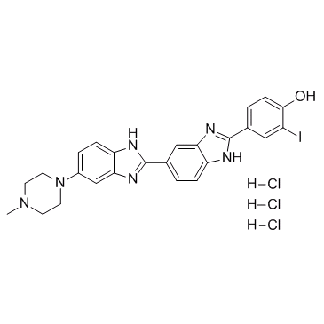
Hoechst 33342 analog 2 trihydrochloride
Le trichlorhydrate de l'analogue 2 de Hoechst 33342 est un anglo-saxon de Hoechst 33342.
-
GC36245
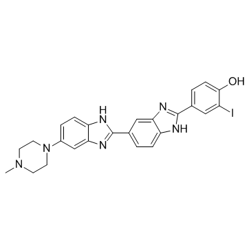
Hoechst 33342 analog 2
Hoechst 33342 analogue 2 est un anglo-saxon de Hoechst 33342, qui est un liant de sillon mineur d'ADN utilisé fluorochrome pour visualiser l'ADN cellulaire.
-
GC36239
Hoechst 33258 analog
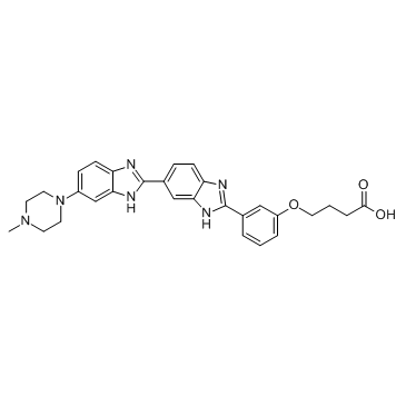
L'analogue Hoechst 33258 fait partie d'une famille de colorants fluorescents bleus utilisés pour colorer l'ADN.
-
GC10939
Hoechst 33342 trihydrochloride
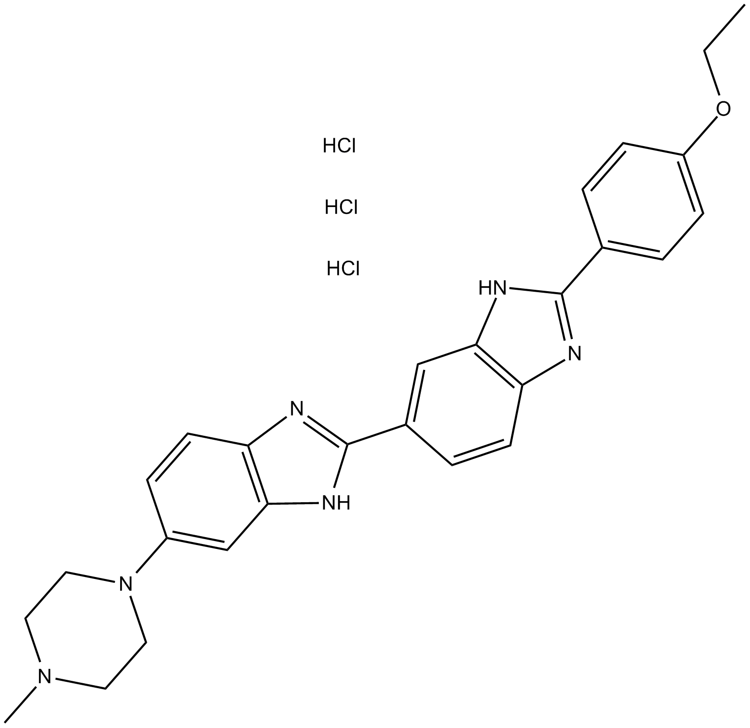
Hoechst 33342 analog 2 is an analog of Hoechst 33342 dye, used for fluorescent staining of cellular DNA. The excitation/emission light is 350/460nm respectively.
-
GC36850
para-iodoHoechst 33258
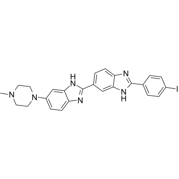
para-iodoHoechst 33258 fait partie d'une famille de colorants fluorescents bleus utilisés pour colorer l'ADN.
-
GC36817
ortho-iodoHoechst 33258
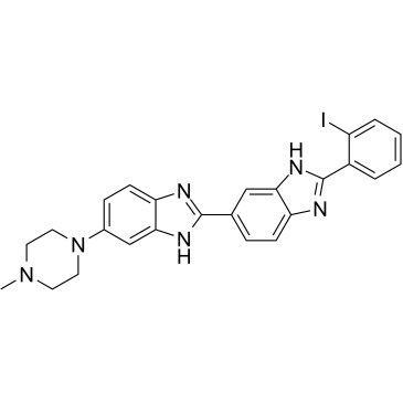
ortho-iodoHoechst 33258 fait partie d'une famille de colorants fluorescents bleus utilisés pour colorer l'ADN.
-
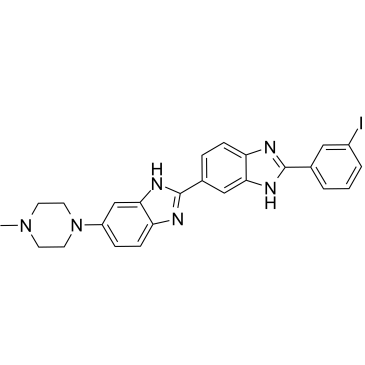
GC36586
meta-iodoHoechst 33258
Les colorants Hoechst font partie d'une famille de colorants fluorescents bleus utilisés pour colorer l'ADN.
-
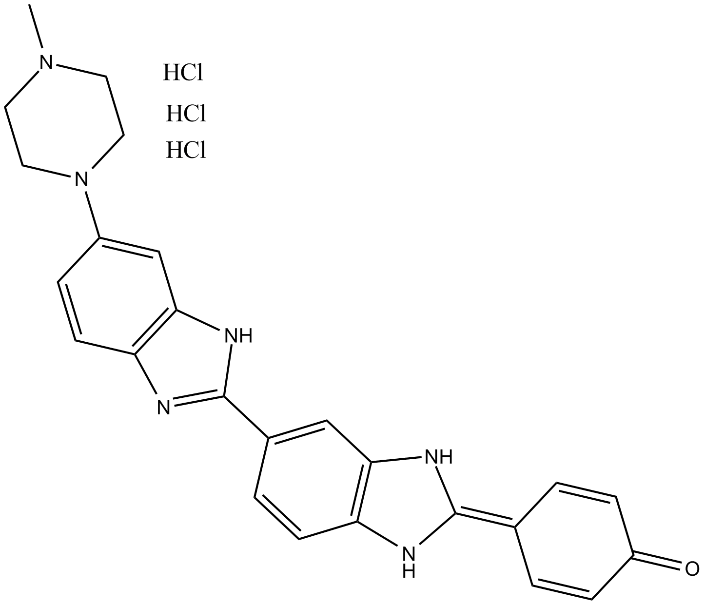
GC12187
Hoechst 33258 trihydrochloride
Hoechst 33258 trihydrochloride is an analog of the nucleic acid stain Hoechst 33258. It emits blue fluorescence after binding to dsDNA, with a maximum excitation/emission light of 365/458 nm.
-
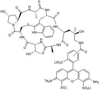
GC50523
Phalloidin-TFAX 488
Green fluorescent cytoskeletal stain; binds F-actin
-
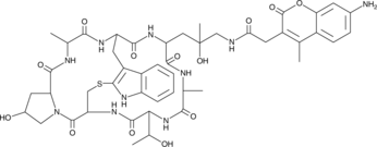
GC44618
Phalloidin-AMCA Conjugate
Phalloidin-AMCA conjugate is a blue fluorophore that specifically labels filamentous actin (F-actin).
-
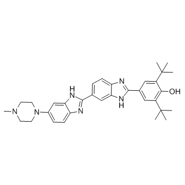
GC36243
Hoechst 33258 analog 6
Hoechst 33258 analogue 6 est un anglo-saxon des colorants Hoechst (Hoechst 33258), qui font partie d'une famille de colorants fluorescents bleus utilisés pour colorer l'ADN.
-
GC19997
FITC-Phalloidin
Phalloidin is a cyclic heptapeptide toxin derived from Amanita phalloides, which selectively binds to filamentous actin F-actin with high affinity (Kd=20nM).

-
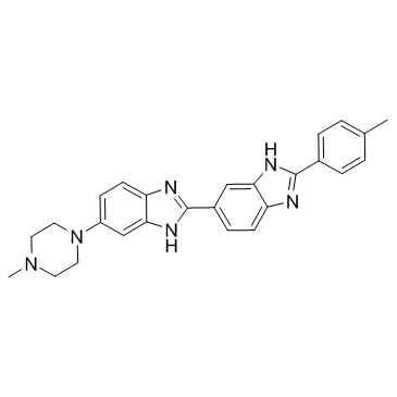
GC36241
Hoechst 33258 analog 3
Hoechst 33258 analogue 3 fait partie d'une famille de colorants fluorescents bleus utilisés pour colorer l'ADN.
-
GC18292
TRITC Phalloidin
TRITC is an orange-red fluorescent probe,Phalloidin, a cyclic heptapeptide toxin derived from Amanita phalloides,

-
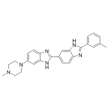
GC36240
Hoechst 33258 analog 2
Hoechst 33258 analogue 2 fait partie d'une famille de colorants fluorescents bleus utilisés pour colorer l'ADN.
-
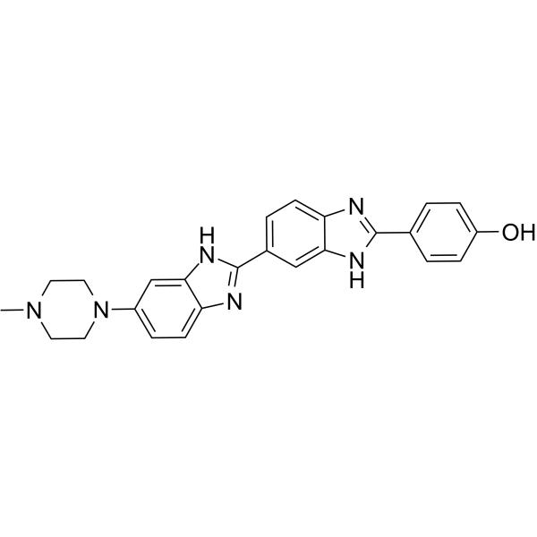
GC64380
Hoechst 33258
The nucleic acid stain Hoechst 33258 (Ex/Em: 352/461 nm) is frequently utilized as a cell-permeable nuclear counterstain that emits a blue fluorescence upon binding to dsDNA.
-
GC62873
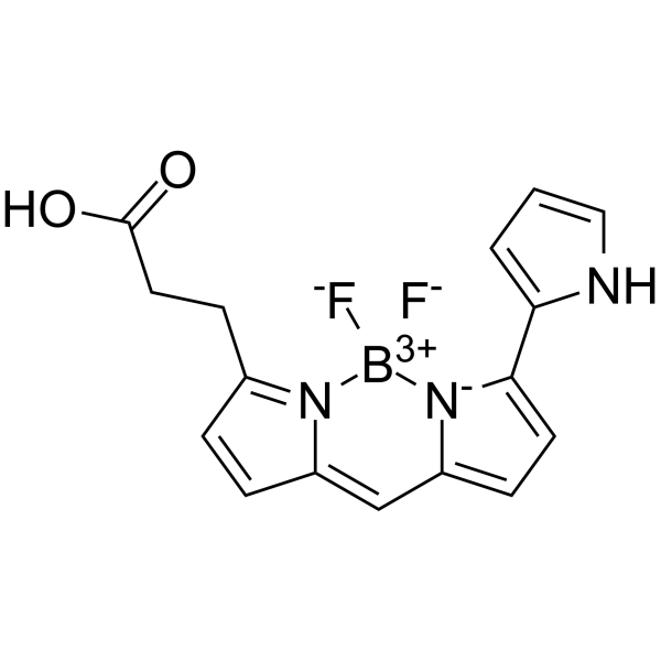
BODIPY 576/589
BODIPY 576/589 est un colorant biologique marqué À grande longueur d'onde.
-
GC45796
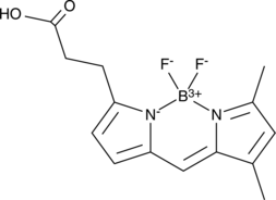
BODIPY 503/512
A lipophilic, amine-reactive fluorescent probe
-
GC45571
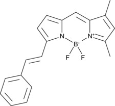
STY-BODIPY
A fluorogenic probe for radical-trapping antioxidant activity
-
GC44574
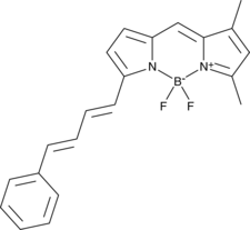
PBD-BODIPY
PBD-BODIPY is a probe for the spectrophotometric measurement of autoxidation reactions.
-
GC42961
BODIPY 558/568 C12
BODIPY 558/568 C12 is a fatty acid-conjugated fluorescent probe for lipid droplets.

-
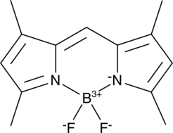
GC42960
BODIPY 505/515
BODIPY 505/515, a lipophilic fluorescence dye, emits fluorescence has been used extensively for lipid droplet labeling (Ex/Em: 505/515 nm).
-
GC42959
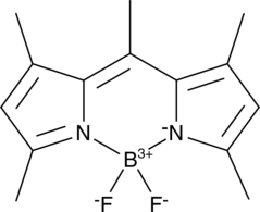
BODIPY 493/503
BODIPY 493/503, a lipophilic fluorescence dye, emits bright green fluorescence has been used extensively for lipid droplet labeling (Ex/Em: 493/503 nm).
-
GC40165
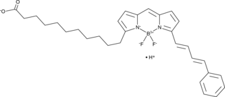
C11 BODIPY 581/591
C11-BODIPY581/591 is an oxidation-sensitive fluorescent fatty acid analogue
-
GC15539
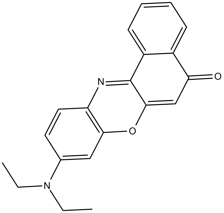
Nile Red
Nile red is a common lipid dye.
-
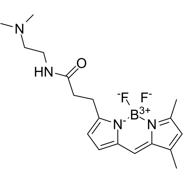
GC64677
Green DND-26
Le vert DND-26 est utilisé pour marquer les lysosomes acides afin de déterminer la distribution cellulaire.
-
GK10021
Deacetylase Inhibitor Cocktail (100× in 70% DMSO)
Deacetylase Inhibitor Cocktail is a synergistic combination of chemicals designed to preserve the acetylation state of proteins.

-
GC20101
Lyso-Tracker Green

-
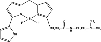
GC19882
Lyso-Tracker Red
Lysosomal red fluorescent probe
-
GC68628
Aflibercept
Aflibercept (VEGF Trap) is a soluble decoy VEGFR composed of Ig domains of VEGFR1 and VEGFR2 fused to the Fc domain of human IgG1. Aflibercept inhibits the VEGF signaling pathway by blocking pathways regulated by VEGF. Aflibercept can be used in research for age-related macular degeneration (AMD) and cardiovascular diseases.

-
GC30132
Thioflavine S (Direct Yellow 7)
La thioflavine S (Direct Yellow 7) est un marqueur histochimique fluorescent des plaques séniles À noyau dense.

-
GC19476
Tetramethylrhodamine methyl ester (perchlorate)
Non-cytotoxic cell-permeant

-
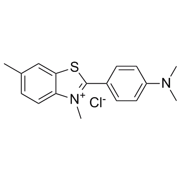
GC30031
Thioflavin T (Basic Yellow 1)
A fluorescent probe for amyloid fibrils
-
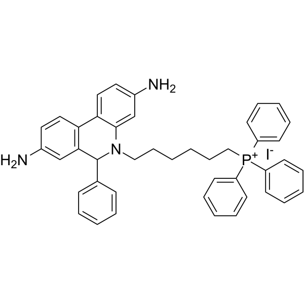
GC68230
MitoSOX Red
MitoSOX Red is a live cell fluorescent probe targeting mitochondria with maximum excitation/emission light of 510/580 nm.
-
GC10894
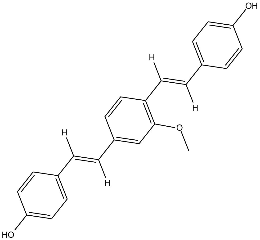
Methoxy-X04
Fluorescent amyloid β (Aβ) probe
-
GC50454
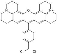
MitoMark Red I
MitoMark Red I est un marqueur mitochondrial fluorescent.
-
GC50453
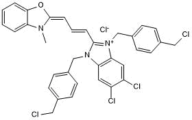
MitoMark Green I
Green fluorescent mitochondrial stain; cell permeable
-
GC64788
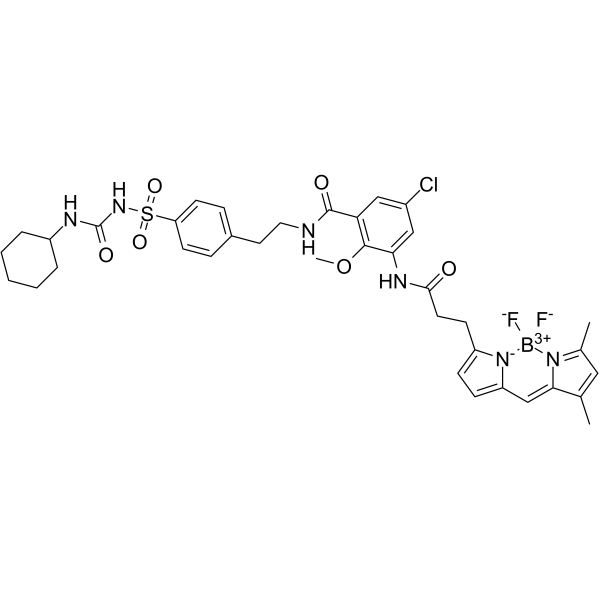
ER-Tracker Green
ER-Tracker Green est un colorant fluorescent spécifique pour le réticulum endoplasmique.
-
GC38730
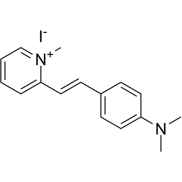
2-Di-1-ASP
Le 2-Di-1-ASP (composé 18a) est un colorant mono-stryryle et largement utilisé comme colorant mitochondrial et sondes fluorescentes de liaison au sillon pour l'ADN double brin.
-
GC30591
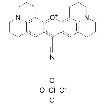
Rhodamine 800
La rhodamine 800 est un colorant fluorescent dans le proche infrarouge.
-
GC33493
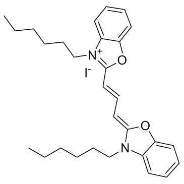
3,3'-Dihexyloxacarbocyanine iodide (DiOC6(3) iodide)
A fluorescent dye
-
GB30168
ER-Tracker Red

-
GC66508
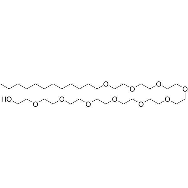
Decaethylene glycol dodecyl ether
L'éther dodécylique de décaéthylène glycol est un lieur PROTAC à base de PEG qui peut être utilisé dans la synthèse des PROTAC.
-
GC43096
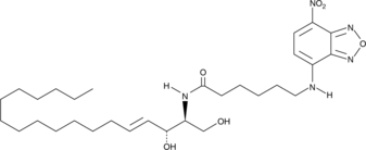
C6 NBD Ceramide (d18:1/6:0)
A biologically active, fluorescent analog of ceramide
-
GC30553
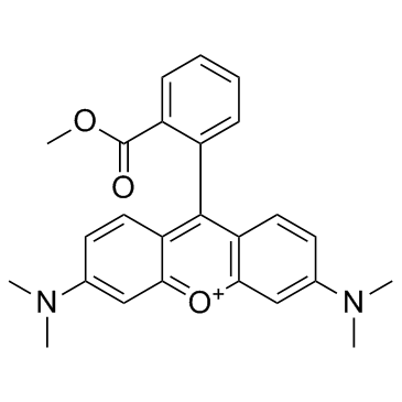
TMRM
Le TMRM est un colorant fluorescent rouge lipophile cationique pénétrant dans les cellules (Λex = 530 nm, Λem = 592 nm).
-
GC19881
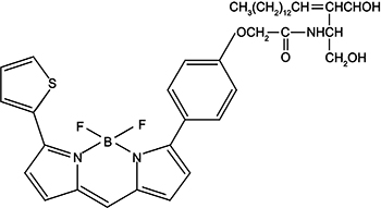
Golgi-Tracker Red
Golgi red fluorescent probe, one of the sphingolipid fluorescent probes, which can be used for Golgi-specific fluorescent staining of living cells.
-
GC12048
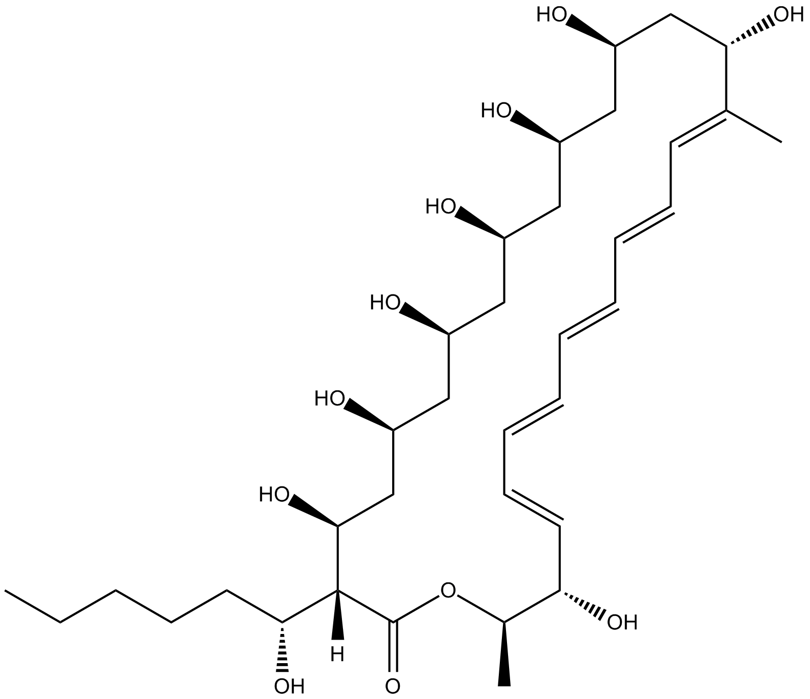
Filipin III
Fluorescing antibiotic used for the detection of lipoproteins
-
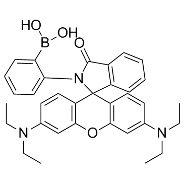
GC30265
ATP-Red 1
ATP-Red 1 est une sonde fluorescente commutable À liaison multisite et peut répondre sélectivement et rapidement aux concentrations intracellulaires d'ATP dans les cellules vivantes.
-
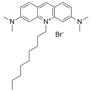
GC30183
Acridine Orange 10-Nonyl Bromide (Nonylacridine orange)
A mitochondrial dye
-
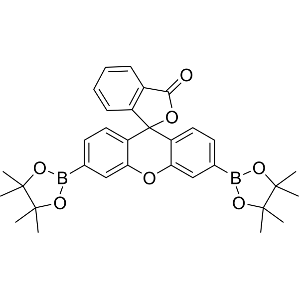
GC69689
Peroxyfluor 1
Peroxyfluor 1 est une sonde H2O2 qui peut pénétrer dans les cellules. Peroxyfluor 1 représente la première génération de sondes fluorescentes vertes.
-
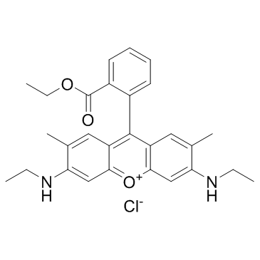
GC30090
Rhodamine 6G (Basic Red 1)
La rhodamine 6G (rouge basique 1) est un analogue de la rhodamine utile dans les tests d'efflux Pgp. Elle peut être utilisée pour caractériser la cinétique de l'efflux médié par MRP1, ainsi que comme colorant laser et sonde mitochondriale potentielle.
-
GC64022
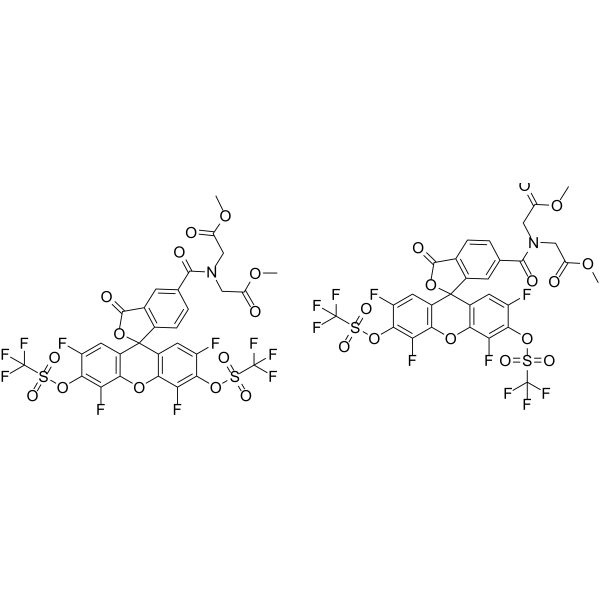
HKSOX-1r (5/6-mixture)
HKSOX-1r (mélange 5/6) est une sonde fluorescente utilisée pour l'imagerie et la détection du superoxyde endogène dans les cellules vivantes et in vivo.
-
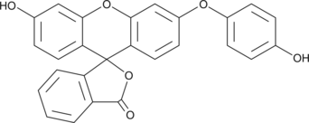
GC43870
HPF
A cell permeable, fluorescent dye for detection of highly reactive oxygen species
-
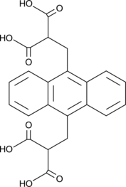
GC42642
9,10-Anthracenediyl-bis(methylene)dimalonic Acid
A singlet oxygen acceptor
-
GC62503
Fengycin
La fengycine est un lipopeptide cyclique utilisé comme fongicide agricole.

-
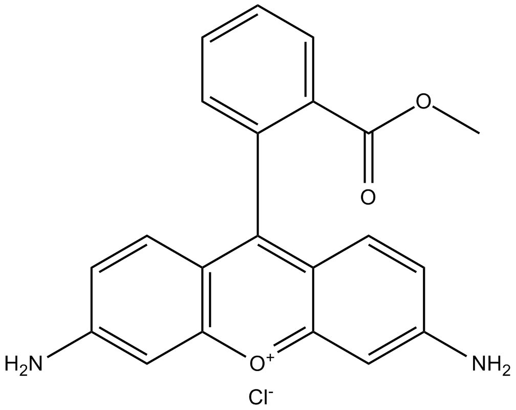
GC15795
Rhodamine 123 (chloride)
Rhodamine 123 is a cell-permeable cationic fluorescent probe that specifically recognizes mitochondrial membrane potential. Its maximum excitation/emission light is 507/529nm.
-
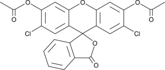
GC42079
2',7'-Dichlorofluorescein diacetate
A cell-permeable fluorogenic probe to quantify ROS and NO
-
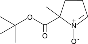
GC41397
BMPO
BMPO is a cyclic nitrone spin trap agent, it is a water-soluble white solid which makes BMPO purification easier than other spin trap agents.
-
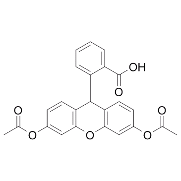
GC33512
Dihydrofluorescein diacetate
Le diacétate de dihydrofluorescéine est une sonde fluorimétrique principalement utilisée pour les mesures de stress oxydatif, À la fois dans les systèmes acellulaires et les modèles cellulaires.
-
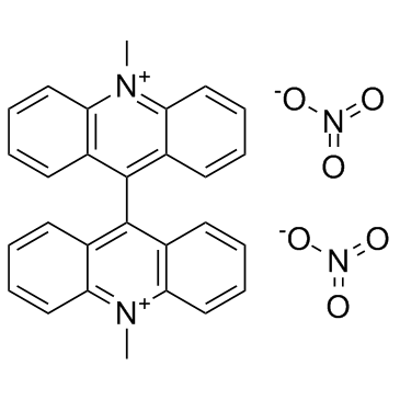
GC33485
Lucigenin (NSC-151912)
A chemiluminescent probe
-
GC30581
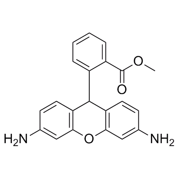
Dihydrorhodamine 123 (DHR 123)
Dihydrorhodamine 123 (DHR 123) is a fluorescent probe primarily used for the detection of reactive oxygen species (ROS). It has an excitation wavelength (λex) of 485 nm and an emission wavelength (λem) of 535 nm.
-
GC30025
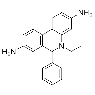
Dihydroethidium (Hydroethidine)
Dihydroethdium(Hydroethidine) (DHE) oxidation is commonly used as a method for monitoring cellular production of "reactive oxygen species (ROS)".
-
GB57947
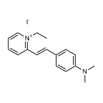
DASPEI
-
GC30006
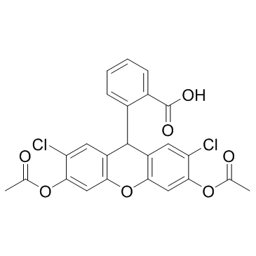
H2DCFDA (DCFH-DA)
H2DCFDA(DCFH-DA) is a redox-sensitive fluorescent probe, which could be used to measure intracellular reactive oxygen species levels
-
GC43372
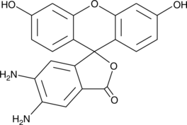
DAF-2
A fluorescent probe used for the detection of NO
-
GC33465
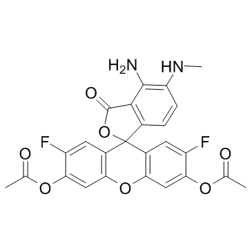
DAF-FM DA (Diaminofluorescein-FM diacetate)
A fluorescent probe used for the detection of NO
-
GC30702
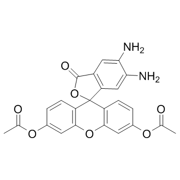
DAF 2DA (DAF-2DA)
A cellpermeable fluorescent probe for NO
-
GC47346
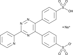
Ferrozine
Ferrozine reacts with divalent iron to form a stable magenta complex species and used for the direct determination of iron in water with maximum absorbance at 562 nm.
-
GC44832
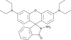
Rhodamine B hydrazide
A fluorescent probe
-
GC20174
Mito-FerroGreen

-
GC20134
FeRhoNox-1(Fe2+ indicaton )
FeRhoNox-1 is a highly sensitive red fluorescent probe used to detect the level of ferrous ions in living cells. The maximum excitation wavelength and emission wavelength after binding ferrous ions are 540/575 nm respectively.

-
GC60434
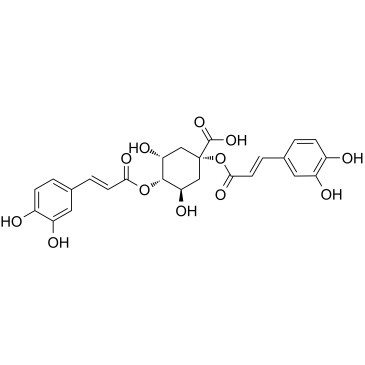
1,4-Dicaffeoylquinic acid
L'acide 1,4-dicaffeoylquinique (1,4-DCQA) est un phénylpropanoÏde de Xanthii fructus, inhibe la production de TNF-α stimulée par le LPS.
-
GC67223
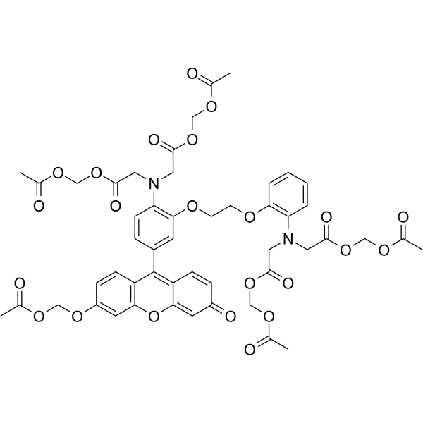
Fluo-8 AM
Fluo-8 AM est une sonde fluorescente de calcium qui peut être chargée dans des protoplastes pour détecter le calcium dans les cellules des tissus de chair de Malus domestica.
-
GC43116
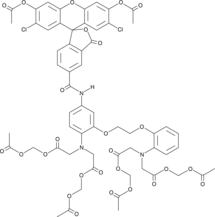
Cal Green™ 1 AM
A cell-permeant fluorescent calcium indicator
-
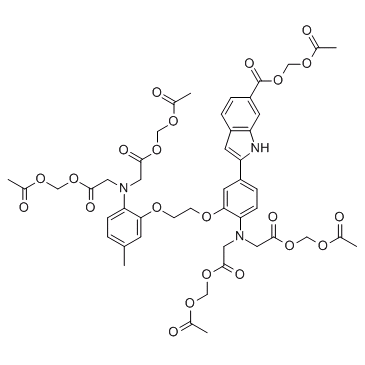
GC33592
Indo-1 AM (Indo-1 Acetoxymethyl ester)
A cell-permeant precursor of Indo-1
-
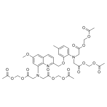
GC33587
Quin-2AM (Quin-2 acetoxymethyl ester)
A cell-permeant precursor of quin-2
-
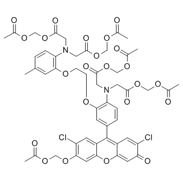
GC33437
Fluo-3AM
Fluo-3AM is one of the most commonly used fluorescent probes to detect intracellular calcium ion concentration.
-
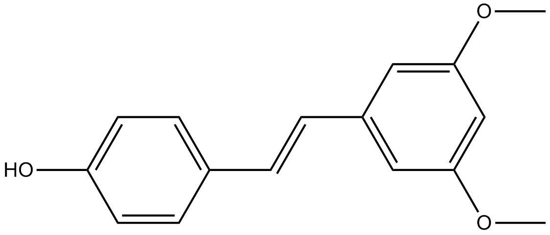
GN10244
Pterostilbene
-
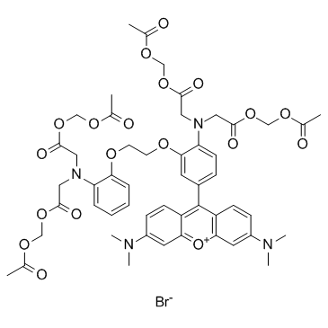
GC30506
Rhod-2 AM
Rhod-2 AM is an acetyl methyl derivative of Rhod-2. Rhod-2 AM can penetrate cell membranes and produce non-permeable Rhod-2 through its lactase cleavage, which can be used to detect Ca2+ levels in cells or tissues. The Ex is 552nm and the Em is 581nm.
-
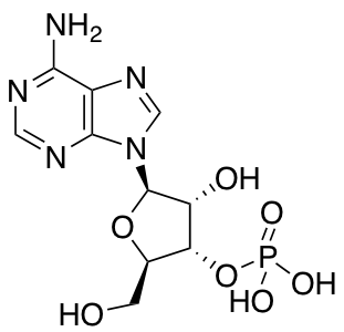
GD01654
Adenosine 3′-monophosphate
-
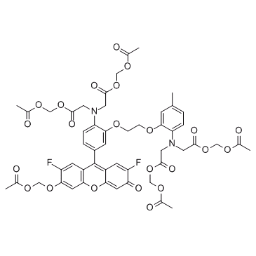
GC30231
Fluo-4 AM
A fluorescent calcium indicator
-
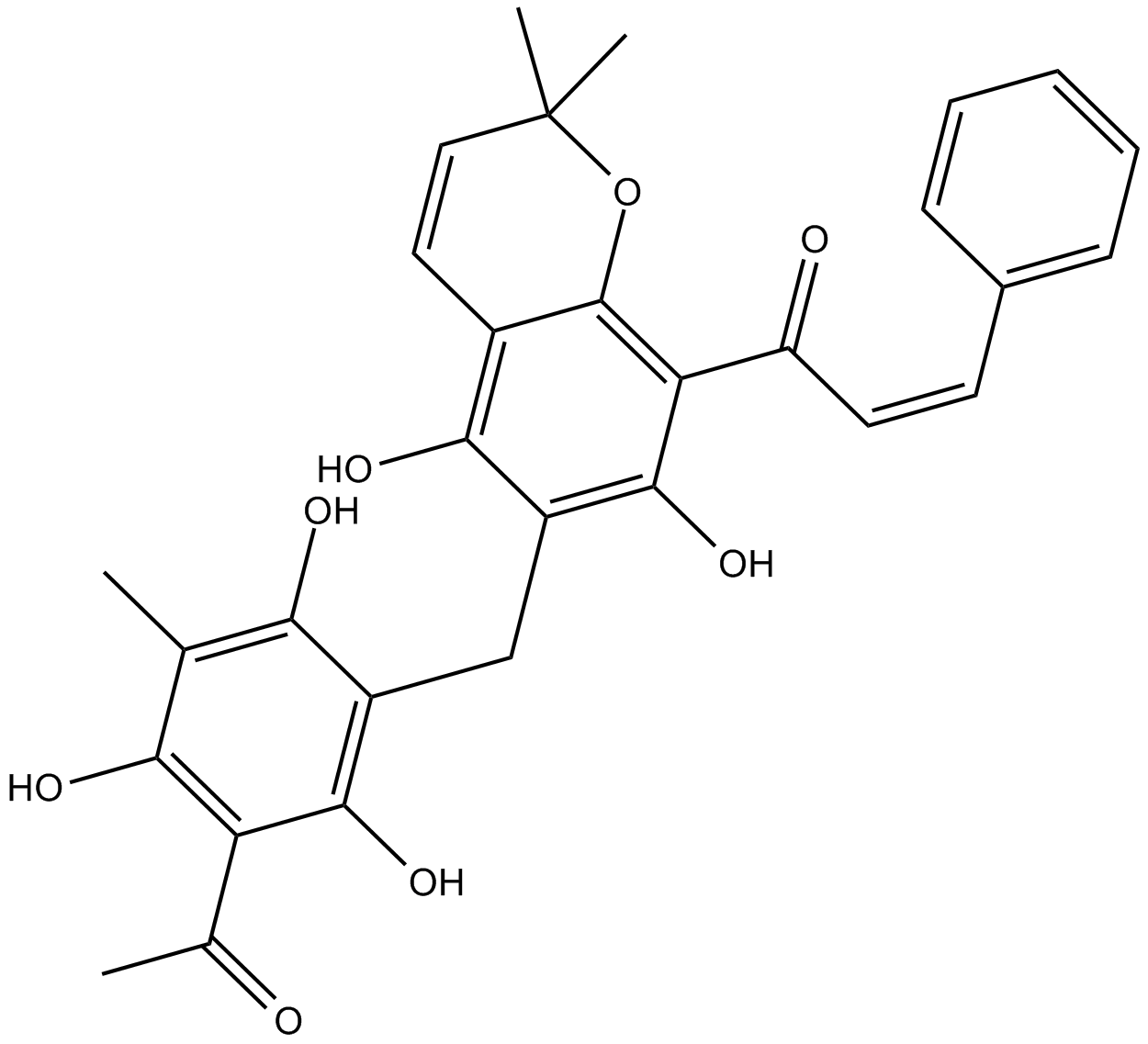
GC10820
Rottlerin
PKC inhibitor
-
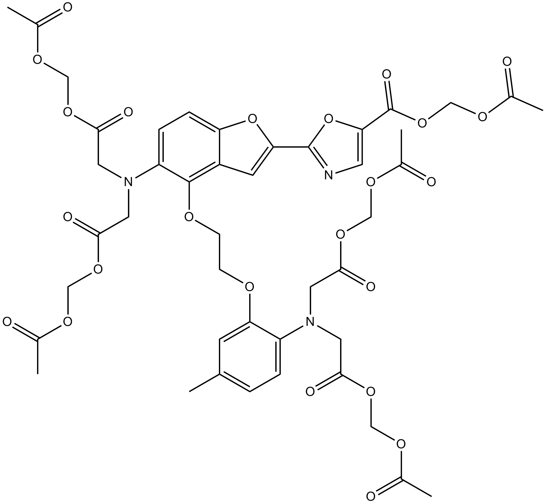
GC16578
Fura-2 AM
Fluorescent Ca2+ indicator
-
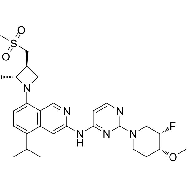
GC63910
BLU-945
Le BLU-945 est un inhibiteur de la tyrosine kinase (TKI) du récepteur du facteur de croissance épidermique (EGFR) puissant, hautement sélectif, réversible et actif par voie orale. Le BLU-945 peut inhiber efficacement l'EGFR avec la mutation par délétion L858R et/ou exon 19, la mutation T790M et la mutation C797S. Le BLU-945 peut être utilisé pour la recherche sur le cancer du poumon, y compris le cancer du poumon non À petites cellules (NSCLC).
-
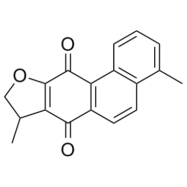
GC34101
Dihydroisotanshinone I
-
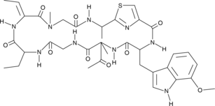
GC45598
Zelkovamycin
An antibiotic
-
GC19097
CHOLESTEROL OXIDASE
10-20u/mg

-
GF03610
RIP2 Kinase Inhibitor 4

-
GD20346
Perfluoro-n-pentane

-
GB60160
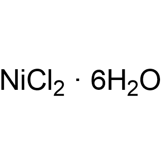
Nickel chloride hexahydrate
-
GC19846
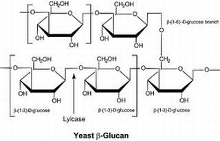
Lysing Enzymes from Arthrobacter luteus
Qualité biotechnologique, 200u/mg
-
GC44961
Succinyl-Coenzyme A (sodium salt)
An intermediate in the citric acid cycle

-
GD08530
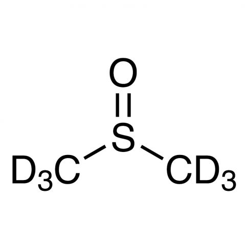
DMSO-d6
-
GC68486
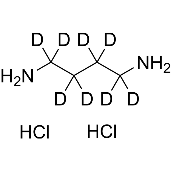
1,4-Diaminobutane-d8 dihydrochloride
1,4-Diaminobutane-d8 (dihydrochloride) est le déutérium substitué de 1,4-Diaminobutane dihydrochloride. Le dihydrochlorure de 1,4-diaminobutane (putrescine) est un métabolite endogène qui peut servir d'indicateur de la pollution causée par le stress au Cr (III) ou Cr (VI) chez les plantes supérieures telles que l'orge et la colza.
-
GC26344
Nuclear and Cytoplasmic Protein Extraction Kit
Nuclear and Cytoplasmic Protein Extraction Kit provides a simple and convenient method for extracting nuclear and cytoplasmic proteins from cultured cells or fresh tissues.

-
GC32199
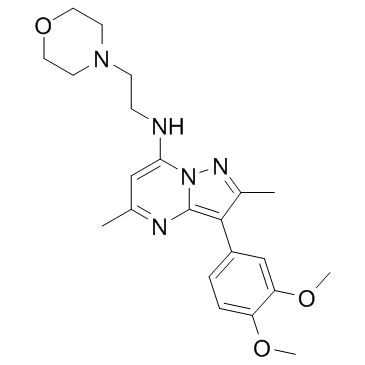
T-00127_HEV1
T-00127_HEV1 est un inhibiteur de la phosphatidylinositol 4-kinase III bêta (PI4KB) avec une IC50 de 60 nM.
-
GC30495
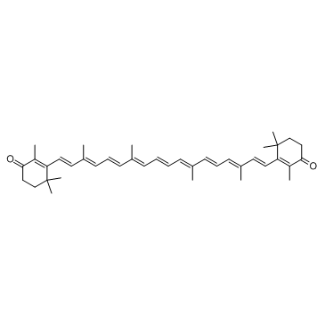
Canthaxanthin (E 161g)
La canthaxanthine (E 161g) est un caroténoÏde rouge-orange avec diverses activités biologiques, telles que des propriétés antioxydantes et antitumorales.
-
GD05924
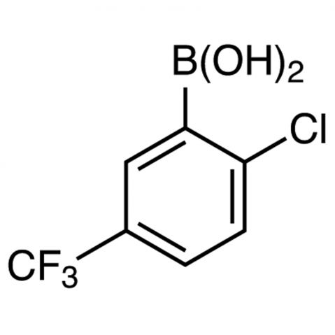
2-Chloro-5-(trifluoromethyl)benzeneboronic acid
- GlpBio - Master of Small Molecules | Compounds - Peptides - Kits.
- Se connecter | S'enregistrer
- sales@glpbio.com
- (909) 407-4943
-
LangueFR - Français
-
Research Areas
-
Signaling Pathways
- Proteases
- Apoptosis
- Chromatin/Epigenetics
- Metabolism
- MAPK Signaling
- Tyrosine Kinase
- DNA Damage/DNA Repair
- PI3K/Akt/mTOR Signaling
- Microbiology & Virology
- Cell Cycle/Checkpoint
- Ubiquitination/ Proteasome
- JAK/STAT Signaling
- TGF-β / Smad Signaling
- Angiogenesis
- GPCR/G protein
- Stem Cell
- Cancer Biology
- Endocrinology and Hormones
- Immunology/Inflammation
- Neuroscience
- Membrane Transporter/Ion Channel
- Other Signal Transduction
- Product Type
-
Signaling Pathways
- Screening Library
- Kits
- Outils
- Ordering
- About us
- Contact us
- Blog


