

FRAX1036 |
Catalog No.GC10251
 One-Click Copy Product Info
|

p21-activated kinase I (PAK1) inhibitor
Products are for research use only. Not for human use. We do not sell to patients.
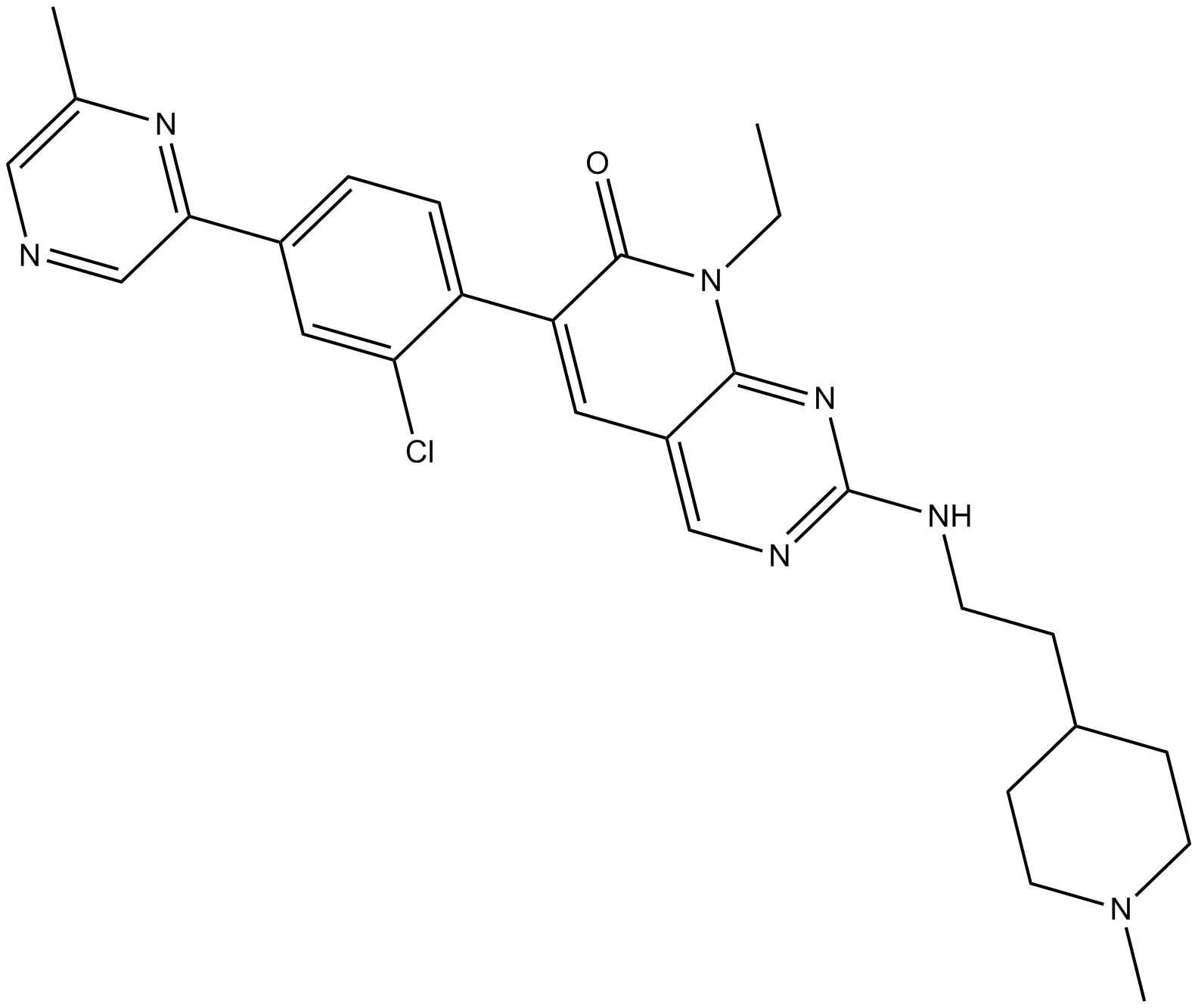
Cas No.: 1432908-05-8
Sample solution is provided at 25 µL, 10mM.
_1-3.$$$39978408@51.jpg');)
_1-9.$$$38538795@65.jpg');)
_1-9.$$$39112701@51.jpg');)
_1-7.$$$37316672@70.jpg');)
_366-372.$$$36198801@70.jpg');)
.$$$38565142@65.jpg');)
_1867-1883.$$$33248023@67.jpg');)
_eads6055.$$$39977546@45.jpg');)
_eadm9805.$$$40080571@45.jpg');)
_eadj3020.$$$39666821@45.jpg');)
_1-20.$$$39757301@28.jpg');)
_1-24.$$$38898113@28.jpg');)
_273-287.$$$36806353@46.jpg');)
_1-16.$$$37156877@44.jpg');)
_1-19.$$$37460805@44.jpg');)
_1291-1307.$$$34518654@46.jpg');)
IC50: N/A
FRAX1036 is a p21-activated kinase I (PAK1) inhibitor.
Breast cancer is a clinically and molecularly heterogeneous disease and plenty of genetic and epigenetic studies of breast tumors has revealed novel putative driver genes, such as p21-activated kinase (PAK)1. As a serine/threonine kinase downstream of small GTP-binding proteins including Rac1 and Cdc42, PAK1 is an critical component of growth factor signaling networks and cellular functions important to tumorigenesis.
In vitro: Previous study demonstrated that the administration of docetaxel with either FRAX1036 or PAK1 small interfering RNA oligonucleotides was able to alter signaling to cytoskeletal-associated proteins dramatically, such as stathmin, and also able to induce microtubule disorganization and cellular apoptosis. In addition, the live-cell imaging data showed that the duration of mitotic arrest mediated by docetaxel could be significantly reduced by the treatment of FRAX1036, which was associated with increased kinetics of apoptosis [1].
In vivo: In previous animal study, the untreated mice bearing KT21 transplants showed ventricular invasion, whereas Ben-Men grew at the injection site. Efficacy results found that the treatment with Frax1036 could lead to a slower tumor growth, with reduction in body mass index (BMI) signals of 37% when compared to vehicle cohort [2].
Clinical trial: Up to now, FRAX1036 is still in the preclinical development stage.
References:
[1] Ong CC et al. Small molecule inhibition of group I p21-activated kinases in breast cancer induces apoptosis and potentiates the activity of microtubule stabilizing agents. Breast Cancer Res.2015 Apr 23;17:59.
[2] Chow HY et al. Group I Paks as therapeutic targets in NF2-deficient meningioma. Oncotarget.2015 Feb 10;6(4):1981-94.
Kinase experiment: | The activity/inhibition of human recombinant PAK1 (kinase domain), PAK2 (full length) or PAK4 (kinase domain) is estimated by measuring the phosphorylation of a FRET peptide substrate (Ser/Thr19) labeled with Coumarin and Fluorescein using Z'-LYTETM assay. The 10 μL assay mixtures containe 50 mM HEPES (pH 7.5), 0.01% Brij-35, 10 mM MgCl2, 1 mM EGTA, 2 μM FRET peptide substrate, and PAK enzyme (20 pM PAK1; 50 pM PAK2; 90 pM PAK4). Incubations are carried out at 22°C in black polypropylene 384-well plates. Prior to the assay, enzyme, FRET peptide substrate and serially diluted test compounds (FRAX1036, etc.) are preincubated together in assay buffer (7.5 μL) for 10 minutes, and the assay is initiated by the addition of 2.5 μL assay buffer containing 4× ATP (160 μM PAK1; 480 μM PAK2; 16 μM PAK4). Following the 60-minute incubation, the assay mixtures are quenched by the addition of 5 μL of Z'-LYTETM development reagent, and 1 hour later the emissions of Coumarin (445 nm) and Fluorescein (520 nm) are determined after excitation at 400 nm. An emission ratio (445 nm/520 nm) is determined to quantify the degree of substrate phosphorylation[1]. |
Cell experiment: | For caspase 3/7 activation apoptosis assays, cells are plated at 10,000 cells/well in 96-well plates for 24 hours prior to treating with DMSO, FRAX1036, and/or docetaxel. Caspase 3/7 reagent is added at a 1:1000 dilution. Cells are imaged at 10× magnification in an IncuCyte Zoom Live-content imaging system at 37°C, 5% CO2. Images are acquired every 2 hours or 4 hours for 36 to 72 hours, two images/well. Data is analyzed using IncuCyte analysis software to detect and quantify green (apoptotic) cells/image. Each condition is performed in triplicate. Averages with SEM at each time point are plotted in Excel. A t-test is performed for the final time point comparing the combination of FRAX1036 and docetaxel with each single agent in Prism. The apoptotic index is calculated from the apoptosis assays by dividing the final apoptotic cell count by the total cell count. Averages with SEM are plotted in Excel, and a t-test is performed comparing the combination of FRAX1036 and docetaxel with each single agent in Prism[1]. |
References: [1]. Ong CC, et al. Small molecule inhibition of group I p21-activated kinases in breast cancer induces apoptosis and potentiates the activity of microtubule stabilizing agents. Breast Cancer Res. 2015 Apr 23;17:59. | |
| Cas No. | 1432908-05-8 | SDF | |
| Chemical Name | 6-(2-chloro-4-(6-methylpyrazin-2-yl)phenyl)-8-ethyl-2-((2-(1-methylpiperidin-4-yl)ethyl)amino)pyrido[2,3-d]pyrimidin-7(8H)-one | ||
| Canonical SMILES | O=C1C(C2=CC=C(C3=NC(C)=CN=C3)C=C2Cl)=CC4=CN=C(NCCC5CCN(C)CC5)N=C4N1CC | ||
| Formula | C28H32ClN7O | M.Wt | 518.05 |
| Solubility | DMF: 1 mg/ml,DMF:PBS (pH 7.2)(1:3): 0.25 mg/ml,Ethanol: 0.33 mg/ml | Storage | Store at -20°C |
| General tips | Please select the appropriate solvent to prepare the stock solution according to the
solubility of the product in different solvents; once the solution is prepared, please store it in
separate packages to avoid product failure caused by repeated freezing and thawing.Storage method
and period of the stock solution: When stored at -80°C, please use it within 6 months; when stored
at -20°C, please use it within 1 month. To increase solubility, heat the tube to 37°C and then oscillate in an ultrasonic bath for some time. |
||
| Shipping Condition | Evaluation sample solution: shipped with blue ice. All other sizes available: with RT, or with Blue Ice upon request. | ||
| Prepare stock solution | |||

|
1 mg | 5 mg | 10 mg |
| 1 mM | 1.9303 mL | 9.6516 mL | 19.3032 mL |
| 5 mM | 386.1 μL | 1.9303 mL | 3.8606 mL |
| 10 mM | 193 μL | 965.2 μL | 1.9303 mL |
Step 1: Enter information below (Recommended: An additional animal making an allowance for loss during the experiment)
 g
μL
g
μL
Step 2: Enter the in vivo formulation (This is only the calculator, not formulation. Please contact us first if there is no in vivo formulation at the solubility Section.)
Calculation results:
Working concentration: mg/ml;
Method for preparing DMSO master liquid: mg drug pre-dissolved in μL DMSO ( Master liquid concentration mg/mL, Please contact us first if the concentration exceeds the DMSO solubility of the batch of drug. )
Method for preparing in vivo formulation: Take μL DMSO master liquid, next addμL PEG300, mix and clarify, next addμL Tween 80, mix and clarify, next add μL saline, mix and clarify.
Method for preparing in vivo formulation: Take μL DMSO master liquid, next add μL Corn oil, mix and clarify.
Note: 1. Please make sure the liquid is clear before adding the next solvent.
2. Be sure to add the solvent(s) in order. You must ensure that the solution obtained, in the previous addition, is a clear solution before proceeding to add the next solvent. Physical methods such as vortex, ultrasound or hot water bath can be used to aid dissolving.
3. All of the above co-solvents are available for purchase on the GlpBio website.
Quality Control & SDS
- View current batch:
- Purity: >99.00% Appearance: A solid
- COA (Certificate of Analysis)
- SDS (Safety Data Sheet)
- Datasheet
Average Rating: 5 (Based on Reviews and 4 reference(s) in Google Scholar.)