

RS 504393 |
Catalog No.GC15489
 One-Click Copy Product Info
|

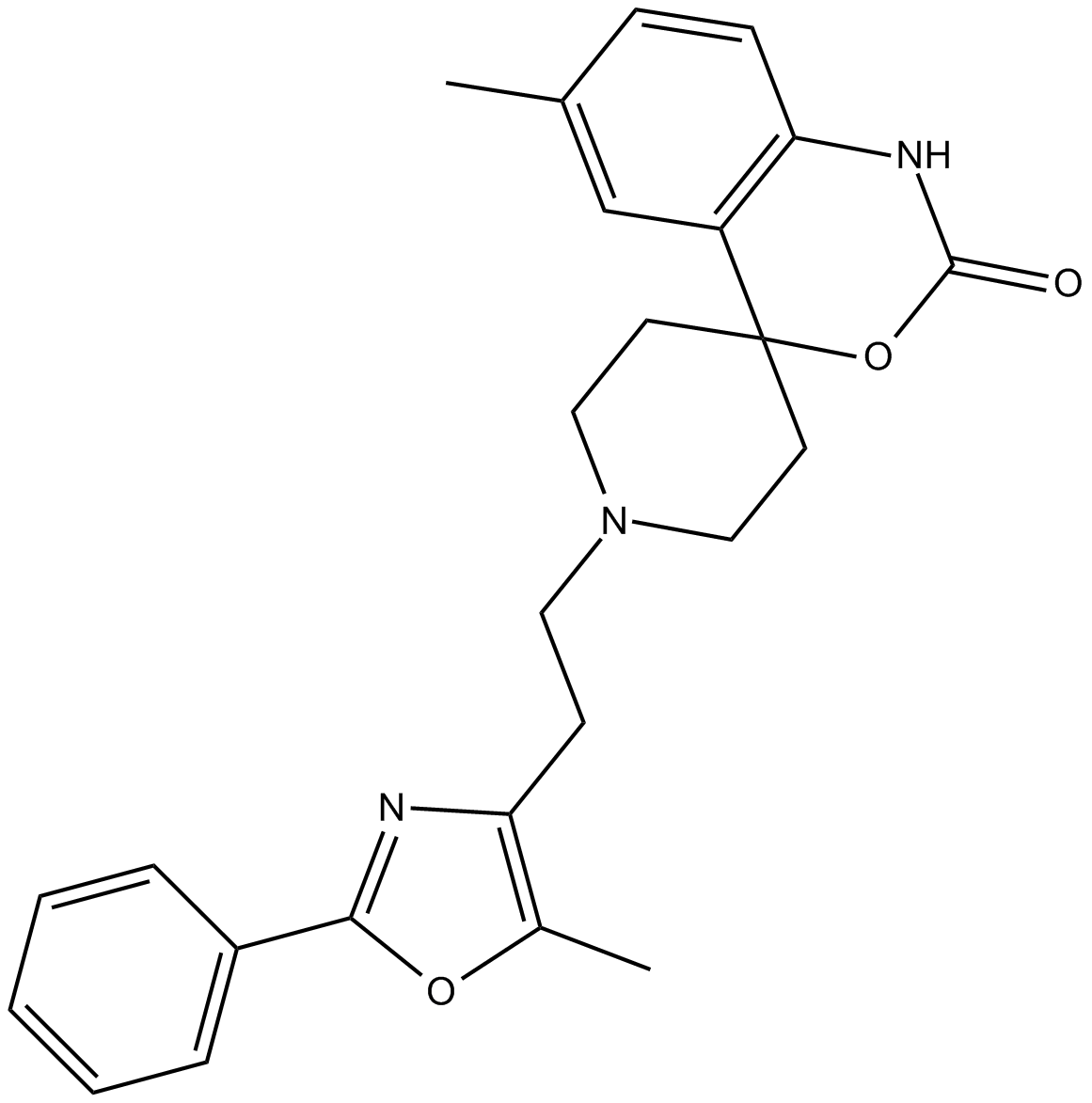
RS 504393 is a selective CCR2 chemokine receptor antagonist, with IC50 values of 89nM and 100μM for human recombinant CCR2 and CCR1 receptors, respectively.
Products are for research use only. Not for human use. We do not sell to patients.
Cas No.: 300816-15-3
Sample solution is provided at 25 µL, 10mM.
_1-3.$$$39978408@51.jpg');)
_1-9.$$$38538795@65.jpg');)
_1-9.$$$39112701@51.jpg');)
_1-7.$$$37316672@70.jpg');)
_366-372.$$$36198801@70.jpg');)
.$$$38565142@65.jpg');)
_1867-1883.$$$33248023@67.jpg');)
_eads6055.$$$39977546@45.jpg');)
_eadm9805.$$$40080571@45.jpg');)
_eadj3020.$$$39666821@45.jpg');)
_1-20.$$$39757301@28.jpg');)
_1-24.$$$38898113@28.jpg');)
_273-287.$$$36806353@46.jpg');)
_1-16.$$$37156877@44.jpg');)
_1-19.$$$37460805@44.jpg');)
_1291-1307.$$$34518654@46.jpg');)
RS 504393 is a selective CCR2 chemokine receptor antagonist, with IC50 values of 89nM and 100μM for human recombinant CCR2 and CCR1 receptors, respectively[1]. CCR2 is a key mediator for inflammatory monocyte activation and migration. RS 504393 can be applied for the treatment of autoimmune arthritis and cancer pain [2].
In vitro, when primary microglia were treated with RS 504393 (10μM) for 1 hour before MCP-1 stimulation, it inhibited microglial activation and reduced the expression of P2X4R in the cultured microglia [3]. Treatment of fibroblast-like synoviocytes (FLS) with RS 504393 (20μM) for 5 minutes inhibited the activation of extracellular signal-regulated kinase 1 (ERK1) and ERK2 in CCL2 culture [4].
In vivo, RS 504393 (4mg/kg/day) was orally administered to mice with osteoarthritis (OA) for 4 weeks, which improved the changes in the cartilage and bones of the mice. RS 504393 could alleviate pain in the early stage of osteoarthritis development, but its therapeutic effect weakened in the later stage of the disease [5]. RS 504393 (4mg/kg/day, 8mg/kg/day) was administered subcutaneously once to mice with bleomycin-induced scleroderma, which reduced dermal fibrosis and dermal thickness in the mice, as well as the number of mast cells and myofibroblasts in the skin and lungs, and the amount of collagen in the skin and lungs. RS 504393 could inhibit pulmonary inflammation and fibrosis caused by bleomycin [6].
References:
[1] Mirzadegan T, et al. Identification of the binding site for a novel class of CCR2b chemokine receptor antagonists: binding to a common chemokine receptor motif within the helical bundle. J Biol Chem. 2000 Aug 18;275(33):25562-71.
[2] Carter P H. Progress in the discovery of CC chemokine receptor 2 antagonists, 2009–2012[J]. Expert opinion on therapeutic patents, 2013, 23(5): 549-568.
[3] Yan Y, Liang Y, Ding T, et al. PI3K/Akt signaling pathway may be involved in MCP-1-induced P2X4R expression in cultured microglia and cancer-induced bone pain rats[J]. Neuroscience letters, 2019, 701: 100-105.
[4] Nanki T, Nagasaka K, Hayashida K, et al. Chemokines regulate IL-6 and IL-8 production by fibroblast-like synoviocytes from patients with rheumatoid arthritis[J]. The Journal of Immunology, 2001, 167(9): 5381-5385.
[5] Longobardi L, Temple J D, Tagliafierro L, et al. Role of the CC chemokine receptor-2 in a murine model of injury-induced osteoarthritis[J]. Osteoarthritis and cartilage, 2017, 25(6): 914-925.
[6] Ishikawa M, Yamamoto T. Antifibrogenic effects of C‐C chemokine receptor type 2 antagonist in a bleomycin‐induced scleroderma model[J]. Experimental Dermatology, 2021, 30(1): 179-184.
| Kinase experiment [1]: | |
|
Preparation Method |
Isolated mast cells were sensitized by incubation with anti-DNP IgE (SPE7; Sigma) in RPMI1640 containing 10ng/mL of murine recombinant IL-3, 10ng/mL of recombinant SCF, and 5% murine serum. The cells were then washed with HBSS containing 10ng/mL of murine recombinant IL-3, 10ng/mL of recombinant SCF, 0.04% BSA, and 10mM HEPES. Transfer the cell suspension to three groups of wells on a 96-well U-shaped bottom plate at a concentration of 2 to 8×10^4 cells/100 microliters, and let it stand at 37°C for 10 minutes. Then, couple it with anti-DNP-IgE antibody first, and then stimulate with DNP-albumin. Add recombinant MCP-1 (1, 10, and 100 pg/mL) and/or RS 504393 (500 nM) simultaneously or at different times.After 45 minutes, the plate was centrifuged at 290g for 5 minutes at 4°C. The β-hexosaminidase activity of the culture supernatant was determined using a published protocol. 13 Fifty-microliter aliquots of the supernatant were placed in wells of another 96-well plate together with 100μL of 2.5mM p-nitrophenyl-N-acetyl β-d glucosaminide (Sigma-Aldrich) solubilized in 0.04M citrate buffer adjusted to pH 4.5 with disodium phosphate. After incubation at 37°C for 90 minutes, the reactions were terminated by addition of 50μL of 0.4M glycin adjusted to pH 10.7 with sodium hydroxide. The colored product was measured at 405nm with a reference filter of 570nm. The relative release of β-hexosaminidase was defined as the activity in the supernatant of the tested cells divided by the activity in the positive control cell supernatant, multiplied by 100. Compound 48/80 stimulus was used for assay control. |
|
Reaction Conditions |
500nM;90min |
|
Applications |
RS 504393 inhibits MCP-1-induced chemotaxis with an IC50 of 330nM. Treatment with RS 504393 significantly inhibits allergen-induced release of β-hexosaminidase. Without the initiation of an allergen, MCP-1 induces degranulation of mast cells, which is completely inhibited by RS 504393. |
| Cell experiment [2]: | |
|
Cell lines |
Microglial cells |
|
Preparation Method |
The spinal microglia cells were isolated from Sprague-Dawley neonatal pups. A mixed glial cell culture was prepared by resuspending the cell suspension in DMEM containing 10% heat-inactivated FBS, 100U/ml penicillin and 0.1mg/ml streptomycin to promote the isolation of microglia from the culture flask. After centrifugation at 800rpm for 8 minutes at 4°C, the cells were resuspended in fresh culture medium for all subsequent experiments. The cells were treated with exogenous recombinant MCP-1 (10, 30 or 100ng/ml) for 30 minutes. The CCR2 antagonist RS 504393 (10μM) and the PI3K inhibitor LY 294002 (10μM) were pre-incubated with the microglia for 20 minutes and 1 hour respectively before MCP-1 (100ng/ml) stimulation, and the Iba1 immunostaining was used for determination. |
|
Reaction Conditions |
10μM; 1h |
|
Applications |
The selective CCR2 antagonist RS 504393 blocked the activation of microglia and reduced the expression of P2X4R in the cultured microglia, as well as inhibiting the expressions of MCP-1 and p-Akt. |
| Animal experiment [3]: | |
|
Animal models |
C57BL/6 |
|
Preparation Method |
Injury-induced-osteoarthritis (OA) was induced in twelve-week-old male C57BL/6 mice (Jackson) by transecting the menisco-tibial ligament. These mice received a continuous 4-week treatment of CCR2 antagonist RS 504393 (4mg/kg/day) dissolved in 0.6% DMSO at different stages of disease progression. The mice underwent pain behavior assessment at 2, 4, 8, and 12 weeks after the DMM surgery and were euthanized. Dissected knees were fixed, μCT scanned, then prepared for histology. Evaluate the condition of cartilage and bone lesions and examine the degeneration of the joints. |
|
Dosage form |
4mg/kg/day for 4 weeks; oral |
|
Applications |
In the early stage of osteoarthritis, the use of the CCR2 antagonist RS 504393 can improve the changes in joint cartilage and bone, and alleviate pain. However, its therapeutic effect is weaker in the later stage of the disease. |
|
References: |
|
| Cas No. | 300816-15-3 | SDF | |
| Chemical Name | 6-methyl-1'-(2-(5-methyl-2-phenyloxazol-4-yl)ethyl)spiro[benzo[d][1,3]oxazine-4,4'-piperidin]-2(1H)-one | ||
| Canonical SMILES | CC1=CC2=C(NC(OC23CCN(CCC4=C(C)OC(C5=CC=CC=C5)=N4)CC3)=O)C=C1 | ||
| Formula | C25H27N3O3 | M.Wt | 417.5 |
| Solubility | DMF: 10 mg/ml,DMF:PBS (pH 7.2) (1:9): 0.1 mg/ml,DMSO: 1 mg/ml,Ethanol: Slightly Soluble | Storage | Store at -20°C |
| General tips | Please select the appropriate solvent to prepare the stock solution according to the
solubility of the product in different solvents; once the solution is prepared, please store it in
separate packages to avoid product failure caused by repeated freezing and thawing.Storage method
and period of the stock solution: When stored at -80°C, please use it within 6 months; when stored
at -20°C, please use it within 1 month. To increase solubility, heat the tube to 37°C and then oscillate in an ultrasonic bath for some time. |
||
| Shipping Condition | Evaluation sample solution: shipped with blue ice. All other sizes available: with RT, or with Blue Ice upon request. | ||
| Prepare stock solution | |||

|
1 mg | 5 mg | 10 mg |
| 1 mM | 2.3952 mL | 11.976 mL | 23.9521 mL |
| 5 mM | 479 μL | 2.3952 mL | 4.7904 mL |
| 10 mM | 239.5 μL | 1.1976 mL | 2.3952 mL |
Step 1: Enter information below (Recommended: An additional animal making an allowance for loss during the experiment)
 g
μL
g
μL
Step 2: Enter the in vivo formulation (This is only the calculator, not formulation. Please contact us first if there is no in vivo formulation at the solubility Section.)
Calculation results:
Working concentration: mg/ml;
Method for preparing DMSO master liquid: mg drug pre-dissolved in μL DMSO ( Master liquid concentration mg/mL, Please contact us first if the concentration exceeds the DMSO solubility of the batch of drug. )
Method for preparing in vivo formulation: Take μL DMSO master liquid, next addμL PEG300, mix and clarify, next addμL Tween 80, mix and clarify, next add μL saline, mix and clarify.
Method for preparing in vivo formulation: Take μL DMSO master liquid, next add μL Corn oil, mix and clarify.
Note: 1. Please make sure the liquid is clear before adding the next solvent.
2. Be sure to add the solvent(s) in order. You must ensure that the solution obtained, in the previous addition, is a clear solution before proceeding to add the next solvent. Physical methods such as vortex, ultrasound or hot water bath can be used to aid dissolving.
3. All of the above co-solvents are available for purchase on the GlpBio website.
Quality Control & SDS
- View current batch:
- Purity: >98.00% Appearance: A solid
- COA (Certificate of Analysis)
- SDS (Safety Data Sheet)
- Datasheet
Average Rating: 5 (Based on Reviews and 30 reference(s) in Google Scholar.)