

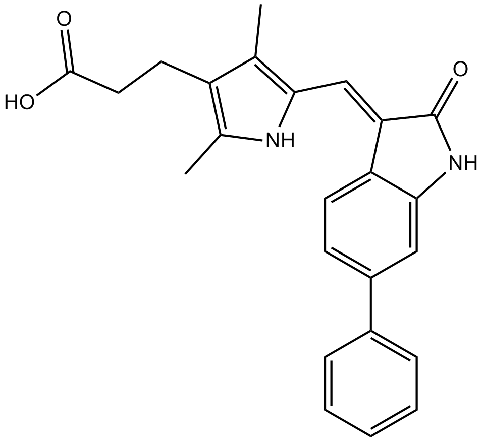
SU 16f (Synonyms: SU16F) |
Catalog No.GC12686
 One-Click Copy Product Info
|

SU 16f is a potent tyrosine kinase inhibitor that inhibits VEGF-R2, FGF-R1, and PDGFRβ, with IC50 values of 0.14, 2.29, and 0.01μM, respectively.
Products are for research use only. Not for human use. We do not sell to patients.
Cas No.: 251356-45-3
Sample solution is provided at 25 µL, 10mM.
_1-3.$$$39978408@51.jpg');)
_1-9.$$$38538795@65.jpg');)
_1-9.$$$39112701@51.jpg');)
_1-7.$$$37316672@70.jpg');)
_366-372.$$$36198801@70.jpg');)
.$$$38565142@65.jpg');)
_1867-1883.$$$33248023@67.jpg');)
_eads6055.$$$39977546@45.jpg');)
_eadm9805.$$$40080571@45.jpg');)
_eadj3020.$$$39666821@45.jpg');)
_1-20.$$$39757301@28.jpg');)
_1-24.$$$38898113@28.jpg');)
_273-287.$$$36806353@46.jpg');)
_1-16.$$$37156877@44.jpg');)
_1-19.$$$37460805@44.jpg');)
_1291-1307.$$$34518654@46.jpg');)
SU 16f is a potent tyrosine kinase inhibitor that inhibits VEGF-R2, FGF-R1, and PDGFRβ, with IC50 values of 0.14, 2.29, and 0.01μM, respectively [1]. SU 16f can inhibit PDGFRβ phosphorylation, resulting in a significant reduction in the phosphorylation of ERK1/2 and STAT3, while the expression of CLDN1 decreased[2]. SU 16f has been widely used as a model compound for molecular docking to analyze the characteristic sites and develop a series of related receptor tyrosine kinase inhibitors[3].
In vitro, SU 16f pretreatment (20μM) for 8 hours significantly inhibited the promoting effect of gastric cancer-derived mesenchymal stem cell (GC-MSC) conditioned medium on the cell proliferation and migration of SGC-7901 cells[4]. SU 16f treatment (5μM; 14 days) eliminated the inhibitory effects of PDGF-BB on BMP2-induced osteogenic differentiation of primary periosteum-derived progenitor cells (PDCs)[5]. Treatment with 10μM SU 16f for 72 hours significantly inhibited the migration of mouse brain vascular pericytes (MBVP) induced by PDGFβ[6].
In vivo, SU 16f administration (2mg/kg/day) via intraperitoneal injection for five consecutive days restored the formation of beige adipose tissue in tamoxifen (TMX)-induced Sma-CreERT2; R26-mTmG male mice exposed to cold environment[7]. Intrathecal injection of a 10μl 3mM SU 16f solution (dissolved in 0.1M PBS containing 3% DMSO) at a rate of 1μl/4s, administered daily for eight consecutive days, blocked the PDGFRβ pathway, inhibited fibrotic scar formation, and promoted axonal regeneration and motor function recovery after spinal cord injury in mice[8].
References:
[1] Sun L, Tran N, Liang C, et al. Design, synthesis, and evaluations of substituted 3-[(3-or 4-carboxyethylpyrrol-2-yl) methylidenyl] indolin-2-ones as inhibitors of VEGF, FGF, and PDGF receptor tyrosine kinases[J]. Journal of medicinal chemistry, 1999, 42(25): 5120-5130.
[2] Pang S, Wu R, Lv W, et al. Use of a pH-responsive imatinib mesylate sustained-release hydrogel for the treatment of tendon adhesion by inhibiting PDGFRβ/CLDN1 pathway[J]. Bioactive Materials, 2024, 38: 124-136.
[3] Kammasud N, Boonyarat C, Tsunoda S, et al. Novel inhibitor for fibroblast growth factor receptor tyrosine kinase[J]. Bioorganic & medicinal chemistry letters, 2007, 17(17): 4812-4818.
[4] Huang F, Wang M, Yang T, et al. Gastric cancer-derived MSC-secreted PDGF-DD promotes gastric cancer progression[J]. Journal of cancer research and clinical oncology, 2014, 140(11): 1835-1848.
[5] Wang X, Matthews B G, Yu J, et al. PDGF modulates BMP2‐induced osteogenesis in periosteal progenitor cells[J]. JBMR plus, 2019, 3(5): e10127.
[6] Li Z, Zheng M, Yu S, et al. M2 macrophages promote PDGFRβ+ pericytes migration after spinal cord injury in mice via PDGFB/PDGFRβ pathway[J]. Frontiers in Pharmacology, 2021, 12: 670813.
[7] Benvie A M, Lee D, Steiner B M, et al. Age-dependent Pdgfrβ signaling drives adipocyte progenitor dysfunction to alter the beige adipogenic niche in male mice[J]. Nature Communications, 2023, 14(1): 1806.
[8] Li Z, Yu S, Liu Y, et al. SU16f inhibits fibrotic scar formation and facilitates axon regeneration and locomotor function recovery after spinal cord injury by blocking the PDGFRβ pathway[J]. Journal of Neuroinflammation, 2022, 19(1): 95.
| Cell experiment [1]: | |
Cell lines | Primary periosteum-derived progenitor cells (PDCs) |
Preparation Method | Primary PDCs were seeded in 12-well plates and cultured in growth medium for 7 days to differentiate into osteoblasts (with 50μg/ml ascorbic acid and 4mM β-glycerophosphate sodium). Starting from the 7th day, recombinant human bone morphogenetic protein 2 (BMP2) and recombinant rat platelet-derived growth factor-BB (PDGF-BB) were added respectively, and the culture was continued for 14 days in the presence or absence of SU 16f (5μM). The growth factor-containing medium was replaced every 2 days, and alkaline phosphatase (ALP) staining was performed. |
Reaction Conditions | 5μM; 14 days |
Applications | SU 16f treatment eliminated the inhibitory effects of PDGF-BB on BMP2-induced osteogenic differentiation of PDCs. |
| Animal experiment [2]: | |
Animal models | Sma-CreERT2; R26-mTmG male mice |
Preparation Method | Sma-CreERT2; R26-mTmG male mice (12 months old) were housed in the animal room, with a light/dark cycle of 14:10 hours, and had free access to food and water. The TMX (50mg/kg) dissolved in sunflower oil was intraperitoneally injected into the mice for two consecutive days, and then the mice were raised at room temperature for 7 days. Subsequently, on each of the following 5 days, the vehicle (5% DMSO) and SU 16f (2mg/kg/day) were injected intraperitoneally, respectively. Then, the mice were placed in a refrigerated box at 6.5°C for 7 days, and the fat tissues of the mice were collected for analysis. |
Dosage form | 2mg/kg/day for 5 days; i.p. |
Applications | SU 16f treatment restored beige fat formation in aged mice exposed to a cold environment. |
References: | |
| Cas No. | 251356-45-3 | SDF | |
| Synonyms | SU16F | ||
| Chemical Name | (E)-3-(2,4-dimethyl-5-((2-oxo-6-phenylindolin-3-ylidene)methyl)-1H-pyrrol-3-yl)propanoic acid | ||
| Canonical SMILES | O=C1NC2=CC(C3=CC=CC=C3)=CC=C2/C1=C\C4=C(C)C(CCC(O)=O)=C(C)N4 | ||
| Formula | C24H22N2O3 | M.Wt | 386.44 |
| Solubility | DMSO: <100mM | Storage | Store at -20°C |
| General tips | Please select the appropriate solvent to prepare the stock solution according to the
solubility of the product in different solvents; once the solution is prepared, please store it in
separate packages to avoid product failure caused by repeated freezing and thawing.Storage method
and period of the stock solution: When stored at -80°C, please use it within 6 months; when stored
at -20°C, please use it within 1 month. To increase solubility, heat the tube to 37°C and then oscillate in an ultrasonic bath for some time. |
||
| Shipping Condition | Evaluation sample solution: shipped with blue ice. All other sizes available: with RT, or with Blue Ice upon request. | ||
| Prepare stock solution | |||

|
1 mg | 5 mg | 10 mg |
| 1 mM | 2.5877 mL | 12.9386 mL | 25.8772 mL |
| 5 mM | 517.5 μL | 2.5877 mL | 5.1754 mL |
| 10 mM | 258.8 μL | 1.2939 mL | 2.5877 mL |
Step 1: Enter information below (Recommended: An additional animal making an allowance for loss during the experiment)
 g
μL
g
μL
Step 2: Enter the in vivo formulation (This is only the calculator, not formulation. Please contact us first if there is no in vivo formulation at the solubility Section.)
Calculation results:
Working concentration: mg/ml;
Method for preparing DMSO master liquid: mg drug pre-dissolved in μL DMSO ( Master liquid concentration mg/mL, Please contact us first if the concentration exceeds the DMSO solubility of the batch of drug. )
Method for preparing in vivo formulation: Take μL DMSO master liquid, next addμL PEG300, mix and clarify, next addμL Tween 80, mix and clarify, next add μL saline, mix and clarify.
Method for preparing in vivo formulation: Take μL DMSO master liquid, next add μL Corn oil, mix and clarify.
Note: 1. Please make sure the liquid is clear before adding the next solvent.
2. Be sure to add the solvent(s) in order. You must ensure that the solution obtained, in the previous addition, is a clear solution before proceeding to add the next solvent. Physical methods such as vortex, ultrasound or hot water bath can be used to aid dissolving.
3. All of the above co-solvents are available for purchase on the GlpBio website.
Quality Control & SDS
- View current batch:
- Purity: >99.50% Appearance: A solid
- COA (Certificate of Analysis)
- SDS (Safety Data Sheet)
- Datasheet
Average Rating: 5 (Based on Reviews and 40 reference(s) in Google Scholar.)