A-443654 |
Catalog No.GC12502 |
A pan Akt inhibitor
Products are for research use only. Not for human use. We do not sell to patients.
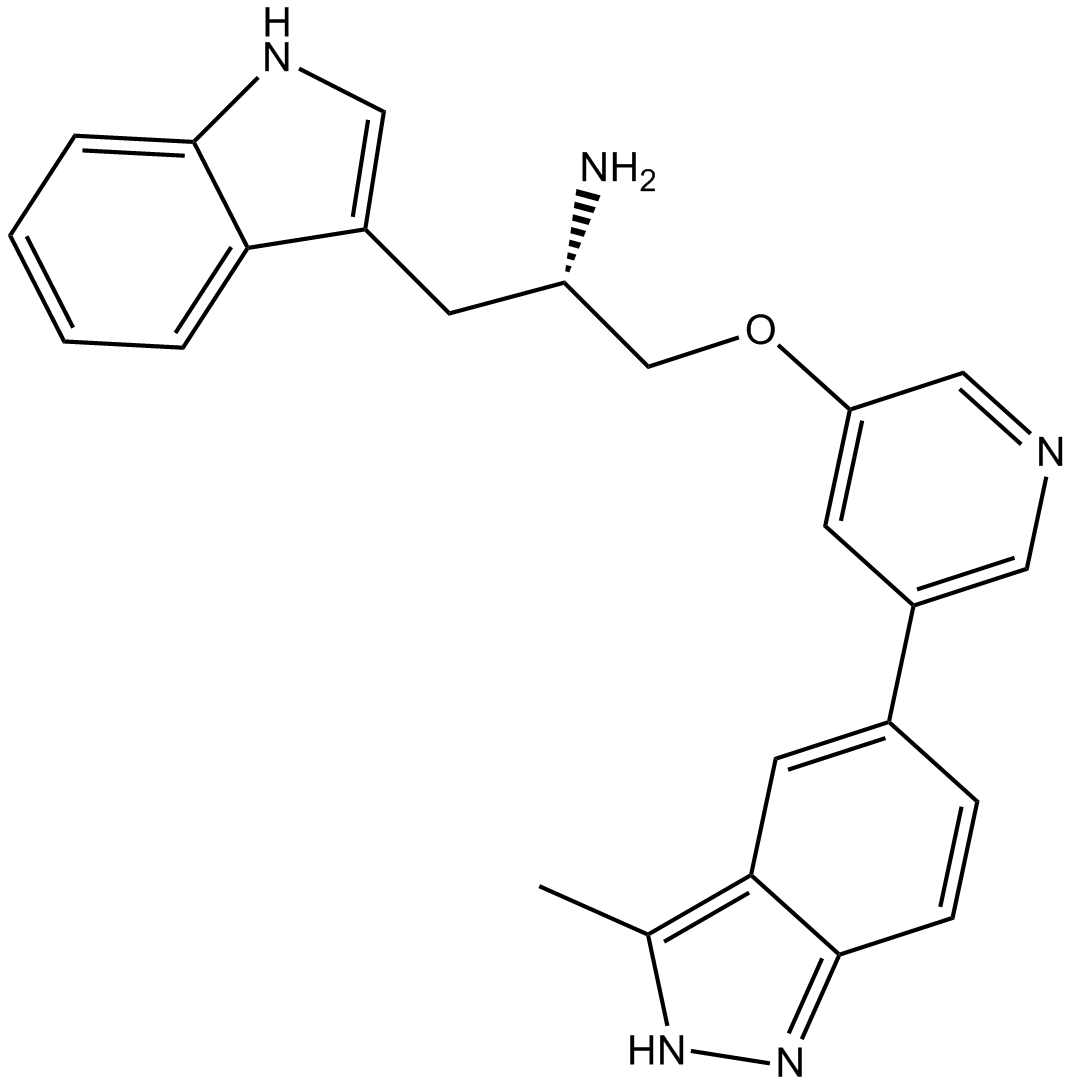
Cas No.: 552325-16-3
Sample solution is provided at 25 µL, 10mM.
A-443654 is a potent and selective inhibitor of Akt with Ki value of 160 pM [1].
The Akt kinases play important roles in cellular signal transduction and take participate in the regulation of cell transformation and tumor progression. The activities of them are usually elevated in human malignancies. A-443654 is a pan inhibitor of Akt1, 2 and 3. It binds to the ATP-binding site of Akt and inhibits the kinase activity reversibly. A-443654 shows selectivity against Akt. The inhibition ability of it for Akt is much higher than that for other kinases. It is found that A-443654 can suppress the phosphorylation of the downstream proteins of Akt while increased the Ser473 and Thr308 phosphorylation of Akt [1].
In murine FL5.12 cells stably transfected with human Akt1, 2 or 3, A-443654 inhibited the phosphorylation of GSK3 dose-dependently. A-443654 at a concentration of 0.6 μM inhibited Akt and induced G2/M accumulation in H1299 cells. In MiaPaCa-2 cells, treatment of A-443654 for 48 h resulted in a suppression of tumor proliferation with EC50 value of 100 nM. In chronic lymphocytic leukemia cells, A-443654 also inhibited the activity of Akt and induced apoptosis with EC50 value of 0.63 μM .Besides that, A-443654 was found to cause decreased phosphorylation of GSK3, FOXO3, TSC2 and mTOR in MiaPaCa-2 cells. It is also reported that A-443654 was effective in blocking the phosphorylation of 4EBP-1 and S6 protein in both T47D and LNCaP cells [1, 2, 3 and 4].
A-443654 is not oral available. In mice model with human MiaPaCa-2 pancreatic cell xenograft, the administration of A-443654 at a dose of 7.5mg/kg/d significantly inhibited tumor growth. A-443654 also inhibited tumor growth in 3T3 murine fibroblast model expressing active Akt. When using the combination of A-443654 and rapamycin in mice model with MiaPaCa-2 pancreatic cancer xenograft, the administration showed more efficacy than each monotherapy [1].
References:
1.Luo Y, Shoemaker A R, Liu X, et al. Potent and selective inhibitors of Akt kinases slow the progress of tumors in vivo. Molecular cancer therapeutics, 2005, 4(6): 977-986.
2.Liu X, Shi Y, Woods K W, et al. Akt inhibitor a-443654 interferes with mitotic progression by regulating aurora a kinase expression. Neoplasia, 2008, 10(8): 828-837.
3.Merce de Frias M, Iglesias-Serret D, Cosialls A M, et al. Akt inhibitors induce apoptosis in chronic lymphocytic leukemia cells. haematologica, 2009, 94(12): 1698-1707.
4.Cherrin C, Haskell K, Howell B, et al. An allosteric Akt inhibitor effectively blocks Akt signaling and tumor growth with only transient effects on glucose and insulin levels in vivo. Cancer biology & therapy, 2010, 9(7): 493-503.
Cell experiment: | The cells on 96-well plates are gently washed with 200 μL of PBS. Alamar Blue reagent is diluted 1:10 in normal growth media. The diluted Alamar Blue reagent (100 μL) is added to each well on the 96-well plates and incubated until the reaction is complete as per manufacturer's instructions. Analysis is done using an fmaxFluorescence Microplate Reader, set at the excitation wavelength of 544 nm and emission wavelength of 595 nm. Data are analyzed using SOFTmax PRO software provided by the manufacturer. |
Animal experiment: | Immunocompromised male scid mice are 6 to 8 weeks of age. The 3T3-Akt1 cell line is developed and characterized in our laboratory. The 1×10⁶ 3T3-Akt1 or 2×10⁶ MiaPaCa-2 and PC-3 cells in 50% Matrigel are inoculated s.c. into the flank. For early treatment studies, mice are randomLy assigned to treatment groups and therapy is initiated the day after inoculation. Ten animals are assigned to each group, including controls. For established tumor studies, tumors are allowed to reach a designated size and mice are assigned to treatment groups of equal tumor size (n=10 mice per group). Tumor size is evaluated by twice weekly measurements with digital calipers. Tumor volume is estimated using the formula: V=L×W2/2. A-443654 is given s.c. in a vehicle of 0.2% HPMC. A-674563 is given orally in a vehicle of 5% dextrose. |
References: [1]. Luo Y, et al. Potent and selective inhibitors of Akt kinases slow the progress of tumors in vivo. Mol Cancer Ther. 2005 Jun;4(6):977-86. |
Cas No. | 552325-16-3 | SDF | |
Chemical Name | (2S)-1-(1H-indol-3-yl)-3-[5-(3-methyl-2H-indazol-5-yl)pyridin-3-yl]oxypropan-2-amine | ||
Canonical SMILES | CC1=C2C=C(C=CC2=NN1)C3=CC(=CN=C3)OCC(CC4=CNC5=CC=CC=C54)N | ||
Formula | C24H23N5O | M.Wt | 397.48 |
Solubility | ≥ 19.85 mg/mL in DMSO, ≥ 44.3 mg/mL in EtOH | Storage | Store at -20°C |
General tips | Please select the appropriate solvent to prepare the stock solution according to the
solubility of the product in different solvents; once the solution is prepared, please store it in
separate packages to avoid product failure caused by repeated freezing and thawing.Storage method
and period of the stock solution: When stored at -80°C, please use it within 6 months; when stored
at -20°C, please use it within 1 month. To increase solubility, heat the tube to 37°C and then oscillate in an ultrasonic bath for some time. |
||
Shipping Condition | Evaluation sample solution: shipped with blue ice. All other sizes available: with RT, or with Blue Ice upon request. |
Prepare stock solution | |||
![]() |
1 mg | 5 mg | 10 mg |
1 mM | 2.5158 mL | 12.5792 mL | 25.1585 mL |
5 mM | 0.5032 mL | 2.5158 mL | 5.0317 mL |
10 mM | 0.2516 mL | 1.2579 mL | 2.5158 mL |
Step 1: Enter information below (Recommended: An additional animal making an allowance for loss during the experiment)
Step 2: Enter the in vivo formulation (This is only the calculator, not formulation. Please contact us first if there is no in vivo formulation at the solubility Section.)
Calculation results:
Working concentration: mg/ml;
Method for preparing DMSO master liquid: mg drug pre-dissolved in μL DMSO ( Master liquid concentration mg/mL, Please contact us first if the concentration exceeds the DMSO solubility of the batch of drug. )
Method for preparing in vivo formulation: Take μL DMSO master liquid, next addμL PEG300, mix and clarify, next addμL Tween 80, mix and clarify, next add μL ddH2O, mix and clarify.
Method for preparing in vivo formulation: Take μL DMSO master liquid, next add μL Corn oil, mix and clarify.
Note: 1. Please make sure the liquid is clear before adding the next solvent.
2. Be sure to add the solvent(s) in order. You must ensure that the solution obtained, in the previous addition, is a clear solution before proceeding to add the next solvent. Physical methods such as vortex, ultrasound or hot water bath can be used to aid dissolving.
3. All of the above co-solvents are available for purchase on the GlpBio website.
Quality Control & SDS
- View current batch:
- Purity: >99.00%
- COA (Certificate Of Analysis)
- SDS (Safety Data Sheet)
- Datasheet
Average Rating: 5
(Based on Reviews and 30 reference(s) in Google Scholar.)GLPBIO products are for RESEARCH USE ONLY. Please make sure your review or question is research based.
Required fields are marked with *