


3-TYP
3-TYP that is also known as 3-(1H-1,2,3-triazol-4-yl) pyridine, is a small molecule. Its molecular structure is presented in Figure 1. It effectively and selectively modulates i.e., inhibits the Sirt3 protein, a mitochondrial enzyme having roles in the regulation of a vast spectrum of the metabolic processes (a collective term for catabolism and anabolism) thus contributes to the body’s overall homeostasis. Sirt3 protein recovers the cells from the oxidative stress, which is essentially the imbalance in the concentration of free radicals (oxidative in nature) and antioxidants, primarily due to the imbalance in the oxidation reduction (Redox) reactions taking place in the cell and its micro environment. Thus, it maintains the homeostasis at a cell level. Along with that Sirt3 protein also plays regulatory role in the normal physiological processes like aging, suppressing the production of tumors and repairing of DNA.
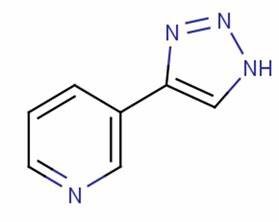
Figure 1 The Structural Formula of 3-TYP is depicted. Another name of which is 3-(1H-1,2,3-triazol-4-yl) pyridine.
As Sirt3 protein is a deacetylase, which means it catalyses the removal of acetyl group, (also called as deacetylation) from the substrates. This catalysis is one of the necessary post transcriptional modifications (critical to the normal functionality of protein/ substrates) which is nicotinamide adenine dinucleotide (NAD+) dependent in nature. It has been shown by a research study that almost 84 proteins are functionally regulated by Sirt3 protein in the mitochondria. The inhibition of Sirt3 protein, is characterized by hyperacetylation that is a condition in which acetyl group remains attached to the substrates, and thus rendering the protein substrates, primarily histones, inactive, which has their protective roles in the condition of oxidative stress. Hyperacetylation also deprives numerous other protein of their respective roles.
In addition, Sirt3 protein also induces the adipogenesis i.e., differentiation of the adipocytes. In a research study, 3-TYP has found to retards the process of adipogenesis in the 3T3-L1 by significantly downregulating the expression of adipocyte marker genes, i.e., peroxisome proliferator-activated receptor gamma (PPARγ) and CCAAT/enhancer binding protein (C/EBPα) as graphically presented in Figure 1. Moreover, it also significantly downregulates the expression of lipolysis marker genes, i.e., adipose triglyceride lipase (ATGL), and hormone-sensitive lipase (HSL), as well as glucose transport (GLUT4) as graphically presented in Figure 2. Furthermore, it also significantly upregulates the genes involved in the synthesis of specialized cytokines by the adipocytes, which are also known as adipokines. These upregulated specialized cytokines include Interleukin 6 (IL 6), Resistin and Tumor Necrosis Factor alpha (TNF-α), as graphically presented in Figure 3. In another research study, high levels of Interleukin 6 (IL 6) and Tumor Necrosis Factor alpha (TNF-α) have been found to be tremendously increased due to the presence of 3-TYP (Shi et al., 2022).
Along with that, it has also been independently involved in the inhibition in the intake of the glucose and free fatty acids in the 3T3-L1 cells as graphically indicated in Figure 4 and Figure 5, respectively. 3-TYP has particularly and majorly been used in designing an experimental control, in the research studies which aimed at highlighting the roles of Sirt3 protein (Lee et al., 2022).
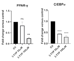
Figure 2 The significantly down regulated expression of the adipocyte marker genes, i.e., peroxisome proliferator-activated receptor gamma (PPARγ) and CCAAT/enhancer binding protein (C/EBPα), by 3-TYP as compared to the control (black bars) especially at the 100 micro-Molar concentration.
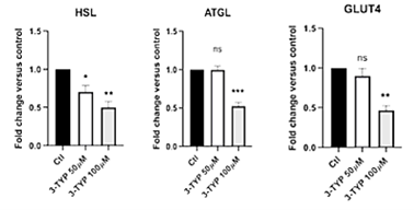
Figure 3 The Significantly down regulated expression of lipolysis marker genes, i.e., adipose triglyceride lipase (ATGL), and hormone-sensitive lipase (HSL), as well as glucose transport (GLUT4), by 3-TYP as compared to the control (black bars) especially at the 100 micro Molar concentration.
Figure 4 The Significantly up regulated expression of adipokines, i.e., Interleukin 6 (IL 6), Resistin and Tumor Necrosis Factor alpha (TNF-α), by 3-TYP as compared to the control (black bars)
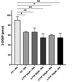
Figure 5 The significantly inhibited uptake of Glucose in the Adipocytes by 3-TYP as compared to the control (white bar graph)
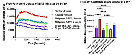
Figure 6 significantly inhibited uptake of Free Fatty Acids in the Adipocytes by 3-TYP as compared to the control (purple and red bars in the graph, without and with insulin, respectively)
In a research study on murine model having thioacetamide (TAA) induced Acute Liver Failure (ALF), the negative effects on the liver by 3-TYP are highlighted. The most prominent and statistically significant negative effects were being the extensive level of necrosis (also called as cell murder, a type of cell death) of the hepatocytes/ liver cells, the abnormal abundance of inflammatory cells (causing inflammation) and the two liver enzymes, i.e., Aspartate aminotransferase (AST) and alanine aminotransferase (ALT). The latter is being graphically presented in Figure 6. In the same study, the level of liver necrosis was so extensive that the survival rates were dropped to the half as compared to the control, this is graphically shown in Figure 7.
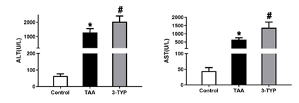
Figure 7 The statistically significant abnormal abundance of two liver enzymes, i.e., Aspartate aminotransferase (AST) and alanine aminotransferase (ALT) as compared with their respective experimental controls (white bars in both of the graphs), that is caused by 3-TYP in the Acute Liver Failure (ALF) model of murine.
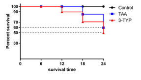
Figure 8 The statistically significantly lowered survival rate, approximately to the 50% caused by 3-TYP when compared to the experimental control (black line in the graph) in the murine based study model of Acute Liver Failure (ALF)
High level of expression of the proteins involved in the inflammatory pathways, especially MAPK pathway and NF-κB pathway, i.e., p-P38, p-ERK1/2, and p-JNK, is caused by 3-TYP in the Acute Liver Failure (ALF) murine model, as graphically presented in Figure 8. This indicates the 3-TYP particularly promotes these pathways which cause inflammation.
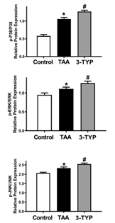
Figure 9 The statistically significant high level expression of protein, involved in the inflammatory pathways, namely, p-P38, p-ERK1/2, and p-JNK caused by 3-TYP, as compared to the experimental control (white bar in the graph).
Considering the most of literature so far, 3-TYP may not have any significant and important direct therapeutic applications as it worse the pathological conditions involving imbalanced glucose and fatty acids metabolism, inflammation and necrosis, particularly in liver. But it has been extensively used in the research studies to design an effective control to study the role of Sirt3, which may have several potential therapeutic applications especially in the treatment of those disease, where some kind of metabolic imbalance, inflammation, degeneration and cell death is involved.
In a recent research study, 3-TYP has been used to inhibit Sirt3, accompanied by the application of resveratrol (Res). In this application, the apoptotic role of Res on the cancer study model of human ovarian cells (SKOV3 cells) is found to be significantly enhanced by the Sirt3 inhibiting activity of 3-TYP. This study also highlights the secondary or supportive role of 3-TYP as an indirect enhancer in the therapeutic application against human ovarian cancer (LI, 2020).
In another recent research study regarding the prosthetic bone implant, the most frequently problem regarding the bone implants has been addressed, that is the osteolysis (the dissolution of bone) around the implant by the activity of osteoclasts (bone dissolving cells). In that study, 3-TYP induced Sirt3 inhibition is found to be effective in the inhibition of the production of osteoclasts, in a dose independent manner (Li et al., 2021).
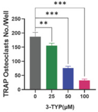
Figure 10 The number of Osteoclasts is significantly reduced. And, it is gradually reduced as a function of concentration, while it is drastically reduced at the higher concentrations


Comments