

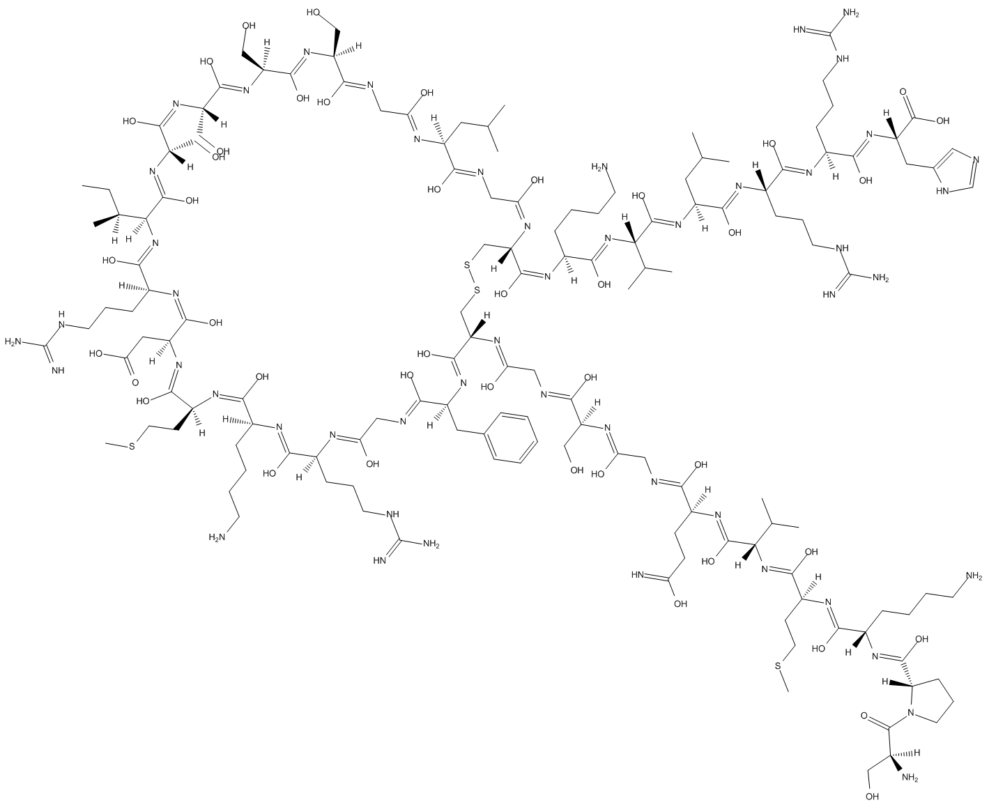
Brain natriuretic peptide (1-32) (human) |
Catalog No.GC17895
 One-Click Copy Product Info
|

Brain natriuretic peptide is a cardiac neurohormone that is secreted by the left ventricle in response to an increase in wall stress.
Products are for research use only. Not for human use. We do not sell to patients.
Cas No.: 124584-08-3
Sample solution is provided at 25 µL, 10mM.
_1-3.$$$39978408@51.jpg');)
_1-9.$$$38538795@65.jpg');)
_1-9.$$$39112701@51.jpg');)
_1-7.$$$37316672@70.jpg');)
_366-372.$$$36198801@70.jpg');)
.$$$38565142@65.jpg');)
_1867-1883.$$$33248023@67.jpg');)
_eads6055.$$$39977546@45.jpg');)
_eadm9805.$$$40080571@45.jpg');)
_eadj3020.$$$39666821@45.jpg');)
_1-20.$$$39757301@28.jpg');)
_1-24.$$$38898113@28.jpg');)
_273-287.$$$36806353@46.jpg');)
_1-16.$$$37156877@44.jpg');)
_1-19.$$$37460805@44.jpg');)
_1291-1307.$$$34518654@46.jpg');)
Brain natriuretic peptide (1-32) (human) is a cardiac hormone produced in the heart, which plays an important physiologic role in cardiorenal homeostasis[1]. Brain natriuretic peptide binds to the natriuretic peptide type A receptor (NPR-A), and via the second messenger cGMP mediates a variety of actions including vasodilation, natriuresis, suppression of renin secretion, lusitropism, and inhibition of fibrosis[2]. Brain natriuretic peptide has been widely used as an indicator of increased ventricular mass and a biomarker of heart failure[3].
In vitro, The treatment with brain natriuretic peptide (1nM) for 3 hours significantly induced the production of reactive oxygen species (ROS) and NO2 in THP-1 cells[4]. The treatment with 100nM Brain natriuretic peptide for 24 hours significantly increased the level of β-catenin in SHSY5Y cells and inhibited the cell death induced by 6-hydroxydopamine[5]. Treatment with 0.8ng/ml of Brain natriuretic peptide for 24 hours significantly reduced the levels of IL-6, TNF-α, and IL-10 in Lipopolysaccharide (LPS)-induced RAW 264.7 cells[6].
In vivo, Brain natriuretic peptide treatment via subcutaneous injection at a dose of 625ng/kg/h for 12 weeks significantly improved the metabolic status and cardiac function of obese diabetic db/db mice, and reduced blood sugar and fat content[7]. Intravenous injection of Brain natriuretic peptide at a dose of 8µg/kg at 30 minutes and 4 hours after pneumatic-induced closed traumatic brain injury (TBI) significantly improved neurological function and alleviated inflammatory responses in mice[8].
References:
[1] Kuwahara K, Nakagawa Y, Nishikimi T. Cutting Edge of Brain Natriuretic Peptide (BNP) Research―The Diversity of BNP Immunoreactivity and Its Clinical Relevance―[J]. Circulation Journal, 2018, 82(10): 2455-2461.
[2] Boerrigter G, Costello-Boerrigter L C, Harty G J, et al. Des-serine-proline brain natriuretic peptide 3–32 in cardiorenal regulation[J]. American journal of physiology-Regulatory, integrative and comparative physiology, 2007, 292(2): R897-R901.
[3] Calzetta L, Orlandi A, Page C, et al. Brain natriuretic peptide: much more than a biomarker[J]. International journal of cardiology, 2016, 221: 1031-1038.
[4] Chiurchiu V, Izzi V, D'Aquilio F, et al. Brain Natriuretic Peptide (BNP) regulates the production of inflammatory mediators in human THP-1 macrophages[J]. Regulatory peptides, 2008, 148(1-3): 26-32.
[5] Giovannini D, Andreola F, Spitalieri P, et al. Natriuretic peptides are neuroprotective on in vitro models of PD and promote dopaminergic differentiation of hiPSCs-derived neurons via the Wnt/β-catenin signaling[J]. Cell death discovery, 2021, 7(1): 330.
[6] Li X, Peng H, Wu J, et al. Brain natriuretic peptide-regulated expression of inflammatory cytokines in lipopolysaccharide (LPS)-activated macrophages via NF-κB and mitogen activated protein kinase (MAPK) pathways[J]. Medical science monitor: international medical journal of experimental and clinical research, 2018, 24: 3119.
[7] Plante E, Menaouar A, Danalache B A, et al. Treatment with brain natriuretic peptide prevents the development of cardiac dysfunction in obese diabetic db/db mice[J]. Diabetologia, 2014, 57(6): 1257-1267.
[8] James M L, Wang H, Venkatraman T, et al. Brain natriuretic peptide improves long-term functional recovery after acute CNS injury in mice[J]. Journal of neurotrauma, 2010, 27(1): 217-228.
| Cell experiment [1]: | |
Cell lines | THP-1 cells |
Preparation Method | THP-1 cells were cultured in RPMI-1640 medium supplemented with 10% fetal bovine serum, 100U/ml penicillin, and 100μg/ml streptomycin at 37°C with 5% CO2 and 95% saturated atmospheric humidity. Cells (1×10⁶ cells/well) were incubated in the dark with 10μM 2′,7′-dichlorofluorescein diacetate (DCF-DA) in serum-free medium for 30min at 37°C. After the incubation with the fluorescent dye, cells were then harvested, and the medium containing the unincorporated dye was eliminated by centrifugation. Cells were then stimulated in serum-free medium with Brain natriuretic peptide (1nM) for 3h. At end timepoint, cells were harvested, resuspended in PBS, and assessed for changes in fluorescence. |
Reaction Conditions | 1nM; 3h |
Applications | Brain natriuretic peptide treatment significantly promoted ROS production in THP-1 cells. |
| Animal experiment [2]: | |
Animal models | Male db/db mice of the C57BL/KsJ-leptdb-leptdb strain |
Preparation Method | Male db/db mice (10 weeks old) were housed under standard laboratory conditions on a 12-hour light/dark cycle (lights on at 7:00 am), constant temperature (23±2°C), and constant humidity (60±5), ad libitum food and water. Mice were randomly assigned (n =10/group) to receive chronic Brain natriuretic peptide treatment at a rate of 625ng/kg/h, or saline vehicle (veh). Treatment was started in 10-week-old mice and continued for 12 weeks using subcutaneous osmotic pumps. Analyze the blood sugar levels and body weight of mice. |
Dosage form | 625ng/kg/h; 12 weeks; s.c. |
Applications | Brain natriuretic peptide treatment reduced blood sugar and body weight in db/db mice. |
References: | |
| Cas No. | 124584-08-3 | SDF | |
| Chemical Name | (1E,3S,4Z,6S,7Z,9S,10Z,12S,13Z,15S,16Z,18S)-18-((1H-imidazol-5-yl)methyl)-1-((4R,5E,8Z,10S,11Z,14Z,16S,17Z,19S,20Z,22S,23E,25S,26Z,28S,29Z,31S,32E,34S,35Z,37S,38E,40S,41Z,43S,44Z,47Z,49S,50Z,52R)-52-((Z)-((1Z,3S,4Z,6S,7Z,9S,10Z,12S,13Z,16Z,18S,19Z)-1-((S) | ||
| Canonical SMILES | CC[C@]([C@@]1([H])/C(O)=N/[C@@](/C(O)=N\[C@@](/C(O)=N/[C@@](/C(O)=N/[C@@](/C(O)=N/C/C(O)=N/[C@@](/C(O)=N/C/C(O)=N\[C@@](/C(O)=N\[C@@](/C(O)=N/[C@@](/C(O)=N/[C@@](/C(O)=N/[C@@](/C(O)=N/[C@@](/C(O)=N/[C@@](C(O)=O)([H])CC2=CN=CN2)([H])CCCNC(N)=N)([H])CCCNC(N | ||
| Formula | C143H244N50O42S4 | M.Wt | 3464.05 |
| Solubility | Soluble to 24mg/ml in sterile water | Storage | -20°C, protect from light |
| Shipping Condition | Evaluation sample solution: shipped with blue ice. All other sizes available: with RT, or with Blue Ice upon request. | ||
| Prepare stock solution | |||

|
1 mg | 5 mg | 10 mg |
| 1 mM | 288.7 μL | 1.4434 mL | 2.8868 mL |
| 5 mM | 57.7 μL | 288.7 μL | 577.4 μL |
| 10 mM | 28.9 μL | 144.3 μL | 288.7 μL |
Step 1: Enter information below (Recommended: An additional animal making an allowance for loss during the experiment)
 g
μL
g
μL
Step 2: Enter the in vivo formulation (This is only the calculator, not formulation. Please contact us first if there is no in vivo formulation at the solubility Section.)
Calculation results:
Working concentration: mg/ml;
Method for preparing DMSO master liquid: mg drug pre-dissolved in μL DMSO ( Master liquid concentration mg/mL, Please contact us first if the concentration exceeds the DMSO solubility of the batch of drug. )
Method for preparing in vivo formulation: Take μL DMSO master liquid, next addμL PEG300, mix and clarify, next addμL Tween 80, mix and clarify, next add μL saline, mix and clarify.
Method for preparing in vivo formulation: Take μL DMSO master liquid, next add μL Corn oil, mix and clarify.
Note: 1. Please make sure the liquid is clear before adding the next solvent.
2. Be sure to add the solvent(s) in order. You must ensure that the solution obtained, in the previous addition, is a clear solution before proceeding to add the next solvent. Physical methods such as vortex, ultrasound or hot water bath can be used to aid dissolving.
3. All of the above co-solvents are available for purchase on the GlpBio website.
Quality Control & SDS
- View current batch:
- Purity: >98.00% Appearance: A solid
- COA (Certificate of Analysis)
- SDS (Safety Data Sheet)
- Datasheet
Average Rating: 5 (Based on Reviews and 6 reference(s) in Google Scholar.)